

Napsin A as a marker of clear cell ovarian carcinoma
- PMID: 24191930
- PMCID: PMC4228360
- DOI: 10.1186/1471-2407-13-524
Napsin A as a marker of clear cell ovarian carcinoma
Abstract
Background: Clear cell carcinomas are aggressive tumors with a distinct biologic behaviour. In a genome-wide screening for genes involved in chemo-resistance, NAPA was over-expressed in cisplatin-resistant cells. The NAPA (protein) Napsin A was described to promote resistance to cisplatin by degradation of the tumor suppressor p53.
Methods: Totally 131 patients were included in this study all in FIGO-stages I-II; 16 were clear cell tumors which were compared with 40 Type I tumors and 75 type II tumors according to the markers Napsin A, p21, p53 and p27 and some clinical features. For detection of the markers tissue microarrays and immunohistochemistry were used.
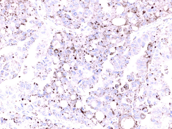
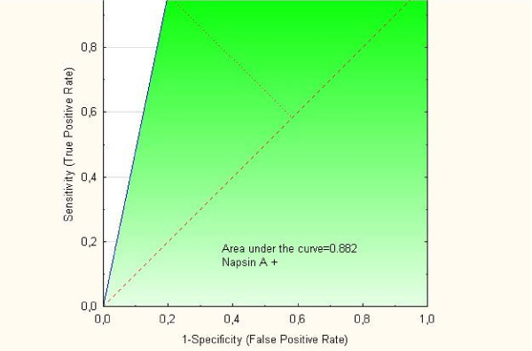
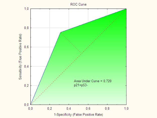
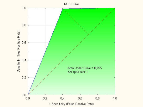
Results: Positivity for Napsin A was detected in 12 (80%) out of the 15 clear cell tumors available for analysis compared with 3 (4%) out of the Type I and II tumors in one group (p<0.001). Differences in p21 status, p53 status, and p21+p53- status were striking when clear cell tumors were compared with Type I, Type II, and Type I and II tumors in one group, respectively. The p21+p53-status was associated to positive staining of Napsin A (p=0.0015) and clear cell morphology (p=0.0003). In two separate multivariate logistic regression analyses with Napsin A as endpoint both clear cell carcinoma with OR=153 (95% C.I. 21-1107); (p<001) and p21+p53- status with OR=5.36 (95% C.I. 1.6-17.5); (p=0.005) were independent predictive factors. ROC curves showed that AUC for Napsin A alone was 0.882, for p21+p53- it was 0.720 and for p21+p53-Napsin A+AUC was 0.795. Patients with clear cell tumors had lower (p=0.013) BMI than Type I patients and were younger (p=0.046) at diagnosis than Type II patients. Clear cell tumors had a higher frequency (p=0.039) of capsule rupture at surgery than Type I and II tumors.
Conclusions: Positivity of Napsin A in an epithelial ovarian tumor might strengthen the morphological diagnosis of clear cell ovarian carcinoma in the process of differential diagnosis between clear cell ovarian tumors and other histological subtypes.
Figures
Similar articles
-
Association of p21, p21 p27 and p21 p53 status to histological subtypes and prognosis in low-stage epithelial ovarian cancer.Cancer Genomics Proteomics. 2013 Jan-Feb;10(1):27-34. Cancer Genomics Proteomics. 2013. PMID: 23382584
-
Differences in Clinical and Biological Features Between Type I and Type II Tumors in FIGO Stages I-II Epithelial Ovarian Carcinoma.Int J Gynecol Cancer. 2015 Sep;25(7):1239-47. doi: 10.1097/IGC.0000000000000484. Int J Gynecol Cancer. 2015. PMID: 26035126 Free PMC article.
-
Prognostic impact of concomitant p53 and PTEN on outcome in early stage (FIGO I-II) epithelial ovarian cancer.Int J Gynecol Cancer. 2011 Aug;21(6):1024-31. doi: 10.1097/IGC.0b013e31821dc906. Int J Gynecol Cancer. 2011. PMID: 21792012
-
Comparative analysis of Napsin A, alpha-methylacyl-coenzyme A racemase (AMACR, P504S), and hepatocyte nuclear factor 1 beta as diagnostic markers of ovarian clear cell carcinoma: an immunohistochemical study of 279 ovarian tumours.Pathology. 2015 Feb;47(2):105-11. doi: 10.1097/PAT.0000000000000223. Pathology. 2015. PMID: 25551297
-
Identification of biomarkers for the diagnosis and targets for therapy in patients with clear cell ovarian cancer: a systematic literature review.Carcinogenesis. 2022 Apr 25;43(3):183-189. doi: 10.1093/carcin/bgac012. Carcinogenesis. 2022. PMID: 35104328 Free PMC article.
Cited by
-
Diagnostic value of dual detection of hepatocyte nuclear factor 1 beta (HNF-1β) and napsin A for diagnosing ovarian clear cell carcinoma.Int J Clin Exp Pathol. 2015 Jul 1;8(7):8305-10. eCollection 2015. Int J Clin Exp Pathol. 2015. PMID: 26339401 Free PMC article.
-
Effective Disease Control After Combinatorial Treatment with a PD-1 Antibody and an mTOR Inhibitor for Recurrent Ovarian Clear Cell Carcinomas: A Case Report and Literature Review.Onco Targets Ther. 2021 Dec 9;14:5429-5434. doi: 10.2147/OTT.S333029. eCollection 2021. Onco Targets Ther. 2021. PMID: 34916808 Free PMC article.
-
Ovarian Seromucinous Borderline Tumor and Clear Cell Carcinoma: An Unusual Combination.Case Rep Obstet Gynecol. 2015;2015:690891. doi: 10.1155/2015/690891. Epub 2015 May 13. Case Rep Obstet Gynecol. 2015. PMID: 26075120 Free PMC article.
-
Diagnostic Accuracy of Alpha-Methylacyl-CoA Racemase Immunohistochemical Expression for the Diagnosis of Ovarian and Endometrial Clear Cell Carcinomas.Iran J Pathol. 2023;18(1):57-63. doi: 10.30699/IJP.2023.556417.2925. Epub 2023 Mar 23. Iran J Pathol. 2023. PMID: 37383161 Free PMC article.
-
The clinical and prognostic correlation of HRNPM and SLC1A5 in pathogenesis and prognosis in epithelial ovarian cancer.PLoS One. 2017 Jun 13;12(6):e0179363. doi: 10.1371/journal.pone.0179363. eCollection 2017. PLoS One. 2017. PMID: 28609484 Free PMC article.
References
-
- Zannoni GF, Morassi F, Prisco MG, De Stefano I, Vellone VG, Arena V, Scambia G, Gallo D. Clinicopathologic and immunohistochemical features of ovarian clear cell carcinomas in comparison with type I and type II tumors. IntJ Gynecol Pathol. 2012;31(6):507–516. doi: 10.1097/PGP.0b013e3182518557. - DOI - PubMed
-
- Zhao C, Wu L, Barner R. Pathogenesis of ovarian clear cell adenofibroma, atypical proliferative (borderline) tumor, and carcinoma: clinicopathologic features of tumors with endometriosis or adenofibromatous components support Two related pathways of tumor development. J Cancer Educ. 2011;2:94–106. - PMC - PubMed
Publication types
MeSH terms
Substances
LinkOut - more resources
Full Text Sources
Other Literature Sources
Medical
Molecular Biology Databases
Research Materials
Miscellaneous
