

Single-step laser-based fabrication and patterning of cell-encapsulated alginate microbeads
- PMID: 24192221
- PMCID: PMC3890439
- DOI: 10.1088/1758-5082/5/4/045006
Single-step laser-based fabrication and patterning of cell-encapsulated alginate microbeads
Abstract
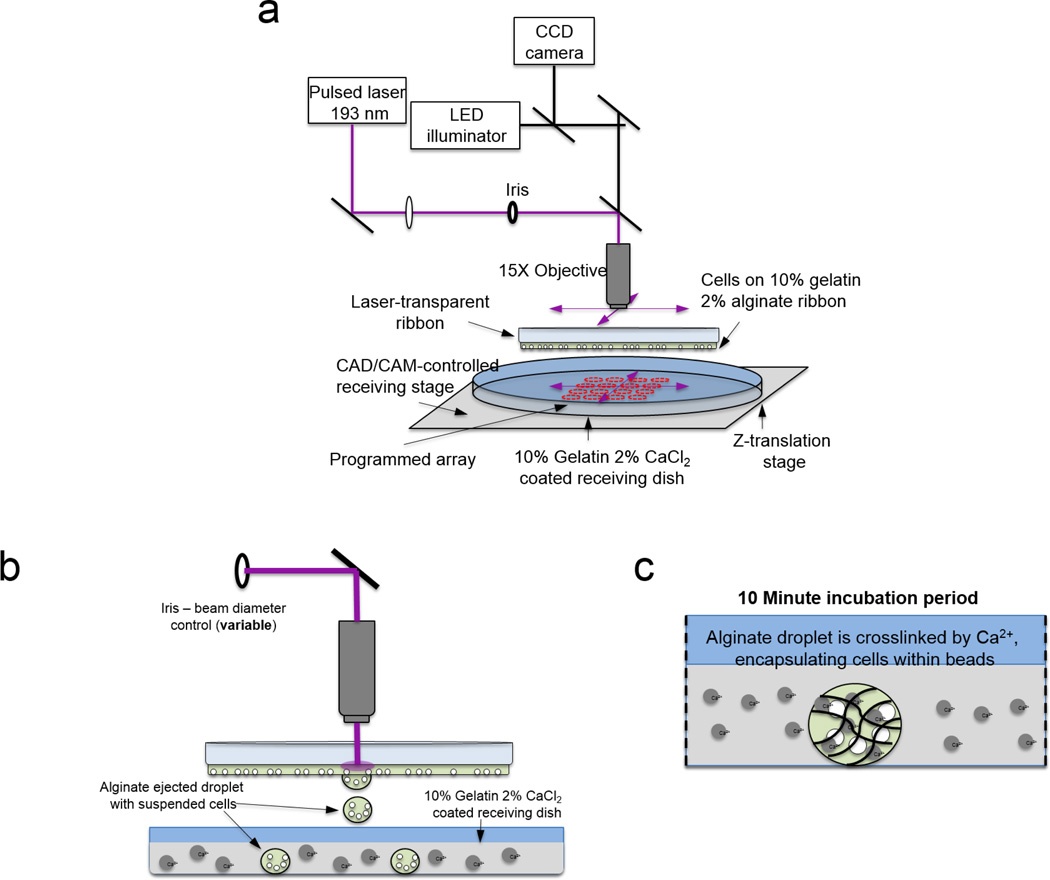
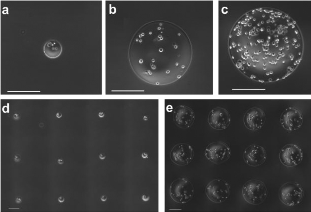
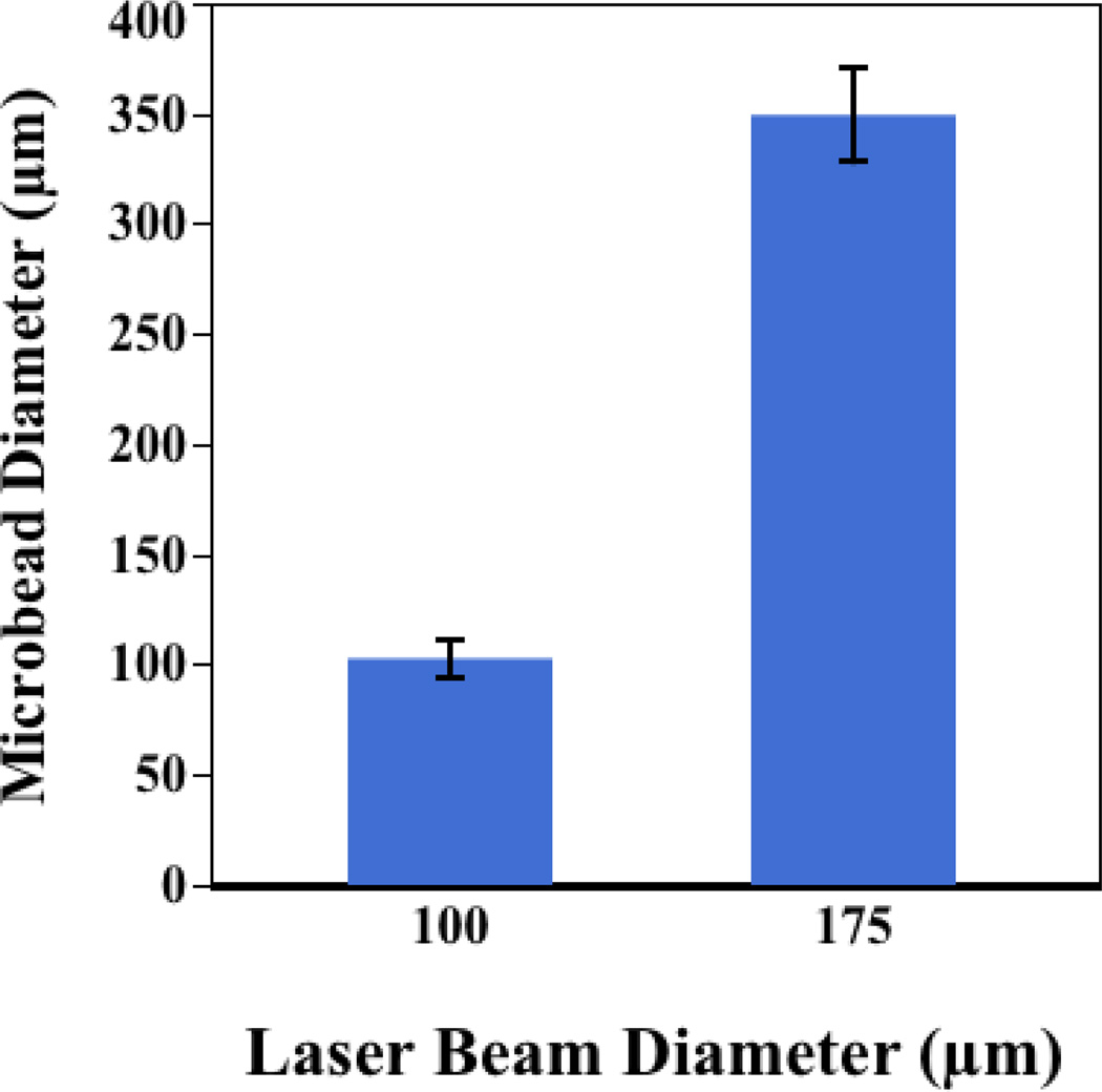
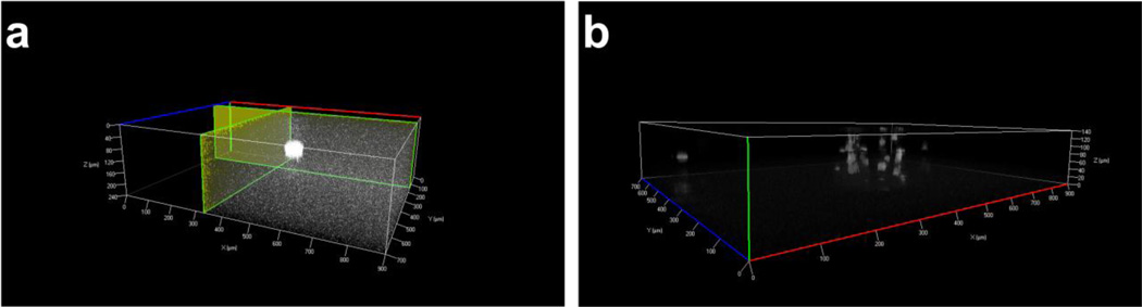
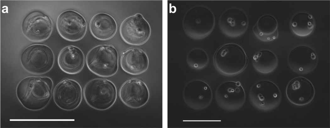
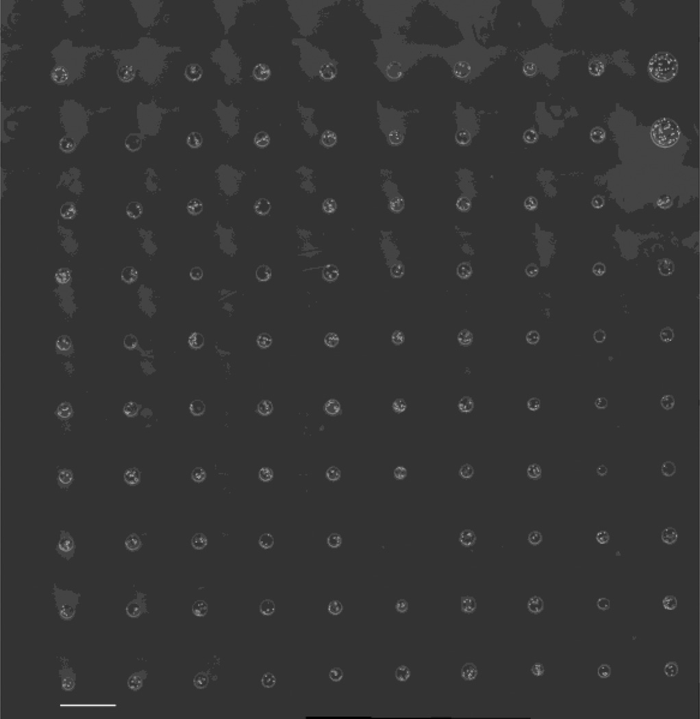
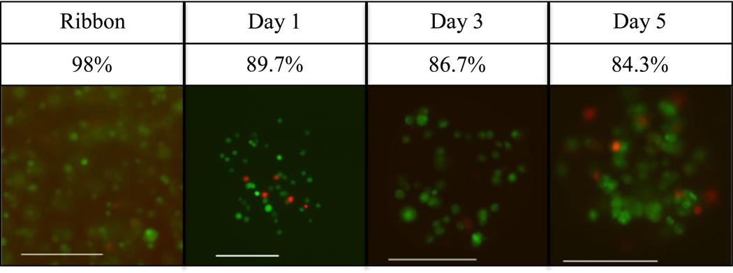
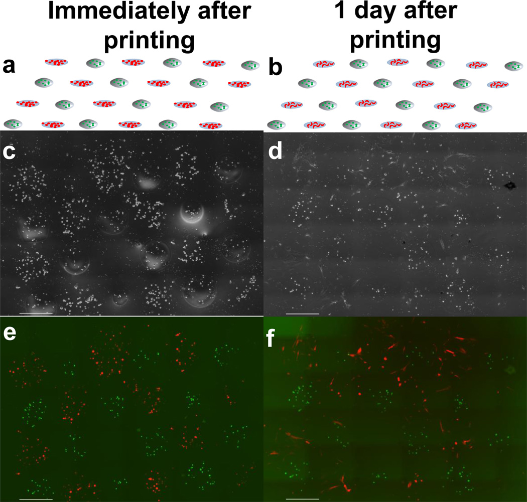
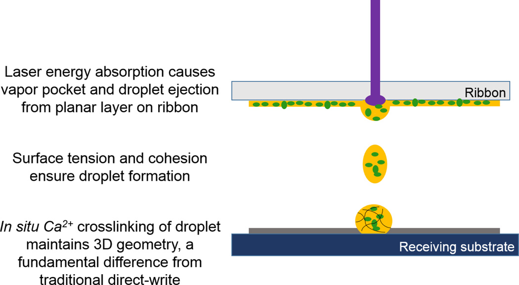
Alginate can be used to encapsulate mammalian cells and for the slow release of small molecules. Packaging alginate as microbead structures allows customizable delivery for tissue engineering, drug release, or contrast agents for imaging. However, state-of-the-art microbead fabrication has a limited range in achievable bead sizes, and poor control over bead placement, which may be desired to localize cellular signaling or delivery. Herein, we present a novel, laser-based method for single-step fabrication and precise planar placement of alginate microbeads. Our results show that bead size is controllable within 8%, and fabricated microbeads can remain immobilized within 2% of their target placement. Demonstration of this technique using human breast cancer cells shows that cells encapsulated within these microbeads survive at a rate of 89.6%, decreasing to 84.3% after five days in culture. Infusing rhodamine dye into microbeads prior to fluorescent microscopy shows their 3D spheroidal geometry and the ability to sequester small molecules. Microbead fabrication and patterning is compatible with conventional cellular transfer and patterning by laser direct-write, allowing location-based cellular studies. While this method can also be used to fabricate microbeads en masse for collection, the greatest value to tissue engineering and drug delivery studies and applications lies in the pattern registry of printed microbeads.
Figures

Similar articles
-
Laser direct-write of single microbeads into spatially-ordered patterns.Biofabrication. 2012 Jun;4(2):025006. doi: 10.1088/1758-5082/4/2/025006. Epub 2012 May 4. Biofabrication. 2012. PMID: 22556116
-
The fast release of stem cells from alginate-fibrin microbeads in injectable scaffolds for bone tissue engineering.Biomaterials. 2011 Oct;32(30):7503-13. doi: 10.1016/j.biomaterials.2011.06.045. Epub 2011 Jul 14. Biomaterials. 2011. PMID: 21757229 Free PMC article.
-
In vitro culture and oxygen consumption of NSCs in size-controlled neurospheres of Ca-alginate/gelatin microbead.Mater Sci Eng C Mater Biol Appl. 2014 Jul 1;40:197-203. doi: 10.1016/j.msec.2014.03.028. Epub 2014 Mar 19. Mater Sci Eng C Mater Biol Appl. 2014. PMID: 24857483
-
The application of an optically switched dielectrophoretic (ODEP) force for the manipulation and assembly of cell-encapsulating alginate microbeads in a microfluidic perfusion cell culture system for bottom-up tissue engineering.Lab Chip. 2012 Mar 21;12(6):1164-73. doi: 10.1039/c2lc21097e. Epub 2012 Feb 10. Lab Chip. 2012. PMID: 22322420
-
Applications of Alginate-Based Bioinks in 3D Bioprinting.Int J Mol Sci. 2016 Nov 25;17(12):1976. doi: 10.3390/ijms17121976. Int J Mol Sci. 2016. PMID: 27898010 Free PMC article. Review.
Cited by
-
Modeling intratumor heterogeneity in breast cancer.Biofabrication. 2024 Dec 19;17(1):10.1088/1758-5090/ad9b50. doi: 10.1088/1758-5090/ad9b50. Biofabrication. 2024. PMID: 39642392 Review.
-
Microcapsules and 3D customizable shelled microenvironments from laser direct-written microbeads.Biotechnol Bioeng. 2016 Oct;113(10):2264-74. doi: 10.1002/bit.25987. Epub 2016 Aug 9. Biotechnol Bioeng. 2016. PMID: 27070458 Free PMC article.
-
Directed self-assembly software for single cell deposition.Int J Bioprint. 2017 Jun 5;3(2):006. doi: 10.18063/IJB.2017.02.006. eCollection 2017. Int J Bioprint. 2017. PMID: 33094190 Free PMC article.
-
Laser-based 3D bioprinting for spatial and size control of tumor spheroids and embryoid bodies.Acta Biomater. 2019 Sep 1;95:357-370. doi: 10.1016/j.actbio.2019.02.014. Epub 2019 Feb 15. Acta Biomater. 2019. PMID: 30776506 Free PMC article.
-
Viscoelastic Properties of Bioprinted Alginate Microbeads Compared to Their Bulk Hydrogel Analogs.J Biomech Eng. 2023 Mar 1;145(3):031002. doi: 10.1115/1.4055757. J Biomech Eng. 2023. PMID: 36149022 Free PMC article.
References
-
- Xie Y, Castracane J. High-voltage, electric field-driven micro/nanofabrication for polymeric drug delivery systems. Engineering in Medicine and Biology. 2009;28:23–30. - PubMed
-
- Gombotz WR, Wee SF. Protein release from alginate matrices. Advanced Drug Delivery Reviews. 2012;64:194–205. - PubMed
-
- Mørch YA, Donati I, Strand BL, Skjåk-Braek G. Effect of Ca2+, Ba2+, and Sr2+ on alginate microbeads. Biomacromolecules. 2006;7:1471–1480. - PubMed
-
- Rokstad AM, Brekke OL, Steinkjer B, Ryan L, Kolláriková G, Strand BL, Skjåk-Bræk G, Lacík I, Espevik T, Mollnes TE. Alginate microbeads are complement compatible, in contrast to polycation containing microcapsules, as revealed in a human whole blood model. Acta Biomaterialia. 2011;7:2566–2578. - PubMed
-
- Robert D, Fayol D, Le Visage C, Frasca G, Brulé S, Ménager C, Gazeau F, Letourneur D, Wilhelm C. Magnetic micro-manipulations to probe the local physical properties of porous scaffolds and to confine stem cells. Biomaterials. 2010;31:1586–1595. - PubMed
Publication types
MeSH terms
Substances
Grants and funding
LinkOut - more resources
Full Text Sources
Other Literature Sources
Research Materials