. 2013 Nov;4(11):1976-85.
doi: 10.18632/oncotarget.1318.

p53 regulates the transcription of the anti-inflammatory molecule developmental endothelial locus-1 (Del-1)

Affiliations

p53 regulates the transcription of the anti-inflammatory molecule developmental endothelial locus-1 (Del-1)

Hyesoon Kim et al. Oncotarget. 2013 Nov.

Abstract

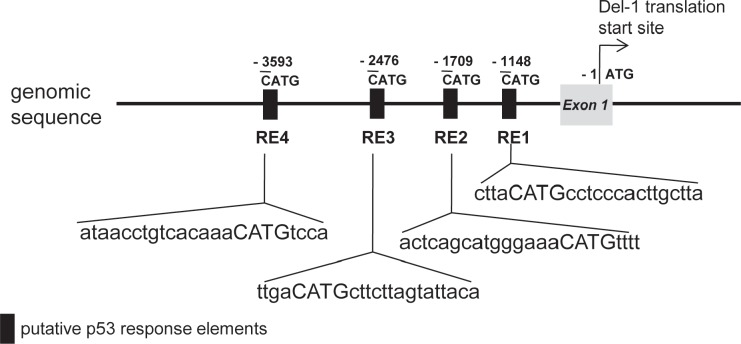
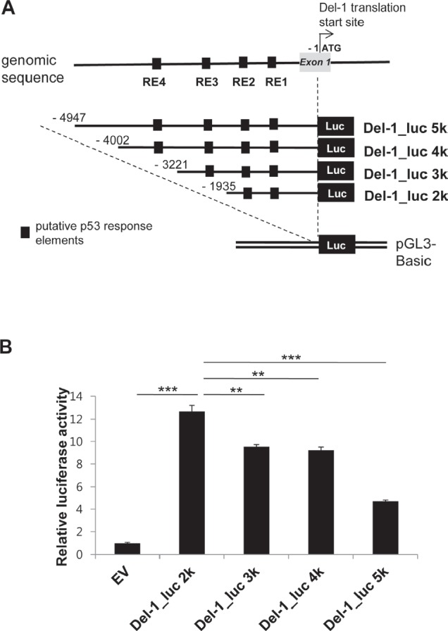
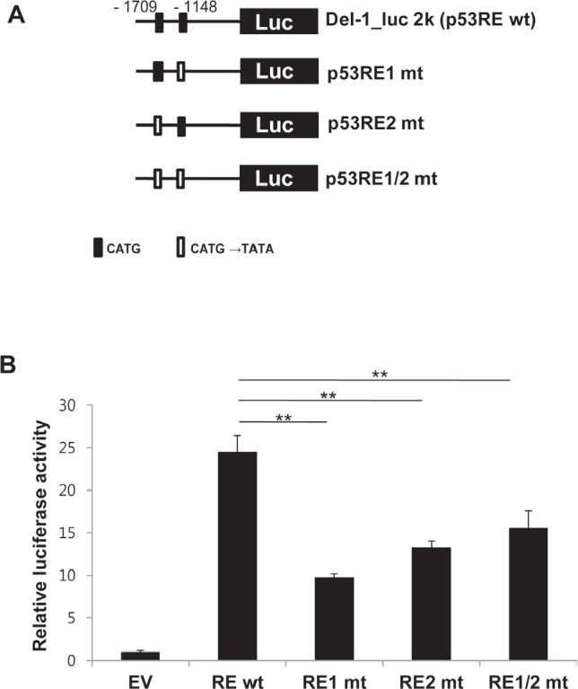
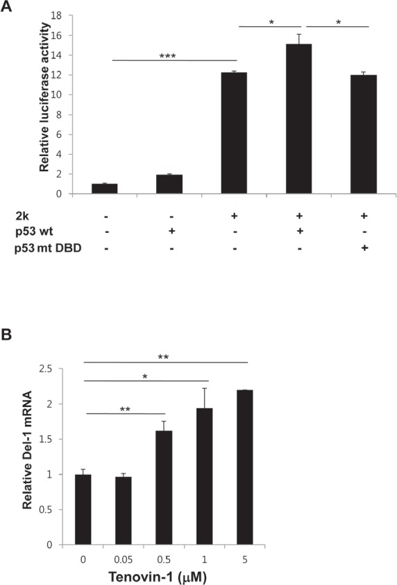
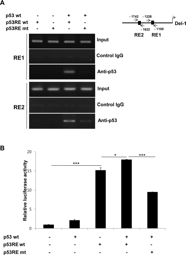
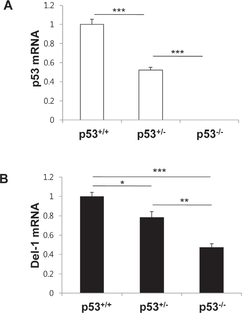
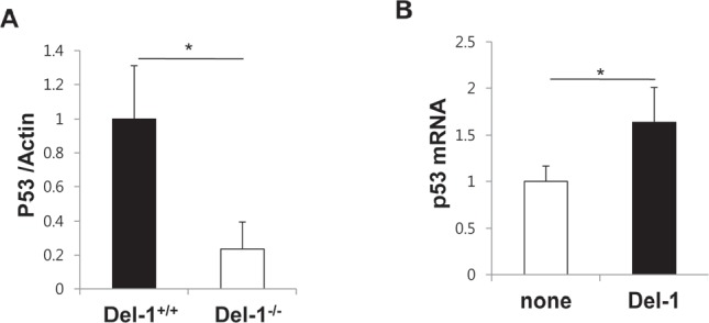
Developmental endothelial locus-1 (Del-1) is an endothelium-derived anti-inflammatory molecule that is downregulated by inflammatory stimuli. Little is known about the molecular mechanisms by which Del-1 transcription is regulated. In the present study, a DNA sequence upstream of the Del-1 gene was analyzed and putative p53 response elements (p53REs) were identified. An approximately 2 kb fragment upstream of the translation start site displayed the highest Del-1 transcriptional activity, and the transcriptional activity of this fragment was enhanced by overexpression of p53. Chemical activation of endogenous p53 elevated the levels of Del-1 mRNA. Site-directed mutagenesis of CATG in the consensus sequences of the 2 kb fragment to TATA significantly reduced the transcription of Del-1. Chromatin immunoprecipitation revealed recruitment of p53 to the p53REs of the Del-1 promoter, resulting in increased Del-1 transcription. Finally, primary endothelial cells isolated from mice with reduced levels of p53 showed a decrease in Del-1 mRNA compared to wild-type endothelial cells. Moreover, Del-1 reciprocally enhanced p53 expression in primary endothelial cells. Thus, these findings suggest that Del-1 is a novel transcriptional target gene of p53.


Conflict of interest statement

The authors declare no conflict of interest.

Figures

Figure 1
Figure 1. Schematic representation of the region upstream of the Del-1 gene
Putative p53 response elements (p53REs) were predicted using the Genometrix program. CATG-containing p53REs in the upstream regions are indicated. The first nucleotide of CATG is numbered relative to the translation start site (TSS) of the Del-1 gene.
Figure 2
Figure 2. Relative transcriptional activity is dependent on the presence of proximal p53 response elements in the upstream region of the Del-1 gene
(A) Schematic diagram of the Del-1 promoter constructs. Nucleotides are indicated relative to the TSS. Upstream fragments of the Del-1 gene were cloned into the pGL3 vector to generate four Del-1 promoter deletion constructs containing multiple putative p53 response elements. (B) Relative luciferase activity of these constructs. The Del-1 promoter constructs shown in (A) were individually transfected into HEK293T cells. Luciferase activity was determined 24 h after transfection and is expressed as the fold activity over that of the empty pGL3 vector (EV). A Renilla luciferase vector was co-transfected for normalization of transfection efficiency. Values are the means ± standard deviations (SD) from triplicate transfections. Data represent four independent experiments. **, p < 0.01; ***, p < 0.001; n.s., non-significant vs. the Del-1_luc 2k construct (i.e., the construct exhibiting the highest activity).
Figure 3
Figure 3. Functional p53 response elements are required to enhance Del-1 transcription
(A) Schematic diagram of wild-type (WT) and mutant Del-1 promoter constructs. The Del-1_luc 2k construct was mutated at the consensus sequence (CATG→TATA) of either or both p53 response elements. (B) The WT and mutant Del-1_luc 2k constructs were independently transfected into HEK293T cells. Luciferase activity was determined 24 h after transfection and is expressed as the fold activity over that of the empty pGL3 vector (EV). Values are means ± SD from triplicate transfections. Data represent four independent experiments. *, p < 0.05; **, p < 0.01; n.s., non-significant vs. the WT Del-1_luc 2k construct.
Figure 4
Figure 4. p53 positively regulates Del-1 transcription
(A) Mouse WT p53 or p53 with mutated DNA binding sites (G239A, R242A, and R243A) was transfected along with the 2k Del-1 promoter construct into HEK293T cells. Luciferase activity was determined 24 h after transfection and is expressed as the fold activity over that of the empty pGL3 vector. Values are means ± standard deviations from triplicate transfections. *, p < 0.05; ***, p < 0.001, vs. the 2k Del-1 promoter construct. (B) Mouse primary endothelial cells were treated with increasing concentrations of tenovin-1 and incubated for 24 h. The Del-1 mRNA level was measured and is expressed as the fold increase over dimethyl sulfoxide (DMSO)-treated cells. Values are means ± SD from triplicate treatments. Data represent three independent experiments. *, p < 0.05; **, p < 0.01 vs. the DMSO-treated cells.
Figure 5
Figure 5. p53 directly binds to p53 response elements to enhance Del-1 transcription
(A) Representative ChIP analysis. HEK293T cells were transfected with a mouse p53-expressing plasmid, together with either the WT or mutant (mutated at both p53REs) 2k Del-1 promoter construct. Nuclear lysates were analyzed 48 h after transfection using a p53 antibody or control IgG and promoter regions containing the p53REs were amplified by PCR. (B) HEK293T cells were transfected with the plasmids in (A). Luciferase activity was determined 24 h after transfection and is expressed as the fold activity over that of the empty pGL3 vector. Values are means ± SD from triplicate transfections. Data represent three independent experiments. *, p < 0.05; ***, p < 0.001 vs. the cells co-transfected with the WT p53 and p53REwt Del-1 constructs.
Figure 6
Figure 6. p53 levels determine Del-1 expression in mouse primary endothelial cells
Mouse p53 (A) and Del-1(B) mRNA levels were analyzed by real-time RT-PCR in primary endothelial cells from p53+/+, p53+/−, and p53−/− mice. The relative Del-1 mRNA levels in cells heterozygous or homozygous null for p53 were compared to those of p53+/+ mice. Values are means ± SD (n = 4–5 mice/group). Data represent three independent experiments. *, p < 0.05; **, p < 0.01; ***, p < 0.001.
Figure 7
Figure 7. Del-1 reciprocally regulates p53 expression in mouse primary endothelial cells
(A) Levels of p53 protein was analyzed by western blotting in primary endothelial cells from Del-1+/+ and Del-1−/− mice. Values are means ± SD (n = 3 mice/group). (B) Transcript levels of p53 were analyzed by real-time RT-PCR. WT primary endothelial cells were incubated in the absence or presence of recombinant Del-1 protein (10 nM) for 24 h. Values are means ± SD (n = 4−5 mice/group). *, p < 0.05.

References

    1. Hidai C, Zupancic T, Penta K, Mikhail A, Kawana M, Quertermous EE, Aoka Y, Fukagawa M, Matsui Y, Platika D, Auerbach R, Hogan BL, Snodgrass R, Quertermous T. Cloning and characterization of developmental endothelial locus-1: an embryonic endothelial cell protein that binds the alphavbeta3 integrin receptor. Genes Dev. 1998;12(1):21–33. - PMC - PubMed
    1. Kitano H, Kokubun S, Hidai C. The extracellular matrix protein Del1 induces apoptosis via its epidermal growth factor motif. Biochem Biophys Res Commun. 2010;393(4):757–761. - PubMed
    1. Rezaee M, Penta K, Quertermous T. Del1 mediates VSMC adhesion, migration, and proliferation through interaction with integrin alpha(v)beta(3) Am J Physiol Heart Circ Physiol. 2002;282(5):H1924–1932. - PubMed
    1. Wang Z, Kundu RK, Longaker MT, Quertermous T, Yang GP. The angiogenic factor Del1 prevents apoptosis of endothelial cells through integrin binding. Surgery. 2012;151(2):296–305. - PubMed
    1. Zhong J, Eliceiri B, Stupack D, Penta K, Sakamoto G, Quertermous T, Coleman M, Boudreau N, Varner JA. Neovascularization of ischemic tissues by gene delivery of the extracellular matrix protein Del-1. J Clin Invest. 2003;112(1):30–41. - PMC - PubMed

Publication types

Substances