. 2014 Jan 17;114(2):257-65.
doi: 10.1161/CIRCRESAHA.114.302734. Epub 2013 Nov 5.

Mitochondrial contagion induced by Parkin deficiency in Drosophila hearts and its containment by suppressing mitofusin

Affiliations

Mitochondrial contagion induced by Parkin deficiency in Drosophila hearts and its containment by suppressing mitofusin

Poonam Bhandari et al. Circ Res. .

Abstract

Rationale: Dysfunctional Parkin-mediated mitophagic culling of senescent or damaged mitochondria is a major pathological process underlying Parkinson disease and a potential genetic mechanism of cardiomyopathy. Despite epidemiological associations between Parkinson disease and heart failure, the role of Parkin and mitophagic quality control in maintaining normal cardiac homeostasis is poorly understood.

Objective: We used germline mutants and cardiac-specific RNA interference to interrogate Parkin regulation of cardiomyocyte mitochondria and examine functional crosstalk between mitophagy and mitochondrial dynamics in Drosophila heart tubes.

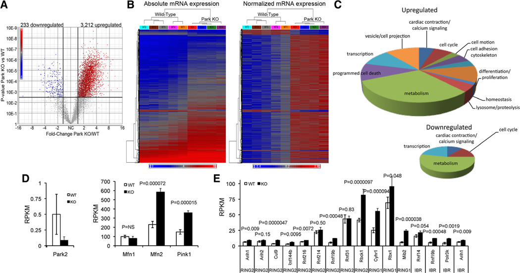
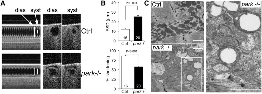
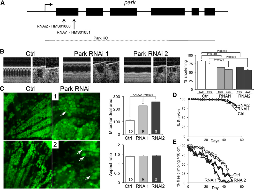
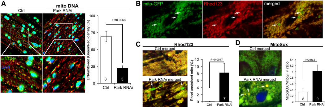
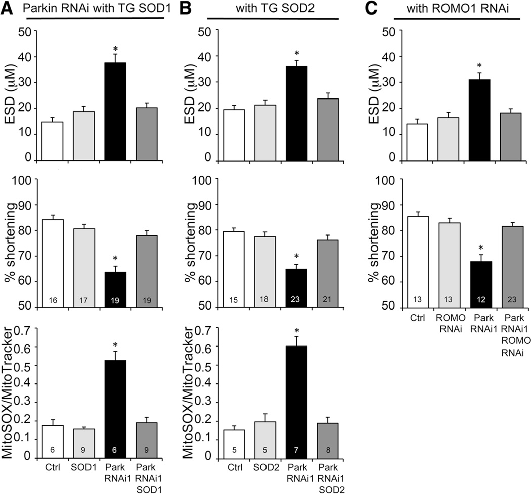
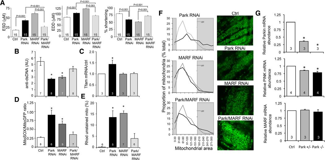
Methods and results: Transcriptional profiling of Parkin knockout mouse hearts revealed compensatory upregulation of multiple related E3 ubiquitin ligases. Because Drosophila lack most of these redundant genes, we examined heart tubes of parkin knockout flies and observed accumulation of enlarged hollow donut mitochondria with dilated cardiomyopathy, which could be rescued by cardiomyocyte-specific Parkin expression. Identical abnormalities were induced by cardiomyocyte-specific Parkin suppression using 2 different inhibitory RNAs. Parkin-deficient cardiomyocyte mitochondria exhibited dysmorphology, depolarization, and reactive oxygen species generation without calcium cycling abnormalities, pointing to a primary mitochondrial defect. Suppressing cardiomyocyte mitochondrial fusion in Parkin-deficient fly heart tubes completely prevented the cardiomyopathy and corrected mitochondrial dysfunction without normalizing mitochondrial dysmorphology, demonstrating a central role for mitochondrial fusion in the cardiomyopathy provoked by impaired mitophagy.

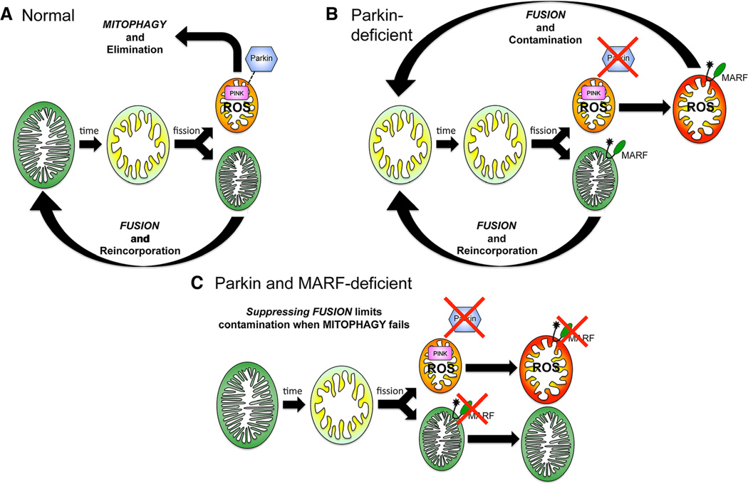
Conclusions: Parkin deficiency and resulting mitophagic disruption produces cardiomyopathy in part by contamination of the cardiomyocyte mitochondrial pool through fusion between improperly retained dysfunctional/senescent and normal mitochondria. Limiting mitochondrial contagion by inhibiting organelle fusion shows promise for minimizing organ dysfunction produced by defective mitophagic signaling.

Keywords: cardiomyopathies; mitochondrial degradation; mitochondrial dynamics.


Figures

Figure 1
Figure 1. Cardiac RNA transcript levels of Parkin pathway factors and Parkin-related E3 ubiquitin ligases in Parkin knockout (KO) mice
A and B, Volcano plot (A) and heat maps (B) of cardiac transcriptional changes induced by germline Parkin gene ablation in mice. C, Gene ontology analysis of up- (top) and downregulated mRNAs in Parkin KO mouse hearts. D, mRNA levels of Parkin and upstream Parkin signaling factors PINK1 and Mfn2; Mfn1 is shown for comparison. E, Transcript levels of Parkin-related E3 ubiquitin ligases of RING2, RING1, and in-between ring families. Gene names are: Arih1, ariadne homolog, ubiquitin-conjugating enzyme E2 binding protein 1; Arih2, ariadne homolog 2; Cul9, cullin 9; Rnf, ring finger protein; Rbck1, RanBP-type and C3HC4-type zinc finger containing 1; Cyhr1, cysteine/histidine-rich 1; Rbx1, ring-box 1; Mib2, mindbomb homolog 2; Polr3k, polymerase (RNA) III (DNA directed) polypeptide K, 12.3 kDa. Values are mean±SEM of RNA sequencing reads per million bases (RPKM) for n=5 wild type (WT) and n=4 germline Parkin null (KO) mice.
Figure 2
Figure 2. Cardiomyopathy and mitochondrial abnormalities in parkin null (−/−) flies
A, Optical coherence tomography (OCT) of heart tube contractions in tincΔ4-Gal4 (Ctrl) and parkin−/− flies 7 days after eclosure. dias indicates diastolic; and syst, systolic. B, Bar graphs showing group mean OCT data for end-systolic dimension (ESD) and percent fractional shortening. C, Transmission electron micrographs showing enlarged mitochondria with disrupted or absent cristae in parkin−/− cardiomyocytes. Note the characteristic hollow donut mitochondrial morphology. White scale bars are 1 µm.
Figure 3
Figure 3. Cardiomyocyte-specific Parkin suppression induces heart failure and mitochondrial abnormalities in Drosophila
A, Parkin targeting by RNAi constructs. B, Optical coherence tomography studies showing hypocontractile heart tubes in tincΔ4-Gal4 UAS-Parkin RNAi fly lines. Bar graphs show group mean fractional shortening data 7 and 28 days after eclosure (n=15–20 per group). C, Cardiomyocyte mitochondria visualized by tincΔ4-Gal4-mitoGFP show organelle enlargement and hollow donut dysmorphology (arrows). Group mean data are to the right (>100 mitochondria analyzed per heart). D, Survival analysis of tincΔ4-Gal4 UAS-Parkin RNAi fly lines (n=150 per group). E, Climbing performance in negative geotaxis test in tincΔ4-Gal4 UAS-Parkin RNAi fly lines (n=150 per group).
Figure 4
Figure 4. Cardiomyocyte mitochondrial dysfunction induced by Parkin suppression
A, Merged confocal analysis of cardiomyocyte mitochondria nucleoids visualized using anti-DNA antibody (green) and cardiomyocyte-specific expression of Mito-DSRed (red). Nuclei visualized with DAPI (blue) were digitally masked and mitochondrial DNA content measured using ImageJ. Quantitative results are to the right. B and C, Depolarization of structurally abnormal mitochondria assessed by rhodamine (Rhod) 123 staining. B, Separate tincΔ4-Gal4–driven mito-GFP (green; left) and Rhod 123 (red; middle) and merged images (right) with representative depolarized (less red staining) mitochondria indicated with arrows. C, Representative merged control and Parkin RNAi mito-GFP/Rhod 123 double-stained cardiomyocytes (left) and group quantitative data (right). Two of several depolarized mitochondria are indicated by arrows. D, Representative merged control and Parkin RNAi mito-GFP/MitoSox double-stained cardiomyocytes (left) and group quantitative data (right). Three of several reactive oxygen species–producing mitochondria are indicated by arrows.
Figure 5
Figure 5. Mitochondrial reactive oxygen species (ROS) production contributes to the cardiomyopathy evoked by Parkin suppression
A and B, Results of optical coherence tomography (OCT) and MitoSOX studies of cardiomyocyte-specific Parkin RNAi flies without (black) and with (dark grey) concomitant cardiomyocyte-specific expression of superoxide dismutase (SOD) 1 (A) or SOD2 (B). End-systolic dimension (ESD) is on top, percent fractional shortening in middle, mitoSOX on bottom. C, OCT studies of cardiac Parkin-deficient flies without (black) and with (dark grey) concomitant RNAi-mediated suppression of mitochondrial ROS modulator 1 (Romo1), a major mitochondrial ROS-producing enzyme. *Significantly different from Ctrl by ANOVA and Bonferroni test.
Figure 6
Figure 6. Interrupting mitochondrial fusion rescues mitochondrial and cardiac dysfunction evoked by Parkin deficiency
A, Results of optical coherence tomography studies of Parkin RNAi-expressing fly heart tubes without and with concomitant RNAi-mediated suppression of cardiomyocyte dMfn/MARF (Parkin/MARF RNAi). EDD indicates end-diastolic dimension; ESD, end-systolic dimension; and MARF, mitochondrial assembly regulatory factor. Normal Ctrl and tincΔ4-Gal4 MARF RNAi results are shown for comparison. B to E, Mitochondrial function studies of the same groups as A; B, mitoDNA content performed as in Figure 4A; C, Tfam mRNA abundance by RT-qPCR; D, Reactive oxygen species production performed as in Figure 4D; E, mitochondrial depolarization assayed as in Figure 4B and 4C. F, Mitochondrial morphology assessed by mito-GFP confocal analysis of the same groups. Group histogram data for mitochondrial area are on the left, with representative micrographs on the right. The grey areas depict abnormally small (left) and abnormally large (right) mitochondria defined as the bottom and top decile of the normal control data. The same control data (grey lines) are shown in all 3 histograms. G, RT-qPCR of Parkin (top), PTEN-inducible kinase 1 (PINK; middle), and mitochondrial assembly regulatory factor (MARF; bottom) mRNA. *Significantly different from Ctrl by ANOVA and Bonferroni test.
Figure 7
Figure 7. Interactive mitochondrial dynamics and quality control pathways as revealed by the present results
Green mitochondria are fully polarized, red mitochondria are fully depolarized, and yellow/orange represents intermediate states.

References

    1. Kitada T, Asakawa S, Hattori N, Matsumine H, Yamamura Y, Minoshima S, Yokochi M, Mizuno Y, Shimizu N. Mutations in the parkin gene cause autosomal recessive juvenile parkinsonism. Nature. 1998;392:605–608. - PubMed
    1. Valente EM, Abou-Sleiman PM, Caputo V, et al. Hereditary early-onset Parkinson’s disease caused by mutations in PINK1. Science. 2004;304:1158–1160. - PubMed
    1. Kitada T, Pisani A, Porter DR, Yamaguchi H, Tscherter A, Martella G, Bonsi P, Zhang C, Pothos EN, Shen J. Impaired dopamine release and synaptic plasticity in the striatum of PINK1-deficient mice. Proc Natl Acad Sci U S A. 2007;104:11441–11446. - PMC - PubMed
    1. Billia F, Hauck L, Konecny F, Rao V, Shen J, Mak TW. PTEN-inducible kinase 1 (PINK1)/Park6 is indispensable for normal heart function. Proc Natl Acad Sci U S A. 2011;108:9572–9577. - PMC - PubMed
    1. Chen Y, Dorn GW., II PINK1-phosphorylated mitofusin 2 is a Parkin receptor for culling damaged mitochondria. Science. 2013;340:471–475. - PMC - PubMed

Publication types

MeSH terms