. 2013 Nov 6:3:3151.
doi: 10.1038/srep03151.

Regulation of epithelial plasticity by miR-424 and miR-200 in a new prostate cancer metastasis model

Affiliations

Regulation of epithelial plasticity by miR-424 and miR-200 in a new prostate cancer metastasis model

Jacqueline Banyard et al. Sci Rep. .

Abstract

Using an in vivo cycling strategy, we selected metastatic cancer cells from the lymph nodes (LN) of mice bearing orthotopic DU145 human prostate tumors. Repeated rounds of metastatic selection (LN1-LN4) progressively increased the epithelial phenotype, resulting in a new model of tumor cell mesenchymal-epithelial transition (MET). DU145-LN4 showed increased cell-cell adhesions, higher expression of multiple epithelial markers, such as E-cadherin, EpCAM and cytokeratin 18, and reduced expression of mesenchymal markers such as vimentin. The MET in DU145-LN4 cells was accompanied by increased expression of the miR-200 family, and antimiRs to miR-200c and miR-141 induced an EMT. MET also correlated with the loss of miR-424. Ectopic transient and stable miR-424 expression induced EMT, with reduced epithelial marker expression and increased cell scattering. Our model provides evidence for spontaneous MET in vivo. We show that this cellular plasticity can be mediated through the combined action of miR-424 and the miR-200 family.


Figures

Figure 1
Figure 1. Selection of DU145 human prostate cancer cells with increased metastatic potential.
(A) Schematic of the experimental approach. DU145 prostate cells were injected orthotopically into the prostate. Lymph nodes were removed and cultured, and selected tumor cells subjected to repeated rounds of orthotopic injection. Illustrations by Kristin Johnson (Vascular Biology Program, Boston Children's Hospital). (B) Photography of gross specimens (tumors and sentinel lymph nodes). DU145 parental cells and DU145-LN sublines (DU145-LN4 shown) were reinjected into the prostate and the prostate and lymph nodes were removed after 5 wks. Scale bar = 1 cm. (C) Representative H&E staining of lymph nodes from mice bearing orthotopic parental DU145 tumors (left panel, P) and DU145-LN4 tumors (center and right panels, LN4). Metastatic nodule indicated by arrowhead in center panel, magnification shown in right panel.
Figure 2
Figure 2. Metastatic DU145-LN4 cells show mesenchymal to epithelial-like changes.
(A) DU145-LN4 cells display more cell–cell clustering as seen by phase microscopy (upper panels), and increased E-cadherin immunostaining (lower panels), scale bar = 50 μm. (B) Microarray analysis shows increased expression of epithelial markers and decreased mesenchymal markers. Heat map diagram shows unsupervised two-way hierarchical clustering of genes and cell lines. Color scale shows relative expression level; red is maximum, blue represents minimum expression level. (C) Western blot confirmed a progressive increase in expression of epithelial markers, and decrease in the mesenchymal marker vimentin, following rounds of metastatic selection from DU145 to DU145-LN4. One membrane was probed for E-cadherin, EpCAM, and GAPDH. Another was probed for vimentin, claudin 7, cytokeratin 18, γ-catenin, and actin. Images were cropped to save space; original images are shown in Supplemental Figure 5.
Figure 3
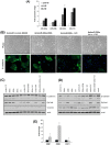
Figure 3. In vivo prostate-cycled DU145 cells do not display the phenotypic and expression changes observed in the metastatic DU145-LN cell lines.
DU145 cells were injected into the mouse prostate, the prostate excised and grown in culture to create DU145-PR1. This process was repeated to create DU145-PR2 and DU145-PR3. (A) Photography of DU145-PR3 gross specimens (tumors and sentinel lymph nodes) after 5 weeks following cell line injection orthotopically into the prostate. Scale bar = 1 cm (B) Phase contrast microscopy shows similar cell phenotype of DU145 parental and DU145-PR3 cells in culture. (C) Western blot analysis of DU145-PR3 and parental DU145 cell lysates indicate that expression of E-cadherin, EpCAM, γ-catenin, vimentin and β-catenin were not changed by in vivo cycling through the prostate, in contrast to the expression changes observed in DU145-LN4 cells. Equal loading shown by actin immunoblots. The same membrane was stripped and reprobed for E-cadherin, EpCAM, γ-catenin, vimentin, and actin. A separate blot was probed for β-catenin and actin. Images were cropped to save space; original images are shown in Supplemental Figure 5. Handwritten pen marks denote molecular weight markers.
Figure 4
Figure 4. Heat map profile of miRNA expression in the DU145-LN cell line model.
RNA collected from DU145, DU145-LN1, DU145-LN2 and DU145-LN4 was analyzed using a miRCURY LNA array (Exiqon). Hierarchical clustering and heat map representation of miRNAs and cell lines. Global color scale shows maximum (red) and minimum (blue) miRNA expression level.
Figure 5
Figure 5. The miR-200 family regulates EMT in metastatic DU145-LN4 cells.
(A) Real time RT-PCR confirmed increased expression of the miR-200 family members; miR-200a, b, c and miR-141 in DU145-LN2 and DU145-LN4 cells, relative to DU145 cells. Mean +/− SEM of triplicates. (B) Treatment of DU145-LN4 cells with a combination of antimiRs to the miR-200a + miR-200b (left center panel) or miR-200c + miR-141 (right center panel) reduced cell clustering and cell-cell interactions, as observed by phase-contrast microscopy (upper panels, scale bar = 100 μm), and E-cadherin immunostaining (lower panels, scale bar = 25 μm) at 14 days. Cells were transfected at day 0 and day 7. (C) Western blot at 7 days (following day 0 transfection) and (D) 14 days (following day 0 and day 7 transfection) showed reduced expression of E-cadherin and EpCAM, and increased vimentin expression following treatment of DU145-LN4 cells with antimiRs 200c + 141. To account for increased antimiR concentration when treated in combination, controls were treated with 200 nM (2X) and 400 nM (4X) non-specific antimiR. Actin shown as protein loading control. The same membrane was stripped and reprobed for each subsequent marker in C or D. Images were cropped to save space; original images are shown in Supplemental Figure 5. (E) Real time RT-PCR amplification of ZEB1, ZEB2 and CDH1 in DU145LN4 cells treated with antimiR-200c + 141 (grey bars), relative to antimiR control (black bars). Data normalized to GAPDH, mean +/− SD.
Figure 6
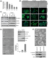
Figure 6. MiRNA-424 regulates the epithelial phenotype.
(A) Progressive downregulation of miR-424 expression in the DU145-LN cell series was measured by real time RT- PCR. (B) DU145-LN4 cells were transfected with two different control premiRs (left and right panels) or premiR-424 (center panel). PremiR-424 induced EMT-like changes, as observed by reduced cell-cell interactions in phase-contrast microscopy, and decreased E-cadherin and EpCAM levels in immunostaining. (C) Western blot analysis of DU145-LN4 cells treated with premiR controls, premiR-424 or mock transfected, collected at 6 and 21 days shows premiR-424 inhibited E-cadherin and EpCAM expression and increased vimentin levels. Actin shown as loading control. (D) Stable overexpression of miR-424 in DU145-LN4 cells resulted in loss of cell-cell contacts as shown by phase contrast microscopy (scale bar 100 μm). DU145-LN4 vector control (v3), and two miR-424-overexpressing DU145-LN4 clones (424-2 and 424-8) shown. Upper right panel shows reduced E-Cadherin, EpCAM and γ-catenin and increased vimentin levels by Western blot. Bar chart of real time PCR data confirmed upregulation of miR-424 level in stable cell lines after normalizing to housekeeping small RNA, SNORD38B. (E) Transient transfection of premiR-424 in epithelial MCF-7 breast cancer cells resulted in increased cell scattering, relative to two individual control premiRNAs, as shown by phase contrast microscopy (scale bar 50 μm). Western blot showed decreased E-Cadherin and EpCAM expression in premiR-424-treated cells. β-actin shown as loading control. Each membrane was stripped and reprobed for each subsequent marker in (C), (D), and (E). Images were cropped to save space; original images are shown in Supplemental Figure 5.
Figure 7
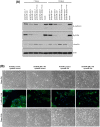
Figure 7. miR-424 and miR-200c/141 act independently and additively to affect cell plasticity.
DU145-LN4 cells were treated with control pre-miR combined with control antimiR, premiR-424 combined with antimiR control, antimiR-200c/141 combined with premiR control, or antimiR-200c/141 combined with premiR-424. (A) Whole cell lysates were collected at 7 and 14 days. Immunoblots showed reduced E-cadherin and EpCAM expression, and increased vimentin levels in premiR-424 or antimiR-200c/141 treated cells. Combining these treatments produced additive inhibition of epithelial markers. The same membrane was stripped and reprobed for each subsequent marker. Images were cropped to save space; original images are shown in Supplemental Figure 5. (B) In parallel with cell lysate collection, treated cells were examined using phase contrast microscopy and E-cadherin immunocytochemistry. Cell scattering was more distinct after premiR-424 treatment at 7 days, compared to antimiR-141/200c. At 14 days we observed additive effects of miR-424 expression and miR-141/200c inhibition, with greater cell scatter and loss of E-cadherin staining.

Similar articles

Cited by

References

    1. Gupta G. P. & Massague J. Cancer metastasis: building a framework. Cell 127, 679–695 (2006). - PubMed
    1. Kalluri R. & Weinberg R. A. The basics of epithelial-mesenchymal transition. J Clin Invest 119, 1420–1428 (2009). - PMC - PubMed
    1. Tarin D., Thompson E. W. & Newgreen D. F. The fallacy of epithelial mesenchymal transition in neoplasia. Cancer Res 65, 5996–6000; discussion 6000-5991; (2005). - PubMed
    1. Thompson E. W., Newgreen D. F. & Tarin D. Carcinoma invasion and metastasis: a role for epithelial-mesenchymal transition? Cancer Res 65, 5991–5995 discussion 5995 (2005). - PubMed
    1. Garber K. Epithelial-to-mesenchymal transition is important to metastasis, but questions remain. J Natl Cancer Inst 100, 232–233 239 (2008). - PubMed

Publication types

MeSH terms