. 2013 Dec 13;288(50):35793-800.
doi: 10.1074/jbc.M113.508333. Epub 2013 Nov 5.

Insights into distinct modulation of α7 and α7β2 nicotinic acetylcholine receptors by the volatile anesthetic isoflurane

Affiliations

Insights into distinct modulation of α7 and α7β2 nicotinic acetylcholine receptors by the volatile anesthetic isoflurane

David D Mowrey et al. J Biol Chem. .

Abstract

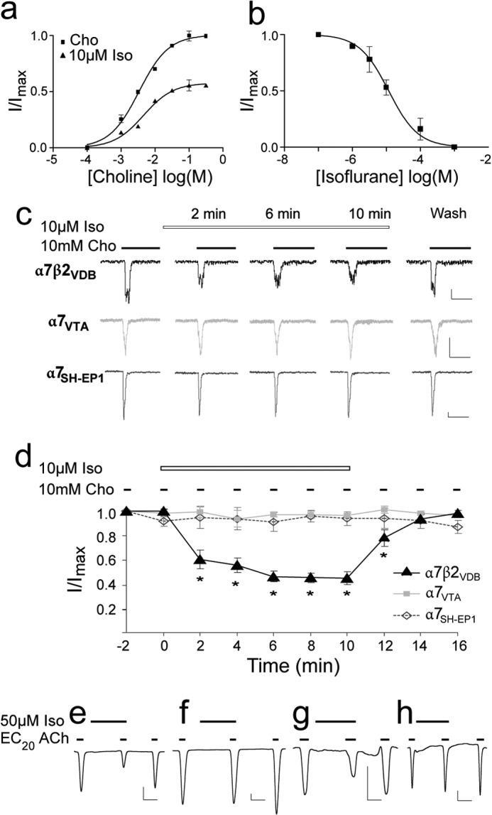
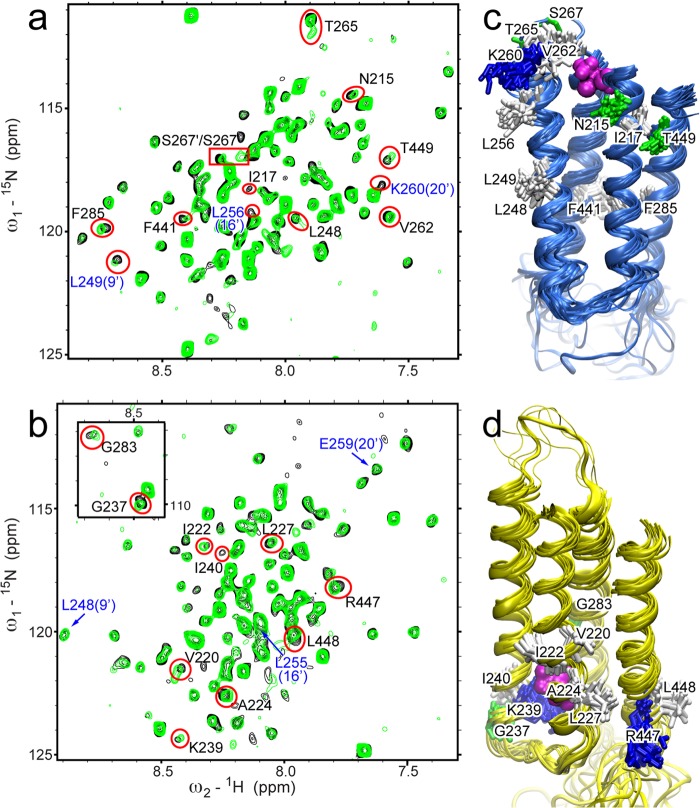
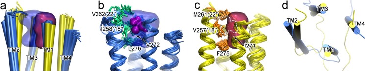
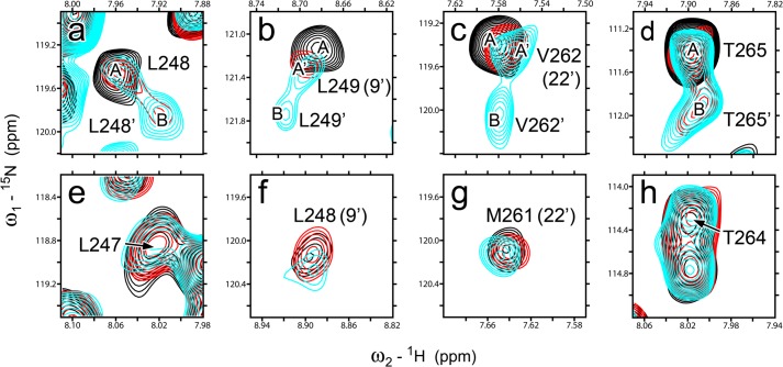
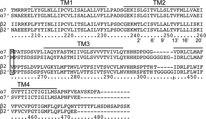
Nicotinic acetylcholine receptors (nAChRs) are targets of general anesthetics, but functional sensitivity to anesthetic inhibition varies dramatically among different subtypes of nAChRs. Potential causes underlying different functional responses to anesthetics remain elusive. Here we show that in contrast to the α7 nAChR, the α7β2 nAChR is highly susceptible to inhibition by the volatile anesthetic isoflurane in electrophysiology measurements. Isoflurane-binding sites in β2 and α7 were found at the extracellular and intracellular end of their respective transmembrane domains using NMR. Functional relevance of the identified β2 site was validated via point mutations and subsequent functional measurements. Consistent with their functional responses to isoflurane, β2 but not α7 showed pronounced dynamics changes, particularly for the channel gate residue Leu-249(9'). These results suggest that anesthetic binding alone is not sufficient to generate functional impact; only those sites that can modulate channel dynamics upon anesthetic binding will produce functional effects.

Keywords: Anesthetics; Cys-loop Receptors; Electrophysiology; Isoflurane; NMR; Nicotinic Acetylcholine Receptors; Protein Dynamics; α7; α7β2.


Figures

FIGURE 1.
FIGURE 1.
Isoflurane inhibited function of the α7β2 but not α7 nAChRs. a, the α7β2 nAChRs expressed in VDB neurons were noncompetitively inhibited by 2 min of preincubation with 10 μm isoflurane (Iso). EC50 of choline (Cho) and Hill coefficients show no significant differences in the absence and presence of isoflurane. b, isoflurane inhibited α7β2 with an IC50 of 11.7 ± 1.6 μm. Fractional currents were obtained from the mean peak currents elicited by 10 mm choline (∼EC70). The error bars are standard errors (n = 6). c, representative whole cell current traces for α7β2 expressed in VDB neurons, native α7 in VTA neurons, and heterologously human α7 nAChRs in the SH-EP1 cells. The vertical and horizontal scales represent 50 pA and 250 ms, respectively. d, normalized mean (± S.E.) peak current responses of α7β2 and α7 expressed in various cells to the prolonged choline stimulation in the presence of 10 μm isoflurane (n = 6). Isoflurane inhibited choline-induced currents in α7β2, but not in α7. e–h, representative current traces for α7β2 (e), α7 (f), the α7-M22′V mutant (g), and the α7β2-V22′M mutant (h) expressed in Xenopus oocytes. The currents were elicited by acetylcholine at the EC20, modulated by isoflurane (50 μm), and recorded by two-electrode voltage clamp at −60 mV. The vertical and horizontal scales represent 25 nA and 1 min, respectively.
FIGURE 2.
FIGURE 2.
Isoflurane binding to the TM domains of β2 and α7. a and b, 1H-15N TROSY-HSQC spectra of β2 (a) and α7 (b) in the presence (green) and absence (black) of 1.3 or 1.6 mm isoflurane, respectively. Residues showing significant changes in chemical shift or peak intensity are labeled and highlighted with red. Residues labeled in blue are pore-lining residues. Full chemical shift assignments for β2 and α7 TM domains are provided in supplemental Figs. S1 and S2, respectively. c, the bundle of 20 NMR structures of the β2 TM domain (Protein Data Bank code 2LM2) mapped with residues highlighted in red in a. d, the bundle of 20 NMR structures of the α7 TM domain (Protein Data Bank code 2MAW) mapped with residues highlighted in red in b. Residues are colored based on residue type: green, polar; white, nonpolar; and blue, basic. Docked isoflurane is shown in magenta.
FIGURE 3.
FIGURE 3.
The intrasubunit cavity at the EC end of the TM domain in β2, but not in α7, can accommodate isoflurane binding. a, alignment of 20 NMR structures with the lowest target function for β2 (blue) and α7 (yellow), and the cavities of β2 (blue) and α7 (red), outlined by grid points present in at least five of the 20 structures. b and c, residues highlighted with the side chain bundles (shown in stick representation) in β2 (b) and α7 (c) have primary responsibility for the different cavity volumes. Note that in β2, the cavity can accommodate isoflurane (purple surface), but the cavity in α7 (dotted outline) cannot do the same. d, the top view of the lowest target function structures of β2 (blue) and α7 (yellow) shows different orientations of TM helices.
FIGURE 4.
FIGURE 4.
Different dynamics responses of β2 and α7 to isoflurane modulation. Overlay of NMR spectra for individual residues in β2 (a–d) and α7 (e–h) in the presence of isoflurane: 0 mm (black), 1.3 mm for β2 and 1.6 mm for α7 (red), and 3.0 mm for β2 and 3.3 mm for α7 (cyan). Note that none of the α7 residues show an additional conformation over the isoflurane concentration range used in the experiments. The peaks representing the different conformations for β2 are labeled A, A′, and B.
FIGURE 5.
FIGURE 5.
Sequence alignments for TM domains of human α7 and β2 nAChRs. Sequences of the constructs used for NMR samples, α7′ and β2′, are aligned with their respective native sequences. Note that only a few terminal and loop residues were changed to increase the stability of NMR samples. The labeled sequence numbers are for the α7 nAChR. The pore lining residues are labeled using the conventional prime numbering. Residues in the box were mutated in the study.

References

    1. Palma E., Maggi L., Barabino B., Eusebi F., Ballivet M. (1999) Nicotinic acetylcholine receptors assembled from the α7 and β3 subunits. J. Biol. Chem. 274, 18335–18340 - PubMed
    1. Khiroug S. S., Harkness P. C., Lamb P. W., Sudweeks S. N., Khiroug L., Millar N. S., Yakel J. L. (2002) Rat nicotinic ACh receptor α7 and β2 subunits co-assemble to form functional heteromeric nicotinic receptor channels. J. Physiol. 540, 425–434 - PMC - PubMed
    1. Liu Q., Huang Y., Xue F., Simard A., DeChon J., Li G., Zhang J., Lucero L., Wang M., Sierks M., Hu G., Chang Y., Lukas R. J., Wu J. (2009) A novel nicotinic acetylcholine receptor subtype in basal forebrain cholinergic neurons with high sensitivity to amyloid peptides. J. Neurosci. 29, 918–929 - PMC - PubMed
    1. Picciotto M. R., Zoli M., Léna C., Bessis A., Lallemand Y., Le Novère N., Vincent P., Pich E. M., Brûlet P., Changeux J. P. (1995) Abnormal avoidance learning in mice lacking functional high-affinity nicotine receptor in the brain. Nature 374, 65–67 - PubMed
    1. Marubio L. M., del Mar Arroyo-Jimenez M., Cordero-Erausquin M., Léna C., Le Novère N., de Kerchove d'Exaerde A., Huchet M., Damaj M. I., Changeux J. P. (1999) Reduced antinociception in mice lacking neuronal nicotinic receptor subunits. Nature 398, 805–810 - PubMed

Publication types

MeSH terms

Associated data