. 2014 Feb;42(3):2085-97.
doi: 10.1093/nar/gkt1001. Epub 2013 Nov 4.

Co-transcriptional production of RNA-DNA hybrids for simultaneous release of multiple split functionalities

Affiliations

Co-transcriptional production of RNA-DNA hybrids for simultaneous release of multiple split functionalities

Kirill A Afonin et al. Nucleic Acids Res. 2014 Feb.

Abstract

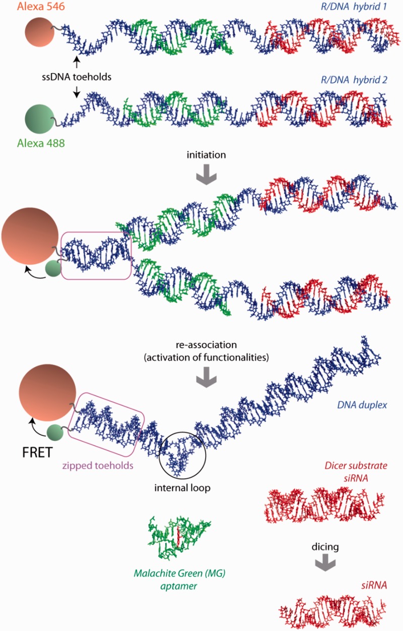
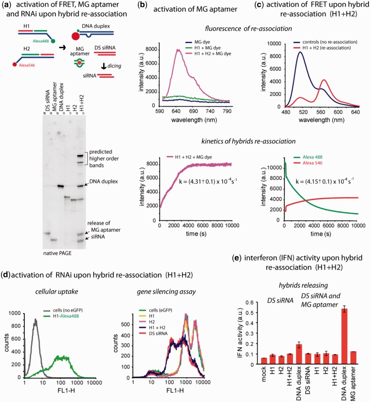
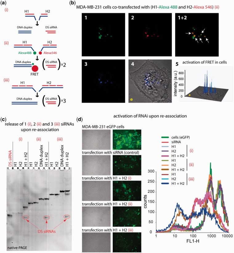
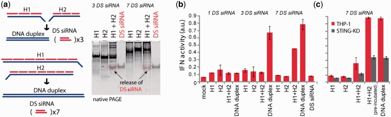
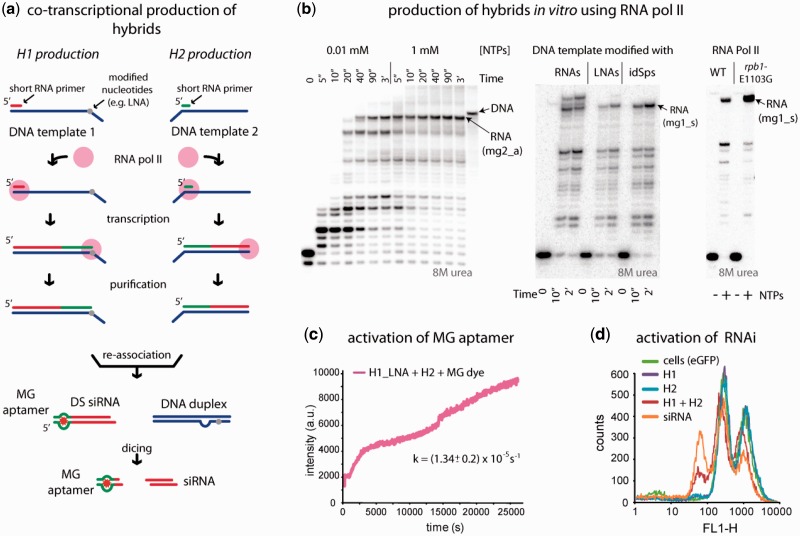
Control over the simultaneous delivery of different functionalities and their synchronized intracellular activation can greatly benefit the fields of RNA and DNA biomedical nanotechnologies and allow for the production of nanoparticles and various switching devices with controllable functions. We present a system of multiple split functionalities embedded in the cognate pairs of RNA-DNA hybrids which are programmed to recognize each other, re-associate and form a DNA duplex while also releasing the split RNA fragments which upon association regain their original functions. Simultaneous activation of three different functionalities (RNAi, Förster resonance energy transfer and RNA aptamer) confirmed by multiple in vitro and cell culture experiments prove the concept. To automate the design process, a novel computational tool that differentiates between the thermodynamic stabilities of RNA-RNA, RNA-DNA and DNA-DNA duplexes was developed. Moreover, here we demonstrate that besides being easily produced by annealing synthetic RNAs and DNAs, the individual hybrids carrying longer RNAs can be produced by RNA polymerase II-dependent transcription of single-stranded DNA templates.


Figures

Figure 1.
Figure 1.
Schematic representation of RNA–DNA hybrid re-association and release of multiple functionalities: FRET response, DS siRNA (in red) and MG RNA aptamer (in green). Three-dimensional (3D) structure of the two-stranded MG aptamer (in green) contains a bound dye (in red). PDB ID: 1f1t. Due to asymmetry of the MG aptamer, the resulting DNA duplex is also asymmetric and contains an internal loop.
Figure 2.
Figure 2.
Release of multiple functionalities (FRET, MG aptamer and DS siRNA) upon re-association of RNA–DNA hybrids. (a) Schematic of hybrid re-association and native PAGE demonstrating the release of DS siRNA and MG aptamer upon re-association of H1(mg1_sDS) and H2(mg2_aDS). Higher order bands on the gel (H1 + H2 lane) are in agreement with the computational predictions and can be attributed to asymmetry of the resulting DNA duplex (36). (b) Static and kinetics fluorescent experiments. Upper panel: activation of MG aptamer during the release. MG by itself is non-fluorescent (blue curve) and the presence of either one of the hybrids does not activate its fluorescence (green curve). However, re-association of two cognate hybrids leads to the release of individual MG aptamer strands, their assembly and further MG uptake leading to the significant increase of its fluorescence (magenta curve). Lower panel: kinetics time trace of the MG aptamer formation during hybrid re-association. (c) FRET activation. Upper panel: activation of FRET during re-association. Emission spectra of control DNA duplexes showing no FRET (blue curve) and re-associated hybrids with increased Alexa546 emission signal (red curve). Lower panel: FRET time traces during re-association of hybrids labeled with Alexa488 and Alexa546. (d) Cell culture experiments. Upper panel: cellular uptake of the fluorescently labeled hybrids. Lower panel: GFP knockdown assays. Three days after the transfection of cells, eGFP expression was statistically analyzed with flow cytometry experiments. As the control, DS siRNA duplexes against eGFP were used. (e) IFN activity was assessed using THP-1 IFN reporter cells that secrete alkaline phosphatase in response to type I IFN. Cells were transfected with hybrids, and culture supernatants were assayed for reporter activity after 24 h.
Figure 3.
Figure 3.
RNA–DNA hybrid re-association with DS siRNA release and intracellular FRET tracking. (a) Schematic of different size hybrids re-association compared in these experiments [(i) H1(sDS) and H2(aDS); (ii) H1(sDS_sDS) and H2 (aDS_aDS); (iii) H1(sDS_sDS_sDS) and H2(aDS_aDS_aDS)]. (b) FRET experiments: cells were co-transfected with cognate hybrids labeled with Alexa 488 and Alexa 546, and images were taken on the next day. (c) Total SYBR Gold staining native PAGE demonstrating the release of DS siRNAs. Due to the full complementarities of the resulting DNA duplexes, there are no higher order bands observed on the gel. (d) GFP knockdown assays. Three days after the transfection of cells with auto-recognizing RNA–DNA hybrids programmed to release one [hybrids (i)], two [hybrids (ii)], and three [hybrids (iii)] DS siRNAs against eGFP, eGFP expression was observed by fluorescence microscopy and statistically analyzed with flow cytometry experiments. As the control, siRNA duplexes against eGFP were used. Image numbers in (b) correspond to: Alexa488 emission (1), Alexa546 emission (2), bleed-through corrected FRET image (3), differential interference contrast image with corrected FRET overlap (4), 3D chart representation of bleed-through corrected FRET image with the yellow dot indicating the correspondence (5).
Figure 4.
Figure 4.
Release of multiple DS siRNA targeting upon re-association of RNA–DNA hybrids. (a) Schematic of hybrid re-association and native PAGE demonstrating the release of three and seven DS siRNA. (b and c) IFN activity assessed using THP-1 IFN reporter cells that secrete alkaline phosphatase in response to type I IFN. Cells were transfected with hybrids, and cell culture supernatants were assayed for reporter activity after 24 h. (b) Cells were transfected with hybrids designed to release varying numbers of DS. (c) THP-1 and THP-1 deficient for STING (in gray) were transfected with hybrids releasing seven DS siRNAs.
Figure 5.
Figure 5.
Co-transcriptional production of RNA-DNA hybrids by yeast RNA Pol II. (a) Schematics of R/DNA hybrid production. (b) Optimization of the full-length RNA synthesis. The RNA primers used for analytical transcription reactions were labeled at the 5′-end by phosphorylation with γ-[32P] ATP. RNA was annealed to DNA, and the elongation complexes were formed as described in Kireeva et al. (18). Transcription was initiated by addition of 1 mM NTPs unless indicated otherwise. Lower bands correspond to intermediate transcription stops. (c and d) Transcription products eluted from the Ni-NTA agarose cartridge containing immobilized Pol II were concentrated by ethanol precipitation, re-dissolved in standard buffer solution and MG2 aptamer (c) and DS siRNA (d) releases were monitored as described above. For (d), the lower relative efficiencies of the functionalities release can be explained by the lower concentrations of the initial hybrids and are consistent with published data (1).

References

    1. Afonin KA, Viard M, Martins AN, Lockett SJ, Maciag AE, Freed EO, Heldman E, Jaeger L, Blumenthal R, Shapiro BA. Activation of different split functionalities on re-association of RNA-DNA hybrids. Nat. Nanotechnol. 2013;8:296–304. - PMC - PubMed
    1. Johnsson N, Varshavsky A. Split ubiquitin as a sensor of protein interactions in vivo. Proc. Natl Acad. Sci. USA. 1994;91:10340–10344. - PMC - PubMed
    1. Cassonnet P, Rolloy C, Neveu G, Vidalain PO, Chantier T, Pellet J, Jones L, Muller M, Demeret C, Gaud G, et al. Benchmarking a luciferase complementation assay for detecting protein complexes. Nat. Methods. 2011;8:990–992. - PubMed
    1. Shekhawat SS, Ghosh I. Split-protein systems: beyond binary protein-protein interactions. Curr. Opin. Chem. Biol. 2011;15:789–797. - PMC - PubMed
    1. Burnett JC, Rossi JJ. RNA-based therapeutics: current progress and future prospects. Chem. Biol. 2012;19:60–71. - PMC - PubMed

Publication types