Preliminary identification of differentially expressed tear proteins in keratoconus
- PMID: 24194634
- PMCID: PMC3816990
Preliminary identification of differentially expressed tear proteins in keratoconus
Abstract
Purpose: To examine the proteins differentially expressed in the tear film of people with keratoconus and normal subjects.
Methods: Unstimulated tears from people with keratoconus (KC) and controls (C) were collected using a capillary tube. Tear proteins from people with KC and controls were partitioned using a novel in-solution electrophoresis, Microflow 10 (ProteomeSep), and analyzed using linear ion trap quadrupole fourier transform mass spectrometry. Spectral counting was used to quantify the individual tear proteins.
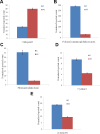
Results: Elevated levels of cathepsin B (threefold) were evident in the tears of people with KC. Polymeric immunoglobulin receptor (ninefold), fibrinogen alpha chain (eightfold), cystatin S (twofold), and cystatin SN (twofold) were reduced in tears from people with KC. Keratin type-1 cytoskeletal-14 and keratin type-2 cytoskeletal-5 were present only in the tears of people with KC.
Conclusions: The protein changes in tears, that is, the decrease in protease inhibitors and increase in proteases, found in the present and other previously published studies reflect the pathological events involved in KC corneas. Further investigations into tear proteins may help elucidate the underlying molecular mechanisms of KC, which could result in better treatment options.
Figures


Similar articles
-
Comparative proteome analysis of the tear samples in patients with low-grade keratoconus.Int Ophthalmol. 2018 Oct;38(5):1895-1905. doi: 10.1007/s10792-017-0672-6. Epub 2017 Aug 7. Int Ophthalmol. 2018. PMID: 28785876
-
Changes in tear protein profile in keratoconus disease.Eye (Lond). 2011 Sep;25(9):1225-33. doi: 10.1038/eye.2011.105. Epub 2011 Jun 24. Eye (Lond). 2011. PMID: 21701529 Free PMC article.
-
Proteases, proteolysis and inflammatory molecules in the tears of people with keratoconus.Acta Ophthalmol. 2012 Jun;90(4):e303-9. doi: 10.1111/j.1755-3768.2011.02369.x. Epub 2012 Mar 13. Acta Ophthalmol. 2012. PMID: 22413749
-
Keratoconus: an inflammatory disorder?Eye (Lond). 2015 Jul;29(7):843-59. doi: 10.1038/eye.2015.63. Epub 2015 May 1. Eye (Lond). 2015. PMID: 25931166 Free PMC article. Review.
-
Tear Levels of Inflammatory Cytokines in Keratoconus: A Meta-Analysis of Case-Control and Cross-Sectional Studies.Biomed Res Int. 2021 Sep 30;2021:6628923. doi: 10.1155/2021/6628923. eCollection 2021. Biomed Res Int. 2021. PMID: 34631885 Free PMC article. Review.
Cited by
-
Pediatric keratoconus: a review of the literature.Int Ophthalmol. 2018 Oct;38(5):2257-2266. doi: 10.1007/s10792-017-0699-8. Epub 2017 Aug 29. Int Ophthalmol. 2018. PMID: 28852910 Free PMC article. Review.
-
What's the situation with ocular inflammation? A cross-seasonal investigation of proteomic changes in ocular allergy sufferers' tears in Victoria, Australia.Front Immunol. 2024 May 24;15:1386344. doi: 10.3389/fimmu.2024.1386344. eCollection 2024. Front Immunol. 2024. PMID: 38855108 Free PMC article.
-
Shotgun Proteomics for the Identification and Profiling of the Tear Proteome of Keratoconus Patients.Invest Ophthalmol Vis Sci. 2022 May 2;63(5):12. doi: 10.1167/iovs.63.5.12. Invest Ophthalmol Vis Sci. 2022. PMID: 35551575 Free PMC article.
-
Targeted Workflow Investigating Variations in the Tear Proteome by Liquid Chromatography Tandem Mass Spectrometry.ACS Omega. 2023 Aug 14;8(34):31168-31177. doi: 10.1021/acsomega.3c03186. eCollection 2023 Aug 29. ACS Omega. 2023. PMID: 37663498 Free PMC article.
-
Inflammatory Biomarkers Profile as Microenvironmental Expression in Keratoconus.Dis Markers. 2016;2016:1243819. doi: 10.1155/2016/1243819. Epub 2016 Aug 3. Dis Markers. 2016. PMID: 27563164 Free PMC article. Review.
References
-
- Kao WW, Vergnes JP, Ebert J, Sundar-Raj CV, Brown SI. Increased collagenase and gelatinase activities in keratoconus. Biochem Biophys Res Commun. 1982;107:929–36. - PubMed
-
- Rehany U, Lahav M, Shoshan S. Collagenolytic activity in keratoconus. Ann Ophthalmol. 1982;14:751–4. - PubMed
-
- Kenney MC, Chwa M, Escobar M, Brown D. Altered gelatinolytic activity by keratoconus corneal cells. Biochem Biophys Res Commun. 1989;161:353–7. - PubMed
-
- Fini ME, Yue BY, Sugar J. Collagenolytic/gelatinolytic metalloproteinases in normal and keratoconus corneas. Curr Eye Res. 1992;11:849–62. - PubMed
Publication types
MeSH terms
Substances
LinkOut - more resources
Full Text Sources
