

3-dimensional physiologic postural range of the mandible: a computerized-assisted technique-a case study
- PMID: 24194764
- PMCID: PMC3806511
- DOI: 10.1155/2013/698397
3-dimensional physiologic postural range of the mandible: a computerized-assisted technique-a case study
Abstract
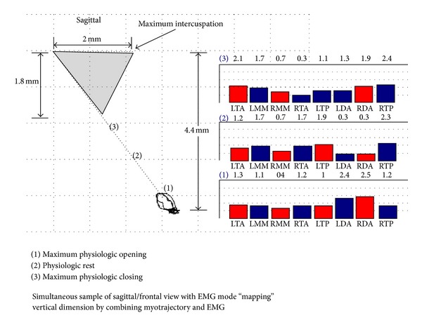
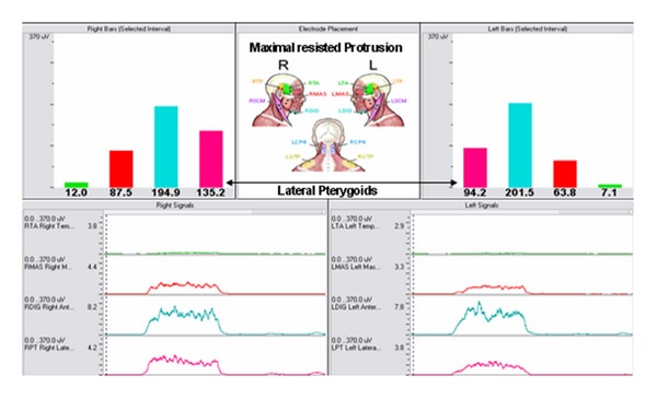
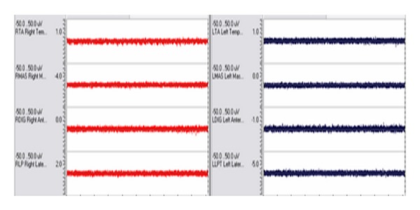
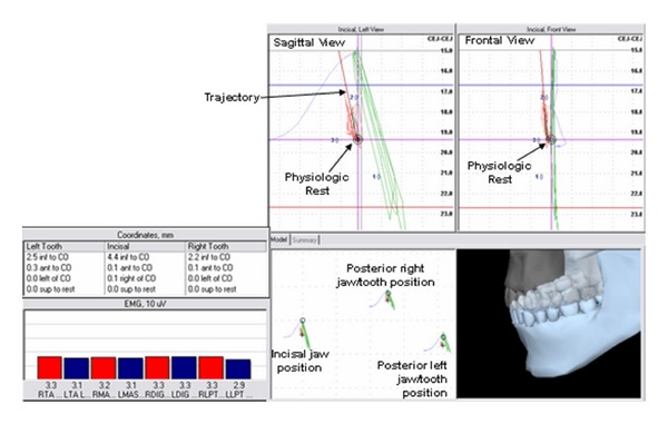
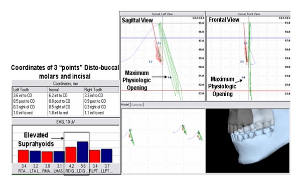
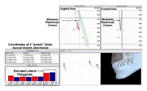
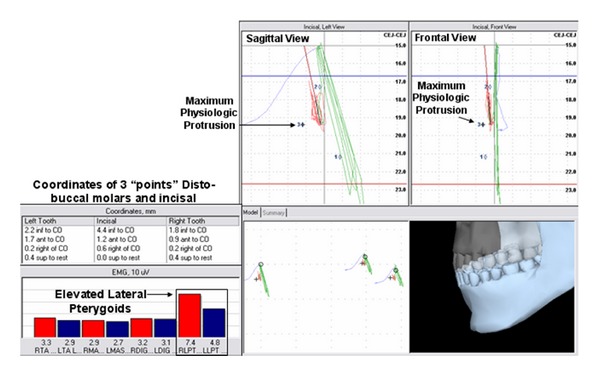
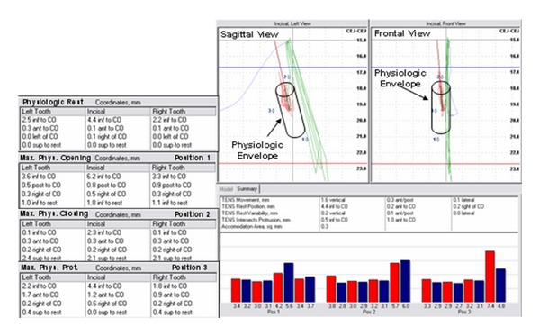
Previous studies demonstrated that while the mandible assumes its resting position in space, antagonistic muscles should assume minimal muscle activity within a spatial range. This zone of mandibular rest has been mapped using physiologic parameters of muscle activity and incisal spatial kinematics. This case study expands on previous research by monitoring incisal and posterior jaw position and includes lateral pterygoid muscle activity, thus allowing for determining the spatial range including additional relevant coordinates and muscle activity. Four positions were evaluated: a maximum physiologic open position, a maximum physiologic closed position, physiologic rest position, and maximum physiologic protrusion position. Within the physiologic zone of rest formed by these 4 positions, the vertical and anterior borders of the envelope of function may be documented for the incisal and posterior mandible in true 3-dimensional fashion to assist the clinician in determining a physiologic interocclusal freeway space and vertical dimension of occlusion. Advantages and limitations are discussed.
Figures
Similar articles
-
Mapping mandibular rest in humans utilizing electromyographic patterns from masticatory muscles.Cranio. 2007 Oct;25(4):264-72. doi: 10.1179/crn.2007.040. Cranio. 2007. PMID: 17983126
-
Muscle activity during mandibular movements in normal and mandibular retrognathic subjects.J Oral Maxillofac Surg. 1997 Mar;55(3):225-33. doi: 10.1016/s0278-2391(97)90530-9. J Oral Maxillofac Surg. 1997. PMID: 9054910
-
A preliminary study on the effect of occlusal vertical dimension increase on mandibular postural rest position.Int J Prosthodont. 1994 May-Jun;7(3):216-26. Int J Prosthodont. 1994. PMID: 7916886
-
The human lateral pterygoid muscle: a review of some experimental aspects and possible clinical relevance.Aust Dent J. 2004 Mar;49(1):2-8. doi: 10.1111/j.1834-7819.2004.tb00042.x. Aust Dent J. 2004. PMID: 15104127 Review.
-
A review of masticatory muscle function.J Prosthet Dent. 1987 Feb;57(2):222-32. doi: 10.1016/0022-3913(87)90151-x. J Prosthet Dent. 1987. PMID: 3550055 Review.
Cited by
-
A Comparative Analysis of Temporomandibular Disorders Using a Jaw Motion Analyzer and Surface Electromyography.Int Dent J. 2025 Jun;75(3):1843-1853. doi: 10.1016/j.identj.2025.03.023. Epub 2025 Apr 26. Int Dent J. 2025. PMID: 40288077 Free PMC article.
References
-
- Cooper BC. Scientific rationale for biomedical instrumentation. In: Hickman DM, Mazzocco MW, Rojas RM, editors. Neuromuscular Dentistry the Next Millennium Anthology. Vol. 5. Seattle, Wash, USA: International College of Cranio-Mandibular Orthopedics (ICCMO); 1999.
-
- Dickerson WD, Chan C, Mazzocco MW. The scientific approach: neuromuscular occlusion. Signature. 2000;7(2, article 14)
-
- Kandell E, Schwartz J, Jessell TM. Principles of Neural Science. New York, NY, USA: McGraw-Hill; 2000.
-
- The American Heritage Stedman's Medical Dictionary. 2nd edition 2004.
-
- Fish F. The functional anatomy of the rest position of the mandible. Dental Practitioner and Dental Record. 1961;2:178–188.
LinkOut - more resources
Full Text Sources
Other Literature Sources
