Characterization of a soluble B7-H3 (sB7-H3) spliced from the intron and analysis of sB7-H3 in the sera of patients with hepatocellular carcinoma
- PMID: 24194851
- PMCID: PMC3806749
- DOI: 10.1371/journal.pone.0076965
Characterization of a soluble B7-H3 (sB7-H3) spliced from the intron and analysis of sB7-H3 in the sera of patients with hepatocellular carcinoma
Abstract
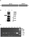
B7-H3 is a recently discovered member of the B7 superfamily molecules and has been found to play a negative role in T cell responses. In this study, we identified a new B7-H3 isoform that is produced by alternative splicing from the forth intron of B7-H3 and encodes the sB7-H3 protein. Protein sequence analysis showed that sB7-H3 contains an additional four amino acids, encoded by the intron sequence, at the C-terminus compared to the ectodomain of 2Ig-B7-H3. We further found that this spliced sB7-H3 plays a negative regulatory role in T cell responses and serum sB7-H3 is higher in patients with hepatocellular carcinoma (HCC) than in healthy donors. Furthermore, we found that the expression of the spliced sb7-h3 gene is higher in carcinoma and peritumor tissues than in PBMCs of both healthy controls and patients, indicating that the high level of serum sB7-H3 in patients with HCC is caused by the increased expression of this newly discovered spliced sB7-H3 isoform in carcinoma and peritumor tissues.
Conflict of interest statement
Figures


Similar articles
-
[Plasma sB7-H3 serves as an assistant diagnostic marker for hepatitis B virus-associated hepatocellular carcinoma].Xi Bao Yu Fen Zi Mian Yi Xue Za Zhi. 2014 Jul;30(7):744-7. Xi Bao Yu Fen Zi Mian Yi Xue Za Zhi. 2014. PMID: 25001942 Chinese.
-
[Expression and clinical significance of soluble B7-H3 in sera of patients with primary hepatocellular carcinoma].Xi Bao Yu Fen Zi Mian Yi Xue Za Zhi. 2013 Jun;29(6):629-32. Xi Bao Yu Fen Zi Mian Yi Xue Za Zhi. 2013. PMID: 23746248 Chinese.
-
Early Detection of Hepatocellular Carcinoma in Patients with Hepatocirrhosis by Soluble B7-H3.J Gastrointest Surg. 2017 May;21(5):807-812. doi: 10.1007/s11605-017-3386-1. Epub 2017 Feb 27. J Gastrointest Surg. 2017. PMID: 28243980
-
B7-H3 in tumors: friend or foe for tumor immunity?Cancer Chemother Pharmacol. 2018 Feb;81(2):245-253. doi: 10.1007/s00280-017-3508-1. Epub 2018 Jan 3. Cancer Chemother Pharmacol. 2018. PMID: 29299639 Review.
-
B7-H3-mediated tumor immunology: Friend or foe?Int J Cancer. 2014 Jun 15;134(12):2764-71. doi: 10.1002/ijc.28474. Epub 2013 Sep 30. Int J Cancer. 2014. PMID: 24013874 Review.
Cited by
-
CD276 as a Candidate Target for Immunotherapy in Medullary Thyroid Cancer.Int J Mol Sci. 2023 Jun 12;24(12):10019. doi: 10.3390/ijms241210019. Int J Mol Sci. 2023. PMID: 37373167 Free PMC article.
-
Molecular Pathways: Targeting B7-H3 (CD276) for Human Cancer Immunotherapy.Clin Cancer Res. 2016 Jul 15;22(14):3425-3431. doi: 10.1158/1078-0432.CCR-15-2428. Epub 2016 May 20. Clin Cancer Res. 2016. PMID: 27208063 Free PMC article.
-
New Emerging Targets in Cancer Immunotherapy: The Role of B7-H3.Vaccines (Basel). 2024 Jan 5;12(1):54. doi: 10.3390/vaccines12010054. Vaccines (Basel). 2024. PMID: 38250867 Free PMC article. Review.
-
Soluble immune checkpoints in cancer: production, function and biological significance.J Immunother Cancer. 2018 Nov 27;6(1):132. doi: 10.1186/s40425-018-0449-0. J Immunother Cancer. 2018. PMID: 30482248 Free PMC article. Review.
-
Global research trends on B7-H3 for cancer immunotherapy: A bibliometric analysis (2012-2022).Hum Vaccin Immunother. 2023 Aug 1;19(2):2246498. doi: 10.1080/21645515.2023.2246498. Hum Vaccin Immunother. 2023. PMID: 37635349 Free PMC article.
References
-
- Greenwald RJ, Freeman GJ, Sharpe AH (2005) The B7 family revisited. Annu Rev Immunol 23: 515–548. - PubMed
-
- Latchman Y, Wood CR, Chernova T, Chaudhary D, Borde M, et al. (2001) PD-L2 is a second ligand for PD-1 and inhibits T cell activation. Nat Immunol 2: 261–268. - PubMed
-
- Yoshinaga SK, Whoriskey JS, Khare SD, Sarmiento U, Guo J, et al. (1999) T-cell co-stimulation through B7RP-1 and ICOS. Nature 402: 827–832. - PubMed
-
- Dong H, Zhu G, Tamada K, Chen L (1999) B7-H1, a third member of the B7 family, co-stimulates T-cell proliferation and interleukin-10 secretion. Nat Med 5: 1365–1369. - PubMed
-
- Kryczek I, Wei S, Zhu G, Myers L, Mottram P, et al. (2007) Relationship between B7-H4, regulatory T cells, and patient outcome in human ovarian carcinoma. Cancer Res 67: 8900–8905. - PubMed
Publication types
MeSH terms
Substances
LinkOut - more resources
Full Text Sources
Other Literature Sources
Medical
Research Materials

