. 2013 Oct 23;8(10):e77282.
doi: 10.1371/journal.pone.0077282. eCollection 2013.

Streptococcus pneumoniae invades erythrocytes and utilizes them to evade human innate immunity

Affiliations

Streptococcus pneumoniae invades erythrocytes and utilizes them to evade human innate immunity

Masaya Yamaguchi et al. PLoS One. .

Erratum in

  • PLoS One. 2014;9(1). doi:10.1371/annotation/de14e562-d899-48da-b677-ca61cca2cf5b

Abstract

Streptococcus pneumoniae, a Gram-positive bacterium, is a major cause of invasive infection-related diseases such as pneumonia and sepsis. In blood, erythrocytes are considered to be an important factor for bacterial growth, as they contain abundant nutrients. However, the relationship between S. pneumoniae and erythrocytes remains unclear. We analyzed interactions between S. pneumoniae and erythrocytes, and found that iron ion present in human erythrocytes supported the growth of Staphylococcus aureus, another major Gram-positive sepsis pathogen, while it partially inhibited pneumococcal growth by generating free radicals. S. pneumoniae cells incubated with human erythrocytes or blood were subjected to scanning electron and confocal fluorescence microscopic analyses, which showed that the bacterial cells adhered to and invaded human erythrocytes. In addition, S. pneumoniae cells were found associated with human erythrocytes in cultures of blood from patients with an invasive pneumococcal infection. Erythrocyte invasion assays indicated that LPXTG motif-containing pneumococcal proteins, erythrocyte lipid rafts, and erythrocyte actin remodeling are all involved in the invasion mechanism. In a neutrophil killing assay, the viability of S. pneumoniae co-incubated with erythrocytes was higher than that without erythrocytes. Also, H2O2 killing of S. pneumoniae was nearly completely ineffective in the presence of erythrocytes. These results indicate that even when S. pneumoniae organisms are partially killed by iron ion-induced free radicals, they can still invade erythrocytes. Furthermore, in the presence of erythrocytes, S. pneumoniae can more effectively evade antibiotics, neutrophil phagocytosis, and H2O2 killing.


Conflict of interest statement

Competing Interests: The authors have declared that no competing interests exist.

Figures

Figure 1
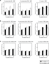
Figure 1. Effects of erythrocytes and iron ions on S. pneumoniae growth.
A. Growth of S. pneumoniae strains R6 and D39, and S. aureus strain Cowan-I in the presence of human erythrocytes with or without an iron chelator. Bacterial cells were incubated for 2, 4, and 6 hours at 37°C in a 5% CO2 atmosphere. B. Growth of S. pneumoniae strains R6 and D39, and S. aureus strain Cowan-I in erythrocyte intracellular solution (erythrocyte lysates without membrane) with or without an iron chelator for 2, 4, and 6 hours at 37°C in a 5% CO2 atmosphere. C. Growth of S. pneumoniae strains R6 and D39, and S. aureus strain Cowan-I in RPMI 1640 medium with or without an iron chelator for 2, 4, and 6 hours at 37°C in a 5% CO2 atmosphere. The experiments were performed 3 times and data shown represent the mean of 3 wells from a representative experiment. S.D. values are represented by vertical lines.
Figure 2
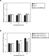
Figure 2. Erythrocyte intracellular solution inhibits pneumococcal growth in presence of erythrocytes with the membrane.
A. Growth of S. pneumoniae strain D39 in RPMI 1640 medium with or without human hemoglobin and/or an iron chelator for 2, 4, and 6 hours at 37°C in a 5% CO2 atmosphere. B. Growth of S. pneumoniae strain D39 in erythrocyte lysates with or without the erythrocyte membrane and/or an iron chelator for 2, 4, and 6 hours at 37°C in a 5% CO2 atmosphere. The experiments were performed 3 times and data shown represent the mean of 3 wells from a representative experiment. S.D. values are represented by vertical lines.
Figure 3
Figure 3. Erythrocytes inhibit pneumococcal growth by reactive oxygen species-related mechanism.
S. pneumoniae cells (∼1×10² CFU, 10 µl) were added to erythrocytes (5×10⁹ cells/ml, 190 µl) with or without 1 mM 2,2′-bipyridyl (iron chelator), 1 mM S-ethyl-ITU (nitric oxide synthase inhibitor), 100 µM EUK8 (synthetic catalytic free radical scavenger), or 150 µM MnTBAP (superoxide dismutase mimetic) for 2 hours at 37°C in a 5% CO2 atmosphere. Next, each mixture was serially diluted and plated on TS blood agar. Following incubation, CFU values were determined. *Significant difference (P<0.005) between mean values, as determined with a Mann-Whitney U-test. The experiments were performed 3 times and data are shown as the mean of 6 wells from a representative experiment. S.D. values are represented by vertical lines.
Figure 4
Figure 4. S. pneumoniae invasion of human erythrocytes.
A. Gram staining of cultures of blood obtained from a patient with invasive pneumococcal pneumonia. A blood sample was obtained from a splenectomized patient with pneumococcal bacteremia and meningitis. We observed that some of the S. pneumoniae cells in the sample adhered to or invaded erythrocytes. B. SEM analysis of S. pneumoniae in blood. S. pneumoniae cells (arrows) were incubated in human whole blood for 30 minutes at 37°C. Strains R6 and D39 adhered to (a, c) and invaded (b, d) erythrocytes in human blood. C. Confocal fluorescence microscopic analysis of S. pneumoniae strains R6 (a, b) and D39 (c, d) incubated with human erythrocytes for 30 minutes at 37°C. (a, c) Erythrocytes were visualized using Alexa Fluor 594 Phalloidin. S. pneumoniae organisms were stained using SYTOX green. (b) Boxed areas from panel (a), along with x–z and y-z projections. (d) 3D analysis of image from panel (c) showing erythrocytes invaded by S. pneumoniae. D. Rate of S. pneumoniae invasion of erythrocytes. The numbers of invaded bacteria were determined as described in the Experimental Procedures section. *Significant difference (P<0.005) between mean values, as determined with a Mann-Whitney U-test. The experiments were performed 3 times and data are shown as the mean of 6 wells from a representative experiment. S.D. values are represented by vertical lines. E. Histopathological examinations of infected mice lung tissues. Tissues were excised from sites of infection after 72 hours, then fixed, embedded in paraffin, and stained with hematoxylin-eosin solution. (a) and (b) were obtained from individual mice. Arrows indicate association of S. pneumoniae with erythrocytes. (c) Numbers of bacteria associated with erythrocytes per field. Data shown represent the mean of 10 fields from a representative mouse. S.D. values are represented by vertical lines.
Figure 5
Figure 5. Involvement of lipid rafts and actin remodeling in erythrocyte invasion by S. pneumoniae.
Erythrocytes were pretreated with or without 5βCD or 20 µM cytochalasin D for 30 minutes at 4°C, then S. pneumoniae cells were added and incubated for 1 hour at 37°C in a 5% CO2 atmosphere. The numbers of invaded bacteria were determined as described in the Experimental Procedures section. The experiments were performed 3 times and data shown represent the mean of 6 wells from a representative experiment. S.D. values are represented by vertical lines. *P<0.005; **P<0.05.
Figure 6
Figure 6. Erythrocytes inhibit killing of S. pneumoniae.
A. Inhibition of killing by neutrophils. S. pneumoniae cells (R6∶2.2×10² CFU/well, D39∶1.4×10² CFU/well) were incubated with human neutrophils (1×10⁵ cells/well), then erythrocytes (5.0×10⁷ cells/well) and/or 10% human blood serum (HBS) or heat-inactivated HBS were added to the mixture. Viable CFU were counted following 1, 2, and 3 hours of incubation. B. Inhibition of killing by H2O2. S. pneumoniae cells were incubated in 0%, 0.03%, or 0.30% H2O2-RPMI 1640, then viable CFU were counted following 1, 2, and 3 hours of incubation. The experiments were performed 3 times and data shown represent the mean of 6 wells from a representative experiment. S.D. values are represented by vertical lines.

References

    1. van der Poll T, Opal SM (2009) Pathogenesis, treatment, and prevention of pneumococcal pneumonia. Lancet 374: 1543–1556. - PubMed
    1. O’Brien KL, Wolfson LJ, Watt JP, Henkle E, Deloria-Knoll M, et al. (2009) Burden of disease caused by Streptococcus pneumoniae in children younger than 5 years: global estimates. Lancet 374: 893–902. - PubMed
    1. Mitchell AM, Mitchell TJ (2010) Streptococcus pneumoniae: virulence factors and variation. Clin Microbiol Infect 16: 411–418. - PubMed
    1. Kyaw MH, Lynfield R, Schaffner W, Craig AS, Hadler J, et al. (2006) Effect of introduction of the pneumococcal conjugate vaccine on drug-resistant Streptococcus pneumoniae . N Engl J Med 354: 1455–1463. - PubMed
    1. Farrell DJ, Klugman KP, Pichichero M (2007) Increased antimicrobial resistance among nonvaccine serotypes of Streptococcus pneumoniae in the pediatric population after the introduction of 7-valent pneumococcal vaccine in the United States. Pediatr Infect Dis J 26: 123–128. - PubMed

Publication types

MeSH terms