. 2013 Oct 23;8(10):e78539.
doi: 10.1371/journal.pone.0078539. eCollection 2013.

Energetic contributions to channel gating of residues in the muscle nicotinic receptor β1 subunit

Affiliations

Energetic contributions to channel gating of residues in the muscle nicotinic receptor β1 subunit

Gustav Akk et al. PLoS One. .

Abstract

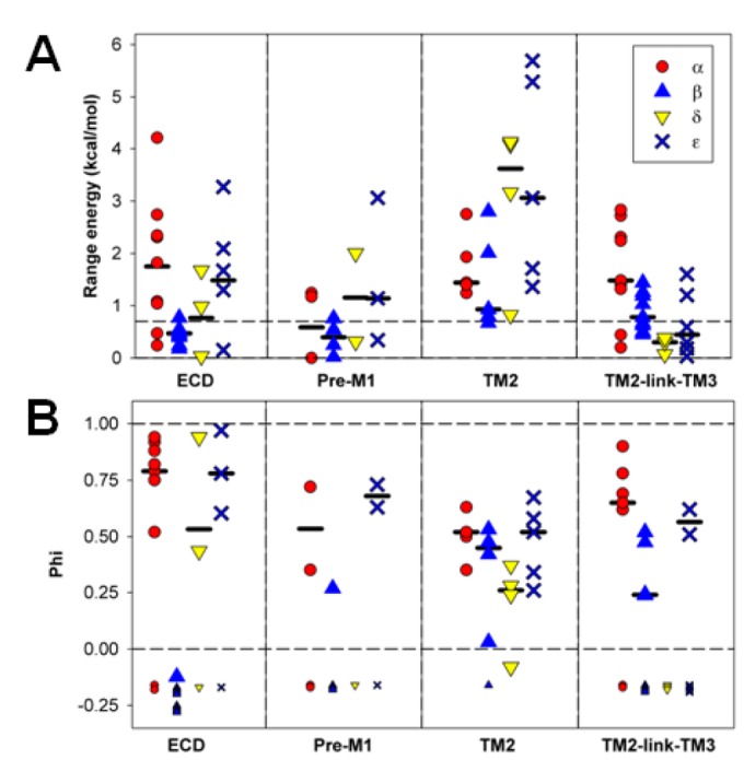
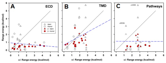
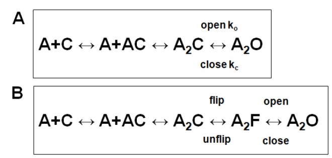
In the pentameric ligand-gated ion channel family, transmitter binds in the extracellular domain and conformational changes result in channel opening in the transmembrane domain. In the muscle nicotinic receptor and other heteromeric members of the family one subunit does not contribute to the canonical agonist binding site for transmitter. A fundamental question is whether conformational changes occur in this subunit. We used records of single channel activity and rate-equilibrium free energy relationships to examine the β1 (non-ACh-binding) subunit of the muscle nicotinic receptor. Mutations to residues in the extracellular domain have minimal effects on the gating equilibrium constant. Positions in the channel lining (M2 transmembrane) domain contribute strongly and relatively late during gating. Positions thought to be important in other subunits in coupling the transmitter-binding to the channel domains have minimal effects on gating. We conclude that the conformational changes involved in channel gating propagate from the binding-site to the channel in the ACh-binding subunits and subsequently spread to the non-binding subunit.


Conflict of interest statement

Competing Interests: The authors have declared that no competing interests exist.

Figures

Figure 1
Figure 1. Schematic summaries of AChR structure.
Panel A shows the locations of relevant regions in the primary sequences. There are 4 transmembrane regions (TM1-TM4). The channel is formed by the TM2 regions (highlighted in red) from the 5 subunits. The ACh-binding loops are in the N-terminal extracellular region (loops A-C form the "principal" side, D-F the "complementary" side). Panel B shows then membrane topology of a subunit (ACh-binding site: green circle, TM2 helix red cylinder). Panel C shows the arrangement of subunits in the pentamer (viewed from the extracellular side; green circles - ACh-binding interfaces). The channel is located in the center of the rosette of subunits. Panel D shows a homology model of the mouse β1 subunit, threaded on the C. elegans GluCl E subunit [16]. The main chain is shown in blue, and the extracellular domain (ECD) and transmembrane domain (TMD) are indicated by brackets. The region coupling the ECD to the TMD is indicated by "coupling," and includes residues in the PreM1, loop 9, the TM2-link-TM3 region and the "principal pathway" (see text). In the ECD mutated residues in "loop 9" are shown in cyan, "Pre M1" in green, K46 in orange, V132 in yellow and other residues in light gray. In the TMD mutated residues in "TM2" are in blue and in the TM2-link-TM3 region in red. Many of the residues studied are located at or near the interface between the ECD and TMD regions of the subunit. Panel E shows the kinetic scheme used to interpret the data. A receptor with a closed channel (C) binds 2 molecules of agonist (A) then the channel opens (O) with an apparent opening rate ko and apparent closing rate kc.
Figure 2
Figure 2. Rate equilibrium free energy relationships.
Panel A shows sample traces of data for wild-type receptors and receptors containing β1(L270A). On the left data at a low concentration (100 μM) of choline are shown, with histograms of the open durations shown below. The fits to the histograms were used to estimate the channel closing rate. The openings are clearly prolonged by the mutation, and the estimates for kc are 2128 s-1 for wild type and 360 s-1 for the mutant. The number of events in the histogram are 506 for β1(L270A) and 695 for wild type. On the right data at a high concentration (20 mM) are shown, with histograms of the closed durations below. The estimates for ko are 44 s-1 for wild type and 153 s-1 for the mutant (estimated from the major, slower component). The number of events in the histogram are 2390 for β1(L270A) and 2259 for wild type. Note that open channel block reduces the channel current amplitude at this high concentration. Panel B shows logarithmic plots of ko on E2 for mutations at 3 positions in the β1 subunit. The lines show the linear regression of log(ko) on log(E2). The range energy is calculated from the range of E2 values and φ is the slope of the linear regression (given as regression value ± SE of fit value). β1(Y149) illustrates a position at which the range energy is small, β1(V266) a position with a linear relationship, and β1(V275) a position at which the slope of the line is poorly defined. The hollow symbol shows data for wild-type receptors. Data points are identified with the residue at the position, and are mean ± SE. Rate-equilibrium plots for all positions are shown in Figure S2.
Figure 3
Figure 3. Range energy at homologous positions in the subunits.
Panel A shows data for positions in the ECD, while panel B shows data for the TMD. The sites are identified by the residue and position in the β1 subunit, and some structural features are shown below (see Text). Residues proposed to be part of the "principal pathway" for coupling binding to gating in the α1 subunit [25] are indicated by stars above the data, and residues lying in a "high range energy column" in the α1 subunit [8] are indicated by plus signs. Results for the β1 subunit are shown by blue triangles (results from the present study shown as filled triangles, while results obtained by others as open triangles). Values for α1 are shown with filled red circles, for δ by yellow inverted triangles and ε by bold crosses. Values are given on a per subunit basis. The dashed line at 0.7 kcal/mol shows the discriminator chosen for the minimal range energy for considering an estimate of φ to be reliable. The values for the present data are shown in Table 1, and all values are shown in Tables S1 and S2.
Figure 4
Figure 4. Summary of results for timing.
Values are shown for φ, in a similar format to that of Figure 3. The small symbols shown below the solid horizontal line indicate positions at which the range energy was less than 0.7 kcal/mol and for which φ could not be reliably calculated. The values for the present data are shown in Table 1, and all values are shown in Tables S1 and S2.
Figure 5
Figure 5. Parameters in subunits divided by structural regions.
The data for positions examined in the present study are summarized in terms of the structural region in which the residue is located. The regions shown are ECD (from β1(V46) to Q184), Pre-M1 (β1(I218) to K221), TM2 (β1(S257) to L270) and TM2-link-TM3 (β1(V275) to I286). The upper panel shows data for range energy, with the median values for the individual subunits indicated by the horizontal bars, while the lower panel shows similar data for φ.
Figure 6
Figure 6. Range energies at homologous positions.
Scatter plots for range energy are shown for positions in the β1, δ or ε subunits (ordinate) plotted against the value for the homologous position in α1 (abscissa). All data shown in Figure 3 are plotted. The regions shown are ECD (β1(V46) to K221), TMD (β1(S257) to I286) and Pathways (see Text, β1(E45), K46, N96, S127, Y149, R220, V266, P276, S279, L280 and P283). Two positions are identified by their locations in the α1 subunit, that have large range energies in the complementary face subunits (δ and ε) but lower values in the β1 subunit. Positions at which all 4 subunits have the same amino acid are shown by triangles, while positions at which one or more differ are shown by circles. Data for the β1 subunit are shown as filled red symbols. The solid lines show the line of equality, the dashed lines show the regression lines (no slope differed significantly from 0).
Figure 7
Figure 7. A kinetic model with additional closed states.
Panel A shows the kinetic scheme used in interpreting our data. Panel B shows an extended scheme incorporating a closed-closed conformational change ("flip") preceeding the channel opening step. In this scheme, A receptor with a closed channel (C) binds 2 molecules of agonist (A) then the receptor enters the flipped state (F) while keeping a closed channel. The channel opens (O) from the flipped state. The scheme is discussed further in the Text.

Similar articles

Cited by

References

    1. Ortells MO, Lunt GG (1995) Evolutionary history of the ligand-gated ion-channel superfamily of receptors. Trends Neurosci 18: 121-127. doi:10.1016/0166-2236(95)93887-4. PubMed: 7754520. - DOI - PubMed
    1. Tasneem A, Iyer LM, Jakobsson E, Aravind L (2005) Identification of the prokaryotic ligand-gated ion channels and their implications for the mechanisms and origins of animal Cys-loop ion channels. Genome Biol 6: R4. doi:10.1186/gb-2005-6-4-p4. PubMed: 15642096. - DOI - PMC - PubMed
    1. Dent JA (2006) Evidence for a diverse Cys-loop ligand-gated ion channel superfamily in early bilateria. J Mol Evol 62: 523-535. doi:10.1007/s00239-005-0018-2. PubMed: 16586016. - DOI - PubMed
    1. Moroni M, Zwart R, Sher E, Cassels BK, Bermudez I (2006) alpha4beta2 nicotinic receptors with high and low acetylcholine sensitivity: pharmacology, stoichiometry, and sensitivity to long-term exposure to nicotine. Mol Pharmacol 70: 755-768. doi:10.1124/mol.106.023044. PubMed: 16720757. - DOI - PubMed
    1. Akk G, Bracamontes J, Steinbach JH (2004) Activation of GABA(A) receptors containing the alpha4 subunit by GABA and pentobarbital. J Physiol 556: 387-399. doi:10.1113/jphysiol.2003.058230. PubMed: 14966300. - DOI - PMC - PubMed

Publication types

LinkOut - more resources