

Pruritic vesicular eruption on the lower legs in a diabetic female
- PMID: 24194986
- PMCID: PMC3806357
- DOI: 10.1155/2013/641416
Pruritic vesicular eruption on the lower legs in a diabetic female
Abstract
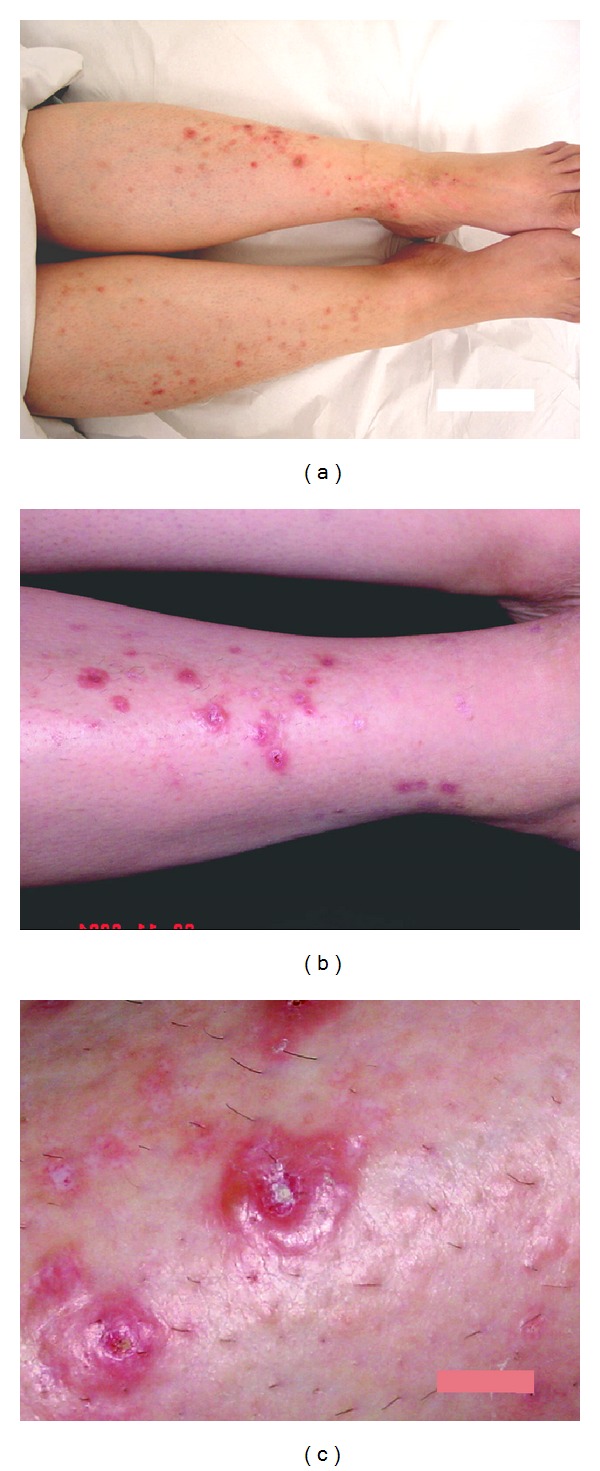
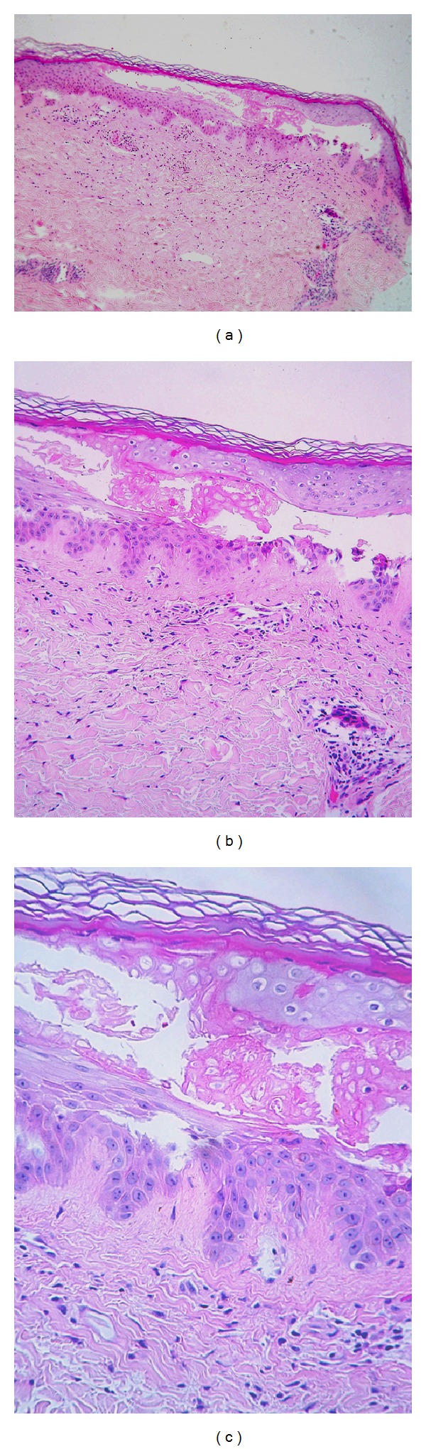
A 50-year-old diabetic female presented with highly pruritic vesicles and excoriated lesions over the anterior aspect of both lower legs. The lesions were recurrent over the last two years. She received a lot of medications with partial response. Hb A1c was 10.8% (normal up to 7%). CBC showed microcytic, hypochromic anemia. Serum zinc, folate, IgE, TSH and T4 were all within normal ranges. Biopsy showed epidermal separation secondary to keratinocyte necrosis and minimal monocytic, perivascular infiltrate. Direct immunofluorescence was negative for intraepidermal and subepidremal deposition of immunoglobulin. The dermis was positive for mucin deposition stainable by both PAS and Alcian blue while it was negative for Congo red and APC immunoperoxidase staining for amyloid material. In conclusion, the case was diagnosed as bullosis diabeticorum by distinctive clinical and pathological features and after exclusion of other possible differentials. Pruritus was partially controlled by topical potent steroid and the case was resolved spontaneously after eight months.
Figures
References
-
- Cantwell AR, Jr., Martz W. Idiopathic bullae in diabetics. bullosis diabeticorum. Archives of Dermatology. 1967;96(1):42–44. - PubMed
-
- Bernstein JE, Medenica M, Soltani K, Griem SF. Bullous eruption of diabetes mellitus. Archives of Dermatology. 1979;115(3):324–325. - PubMed
-
- Goodfield MJ, Millard LG, Harvey L, Jeffcoate WJ. Bullosis diabeticorum. Journal of the American Academy of Dermatology. 1986;15(6):1292–1294. - PubMed
-
- Aye M, Masson EA. Dermatological care of the diabetic foot. American Journal of Clinical Dermatology. 2002;3(7):463–474. - PubMed
LinkOut - more resources
Full Text Sources
Other Literature Sources
