

Solitary fibrous tumor of neck mimicking cold thyroid nodule in 99m tc thyroid scintigraphy
- PMID: 24194989
- PMCID: PMC3806406
- DOI: 10.1155/2013/805745
Solitary fibrous tumor of neck mimicking cold thyroid nodule in 99m tc thyroid scintigraphy
Abstract
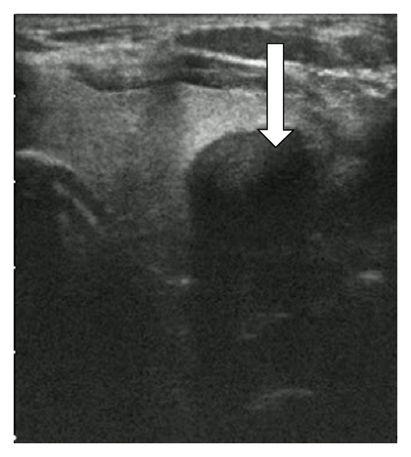
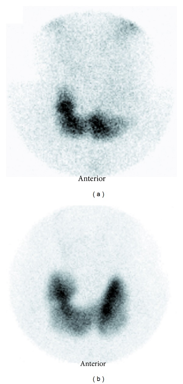
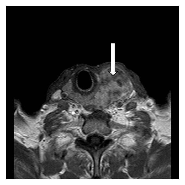
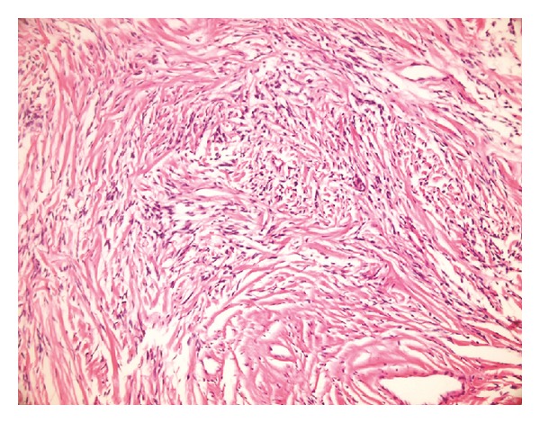
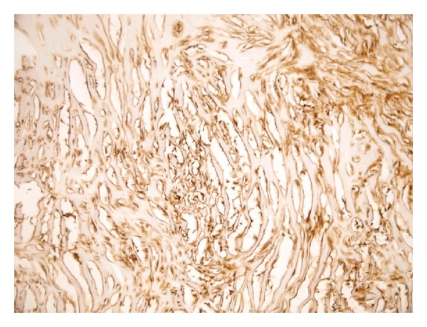
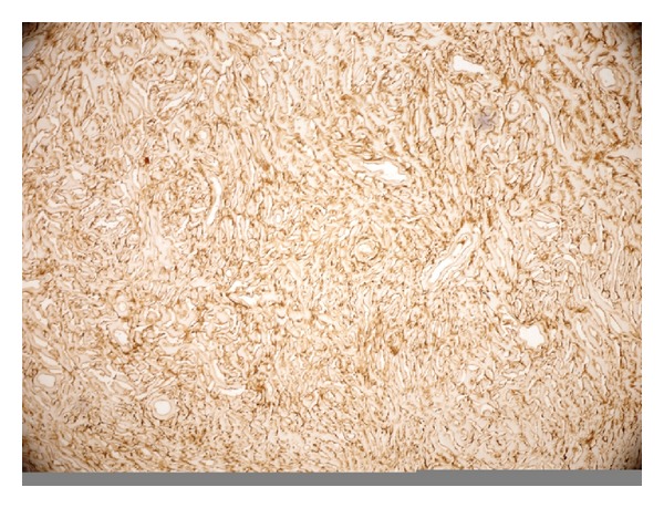
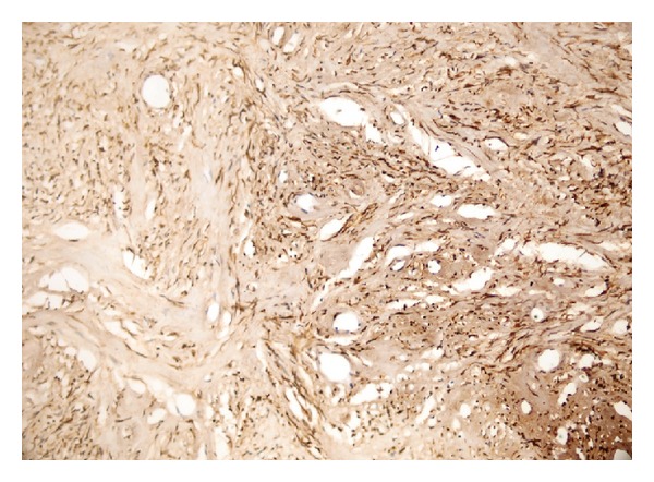
A 68-year-old man had a rapidly growing, painless neck mass, thought to be nodular goiter. Ultrasonography showed a giant, heterogeneous mass occupying the middle and superior poles and protruding outside of the left thyroid lobe. The results of the thyroid function tests were normal. Thyroid scintigraphy revealed a large hypoactive nodule in the left thyroid lobe. Complete surgical removal of tumor was performed and macroscopically demonstrated a well-demarked lesion outside the thyroid gland. Microscopically, the lesion was composed of fibroblast-like spindle cells in a patternless architecture and extensive stromal hyalinization. Immunohistochemistry showed positive reaction for CD34 in spindle cells and diffuse bcl-2 staining. The pathology was confirmed as solitary fibrous tumor. In the follow-up period after surgery, thyroid scintigraphy showed normal left thyroid lobe. Solitary fibrous tumor originated from or associated with thyroid gland is extremely rare. According to our knowledge, this is the first reported solitary fibrous tumor presenting like a cold thyroid nodule. This pathology must be considered for differential diagnosis of neck masses in the thyroid region.
Figures
Similar articles
-
Solitary fibrous tumor arising in an intrathoracic goiter.Thyroid. 2010 Apr;20(4):435-7. doi: 10.1089/thy.2009.0237. Thyroid. 2010. PMID: 20373988
-
The value of Tc-99m tetrofosmin thyroid scintigraphy in patients with nodular goiter.Ann Nucl Med. 1997 Nov;11(4):285-90. doi: 10.1007/BF03165295. Ann Nucl Med. 1997. PMID: 9460519
-
Solitary fibrous tumor of the thyroid gland.Med Mol Morphol. 2014 Jun;47(2):117-22. doi: 10.1007/s00795-013-0056-6. Epub 2013 Sep 8. Med Mol Morphol. 2014. PMID: 24013381
-
Malignant solitary fibrous tumor of the thyroid gland: report of a case and review of the literature.Diagn Cytopathol. 2011 Sep;39(9):694-9. doi: 10.1002/dc.21538. Epub 2010 Dec 31. Diagn Cytopathol. 2011. PMID: 21837658 Review.
-
Rapid recurrence and bilateral lungs, multiple bone metastasis of malignant solitary fibrous tumor of the right occipital lobe: report of a case and review.Diagn Pathol. 2015 Jul 9;10:91. doi: 10.1186/s13000-015-0318-9. Diagn Pathol. 2015. PMID: 26155787 Free PMC article. Review.
Cited by
-
Unveiling the Rarity: A Case Report on Solitary Fibrous Tumor of the Thyroid Gland.Indian J Otolaryngol Head Neck Surg. 2024 Jun;76(3):2798-2804. doi: 10.1007/s12070-024-04498-x. Epub 2024 Jan 26. Indian J Otolaryngol Head Neck Surg. 2024. PMID: 38883480 Free PMC article.
-
Solitary Fibrous Tumor of Head and Neck Region: A Series of Three Cases at an Uncommon Location With a Review of the Literature.Cureus. 2024 Apr 13;16(4):e58213. doi: 10.7759/cureus.58213. eCollection 2024 Apr. Cureus. 2024. PMID: 38741857 Free PMC article.
-
A case report of solitary fibrous tumor of the thyroid gland and literature review.Medicine (Baltimore). 2023 Aug 25;102(34):e34710. doi: 10.1097/MD.0000000000034710. Medicine (Baltimore). 2023. PMID: 37653837 Free PMC article. Review.
References
-
- Klemperer P, Rabin CB. Primary neoplasm of the pleura: a report of five cases. Archives of Pathology & Laboratory Medicine. 1931;11:385–412.
-
- Weidner N. Solitaryn fibrous tumor of the mediastinum. Ultrastructural Pathology. 1991;15(4-5):489–492. - PubMed
-
- Bortolotti U, Calabro F, Loy M, Fasoli G, Altavilla G, Marchese D. Giant intrapericardial solitary fibrous tumor. Annals of Thoracic Surgery. 1992;54(6):1219–1220. - PubMed
-
- Kubota Y, Kawai N, Tozawa K, Hayashi Y, Sasaki S, Kohri K. Solitary fibrous tumor of the peritoneum found in the prevesical space. Urologia Internationalis. 2000;65(1):53–56. - PubMed
-
- Witkin GB, Rosai J. Solitary fibrous tumor of the upper respiratory tract: a report of six cases. American Journal of Surgical Pathology. 1991;15(9):842–848. - PubMed
LinkOut - more resources
Full Text Sources
Other Literature Sources
