

doi: 10.1155/2013/825078.
Epub 2013 Oct 1.
A case of pelvic schwannoma presenting prominent eggshell-like calcification
Affiliations
- PMID: 24194999
- PMCID: PMC3806513
- DOI: 10.1155/2013/825078
Item in Clipboard
A case of pelvic schwannoma presenting prominent eggshell-like calcification
Case Rep Radiol.
2013.
Abstract
Pelvic schwannoma typically forms a large, well-circumscribed mass in the retroperitoneum or presacral area and frequently undergoes cystic degeneration. It appears as a well-demarcated round or oval mass, often showing prominent cystic degeneration and calcification. Characteristics of these calcifications are punctate, mottled, or curvilinear and are seen along the walls of the mass. Herein, we describe a case of schwannoma presenting a huge pelvic mass with unique eggshell-like calcification.
Figures

(a) Contrast-enhanced CT shows a low-density mass containing coarse and round calcifications in early phase (40 s). Cystic lesions are not evident. (b) The mass is slightly enhanced in delayed phase (180 s). (c) 3D-CT shows eggshell-like calcifications within the tumor (arrows).
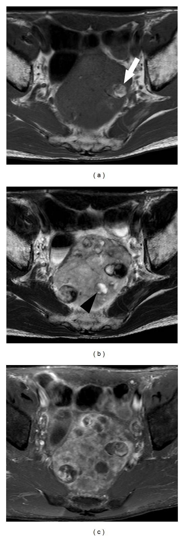
(a) T1-weighted MR image shows a well-defined hypointensity mass with focal high-intensity area (arrow). (b) T2-weighted image shows heterogeneous hyperintensity mass with spotty hyperintensity area (arrowhead). (c) Contrast-enhanced image shows slight enhancement in solid component of the tumor.

(a) Cut surface of the tumor shows encapsulated solid mass containing multiple eggshell-like calcifications. (b) Microscopic image (HE, magnification ×100) shows palisaded arrangement of spindle-shaped cells. (c) Immunohistochemically, tumor cells are positive for S-100 protein. (d) Myxoid degeneration and hyalinization are focally seen in the tumor.
References
-
- Goh BKP, Tan Y-M, Chung Y-FA, Chow PKH, Ooi LLPJ, Wong W-K. Retroperitoneal schwannoma. American Journal of Surgery. 2006;192(1):14–18. - PubMed
-
- Hughes MJ, Thomas JM, Fisher C, Moskovic EC. Imaging features of retroperitoneal and pelvic schwannomas. Clinical Radiology. 2005;60(8):886–893. - PubMed
-
- Sheth S, Horton KM, Garland MR, Fishman EK. Mesenteric neoplasms: CT appearances of primary and secondary tumors and differential diagnosis. Radiographics. 2003;23(2):457–473. - PubMed
-
- Rha SE, Byun JY, Jung SE, Chun HJ, Lee HG, Lee JM. Neurogenic tumors in the abdomen: tumor types and imaging characteristics. Radiographics. 2003;23(1):29–43. - PubMed
-
- Kinoshita T, Naganuma H, Ishii K, Itoh H. CT features of retroperitoneal neurilemmoma. European Journal of Radiology. 1998;27(1):67–71. - PubMed
LinkOut - more resources
Full Text Sources
Other Literature Sources
