

Quantitative assessment of carotid plaque surface irregularities and correlation to cerebrovascular symptoms
- PMID: 24195596
- PMCID: PMC4228278
- DOI: 10.1186/1476-7120-11-38
Quantitative assessment of carotid plaque surface irregularities and correlation to cerebrovascular symptoms
Abstract
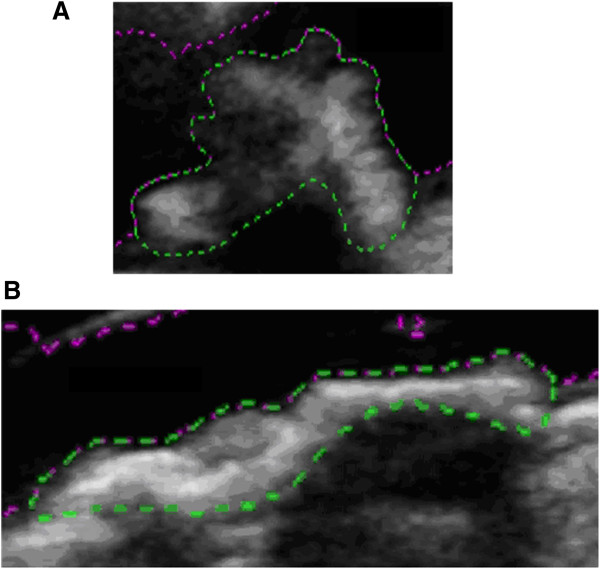
Background: The purpose of this study was to determine whether surface irregularities measured from ultrasound images of carotid artery plaques and quantified using a novel method, correlate with the presence of ipsilateral hemispheric cerebrovascular symptoms.
Methods: A plaque surface irregularity index (SII) was measured in 47 carotid artery plaques (32 subjects, stenosis range 10% -95%, 49% symptomatic) using ultrasound image sequences spanning several cardiac cycles. The differences in the distribution of SII in plaques with ipsilateral hemispheric symptoms versus those without symptoms and the correlation between the SII of plaques and the degrees of stenosis of the corresponding arteries were assessed. Diagnostic performance of plaque SII was evaluated on its own and in combination with the degree of stenosis.
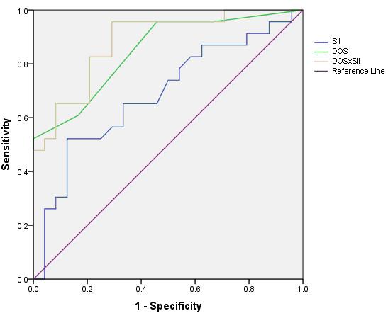
Results: The mean SII was significantly greater for plaques with ipsilateral hemispheric symptoms (1.89 radians/mm) than for asymptomatic plaques (1.67 radians/mm, p = 0.03). There was no statistically significant association between the SII and the degree of stenosis (p = 0.30). SII predicted the presence of cerebrovascular symptoms with an accuracy of 66% (sensitivity 65%, specificity 67%) on its own and with an accuracy of 83% (sensitivity 96%, specificity 71%) in combination with the degree of stenosis.
Conclusions: Quantitative assessment of carotid plaque surface irregularities using a novel SII parameter correlates with the presence ipsilateral hemispheric cerebrovascular symptoms and may increase diagnostic performance beyond that provided by the degree of stenosis.
Figures


References
-
- Leary DH, Holen J, Ricotta JJ, Roe S, Schenk EA. Carotid bifurcation disease: prediction of ulceration with B-mode US. Radiology. 1987;162:523–525. - PubMed
-
- De Bray JM, Baud JM, Dauzat M. Consensus Concerning the Morphology and the Risk of Carotid Plaques. Cerebrovasc Dis. 1997;7:289–296. doi: 10.1159/000108415. - DOI
Publication types
MeSH terms
LinkOut - more resources
Full Text Sources
Other Literature Sources
