. 2013 Dec;27(12):2116-25.
doi: 10.1210/me.2013-1146. Epub 2013 Nov 6.

VDR attenuates acute lung injury by blocking Ang-2-Tie-2 pathway and renin-angiotensin system

Affiliations

VDR attenuates acute lung injury by blocking Ang-2-Tie-2 pathway and renin-angiotensin system

Juan Kong et al. Mol Endocrinol. 2013 Dec.

Abstract

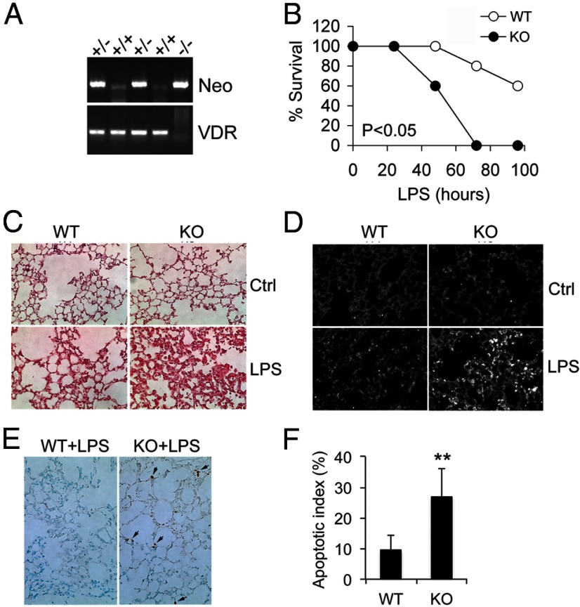
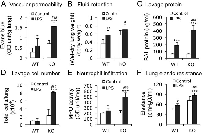
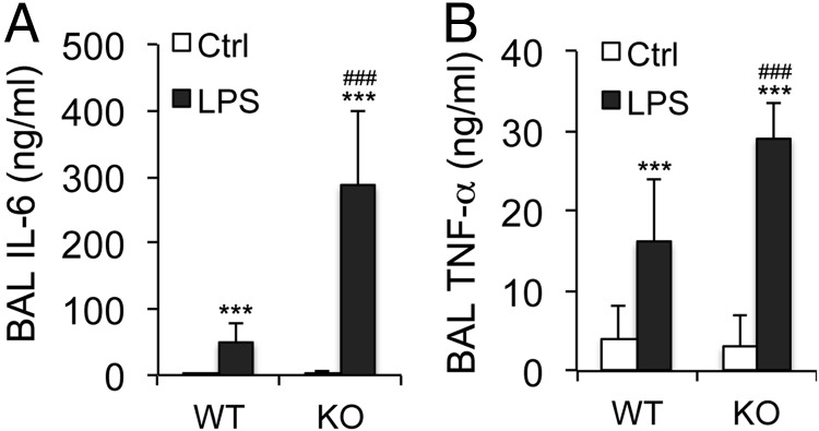
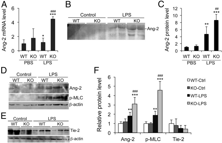
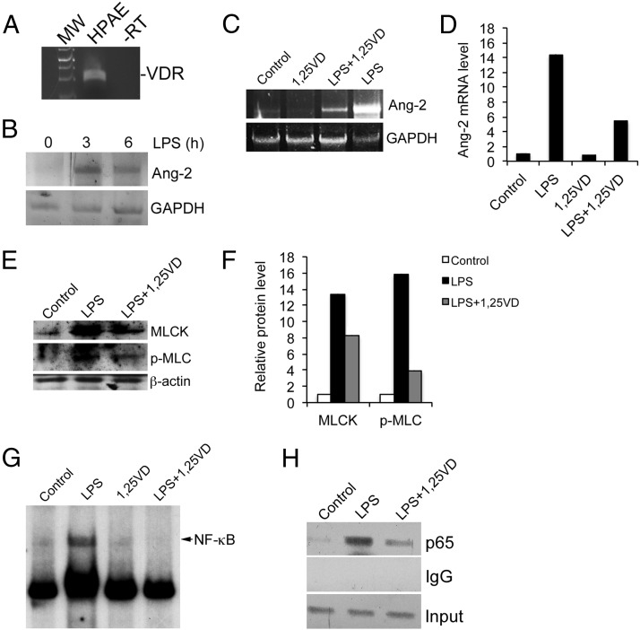
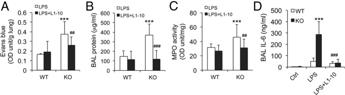
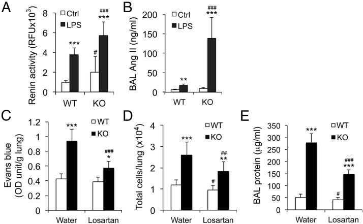
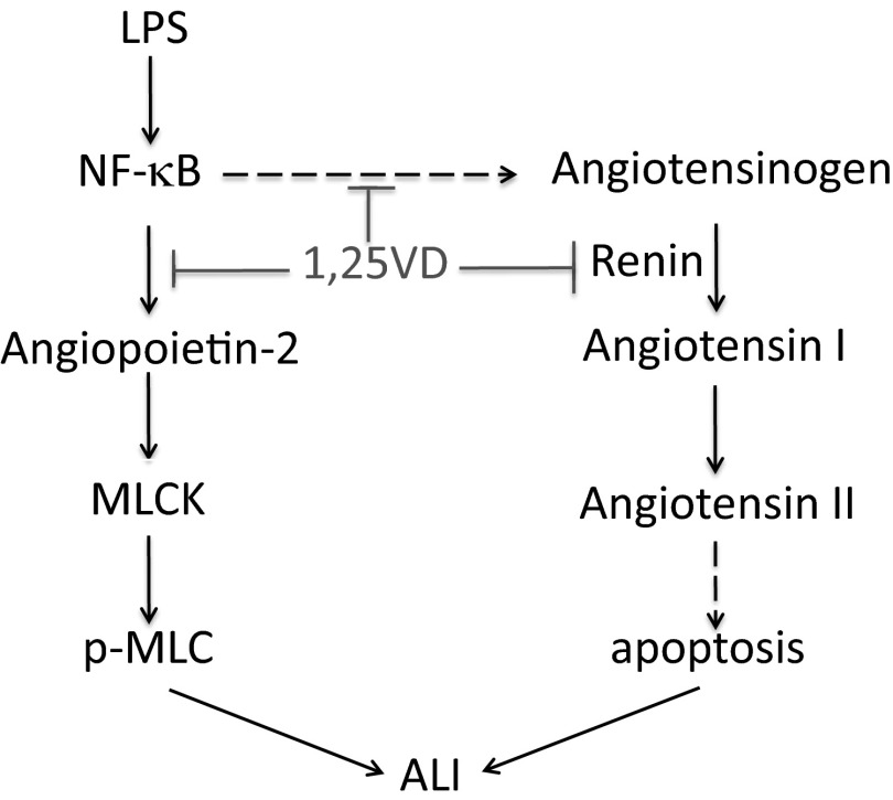
Acute lung injury (ALI) is a hallmark of systemic inflammation associated with high mortality. Although the vitamin D receptor (VDR) is highly expressed in the lung, its role in lung physiology remains unclear. We investigated the effect of VDR deletion on ALI using a lipopolysaccharide (LPS)-induced sepsis model. After LPS challenge VDR-null mice exhibited more severe ALI and higher mortality compared with wild-type (WT) counterparts, manifested by increased pulmonary vascular leakiness, pulmonary edema, apoptosis, neutrophil infiltration, and pulmonary inflammation, which was accompanied by excessive induction of angiopoietin (Ang)-2 and myosin light chain (MLC) phosphorylation in the lung. 1,25-Dihydroxyvitamin D blocked LPS-induced Ang-2 expression by blocking nuclear factor-κB activation in human pulmonary artery endothelial cells. The severity of lung injury seen in VDR-null mice was ameliorated by pretreatment with L1-10, an antagonist of Ang-2, suggesting that VDR signaling protects the pulmonary vascular barrier by targeting the Ang-2-Tie-2-MLC kinase cascade. Severe ALI in VDR-null mice was also accompanied by an increase in pulmonary renin and angiotensin II levels, and pretreatment of VDR-null mice with angiotensin II type 1 receptor blocker losartan partially ameliorated the severity of LPS-induced lung injury. Taken together, these observations provide evidence that the vitamin D-VDR signaling prevents lung injury by blocking the Ang-2-Tie-2-MLC kinase cascade and the renin-angiotensin system.


Figures

Figure 1.
Figure 1.
VDR inactivation leads to severe ALI after LPS challenge. WT and VDR KO mice were treated with saline or LPS (20 mg/kg, ip injection). A, Mouse genotyping. PCR product patterns indicate WT (+/+), heterozygote (+/−), and VDR KO (−/−) genotypes. B, Mouse survival curves after LPS treatment. C, Hematoxylin and eosin staining of lung sections from control (Ctrl) and LPS-treated WT and KO mice. Note the greater thickening of the alveolar interstitial space in LPS-treated KO mice. D, Fluorescent immunostaining of lung sections from control and LPS-treated WT and KO mice with neutrophil-specific monoclonal antibodies 7/4. E and F, TUNEL staining of lung sections from LPS-treated WT and KO mice (E) and semiquantitation of the data (F). Apoptotic index is defined as percent of TUNEL-positive microscopic fields in randomly chosen 50 fields in each mouse. Arrows indicate TUNEL-positive cells. **, P < .01 vs WT.
Figure 2.
Figure 2.
VDR deletion increases pulmonary vascular permeability and impairs lung function. WT and VDR KO mice were treated with saline (control) or LPS (20 mg/kg) via ip injection, and lung analyses were performed after 24 hours. A, Evans blue permeability assays. B, The difference of wet and dry weight of the lung (fluid retention) normalized to body weight. C, Protein concentration in the BAL. D, Cell number in BAL fluid. E, MPO activity in lung lysates. F, Lung elastic resistance. *, P < .05; **, P < .01; ***, P < .001 vs corresponding control of the same genotype; #, P < .05; ###, P < .001 vs LPS-treated WT; n = 5–6 in each genotype.
Figure 3.
Figure 3.
Proinflammatory cytokine levels in BAL fluids. WT and VDR KO mice were treated with saline (control [Ctrl]) or LPS, and levels of IL-6 (A) and TNFα (B) in BAL fluid were quantified by ELISA. ***, P < .001 vs corresponding control of the same genotype; ###, P < .001 vs LPS-treated WT; n = 5–6 in each genotype.
Figure 4.
Figure 4.
VDR signaling attenuates LPS-induced ALI by targeting the Ang-2-Tie-2-MLCK pathway. WT and VDR KO mice were treated with saline (control [Ctrl] or PBS) or LPS (20 mg/kg) by ip injection, and lung analyses were performed at 24 hours. A, Ang-2 mRNA levels quantified by real time RT-PCR; *, P < .05; ***, P < .001 vs corresponding control of the same genotype; ###, P < .001 vs LPS-treated WT; n = 4. B and C, Western blot analysis (B) and densitometric quantitation (C) of Ang-2 protein levels in BAL; **, P < .01; ***, P < .001 vs corresponding control of the same genotype; ###, P < .001 vs LPS-treated WT. D, Western blot analysis of Ang-2 protein and phosphorylated MLC (p-MLC) levels in total lung lysates. E, Western blot analysis of Tie-2 levels in total lung lysates. F, Densitometric quantitation of these Western blot data. **, P < .01; ***, P < .001 vs corresponding control of the same genotype; ###, P < .001 vs LPS-treated WT.
Figure 5.
Figure 5.
1,25(OH)2D3 down-regulates Ang-2 by targeting NF-κB activation in HPAE cells. A, RT-PCR detection of human VDR mRNA transcript in HPAE cells. −RT, minus reverse transcriptase control. B, HPAE cells were treated by LPS for 3 and 6 hours, and Ang-2 mRNA induction was measured by RT-PCR. C and D, HPAE cells were treated with LPS in the presence or absence of 1,25(OH)2D3 (20 nM). Ang-2 mRNA was assessed by RT-PCR at 24 hours (C) and semiquantified by densitometry (D). E and F, HPAE cells were treated with LPS in the presence or absence of 1,25(OH)2D3. MLCK and phosphorylated MLC levels were assessed by Western blotting after 24 hours (E) and semiquantified by densitometry (F). G, EMSA. HPAE cells were treated with LPS in the presence or absence of 1,25(OH)2D3 for 24 hours. Nuclear extracts were prepared for EMSA using 32P-labeled putative κB probe within the first intron of human ANG-2 gene. H, ChIP assay. HPAE cells were treated with LPS in the presence or absence of 1,25(OH)2D3 for 6 hours. ChIP assays were performed using anti-p65 antibodies or nonimmune IgG. PCR amplification was performed using primers flanking the putative κB site within the first intron of human ANG-2 gene. GAPDH, glyceraldehyde-3-phosphate dehydrogenase; MW, molecular weight; −RT, minus reverse transcriptase; 1,25VD, 1,25(OH)2D3; p-MLC, phosphorylated MLC.
Figure 6.
Figure 6.
Blockade of Ang-2 signaling ameliorates lung injury in VDR-null mice. WT and KO mice were pretreated with saline or L1–10 for 2 weeks followed by LPS challenge. Lung damage analyses were performed 24 hours after LPS treatment. A, Evans blue permeability assay. B, Total protein concentrations in the BAL. C, MPO activity in total lung lysates. D, IL-6 levels in the BAL fluid. ***, P < .001 vs LPS-treated WT; ##, P < .01; ###, P < .001 vs LPS-treated KO; n = 5–6 in each genotype. Ctrl, control.
Figure 7.
Figure 7.
VDR signaling attenuates LPS-induced ALI by targeting the RAS. A and B, WT and KO mice were treated with saline (Ctrl) or LPS (20 mg/kg). Renin activity (A) and angiotensin II levels (B) in the BAL fluid were measured at 24 hours. **, P < .01; ***, P < .001 vs control of the same phenotype; #, P < .05; ##, P < .001 vs corresponding WT of the same treatment; n = 5–6 each genotype. C–E, WT and KO mice were fed regular water or losartan-containing water for 2 weeks followed by LPS challenge. Lung damage analyses were performed 24 hours after LPS treatment. C, Evans blue permeability assay. D, Total cell number in the BAL fluid. E, Total protein concentration in the BAL fluid. *, P < .05; **, P < .01; ***, P < .001 vs WT of the same treatment; ##, P < .01; ###, P < .001 vs KO fed regular water; n = 5–6 in each genotype.
Figure 8.
Figure 8.
Proposed mechanism whereby 1,25(OH)2D3-VDR signaling inhibits the Ang-2-Tie-2-MLCK pathway and the RAS cascade to attenuate LPS-induced ALI. Dashed line indicates speculation. p-MLC, phosphorylated MLC; 1,25VD, 1,25(OH)2D3.

References

    1. Beck-Schimmer B, Schimmer RC, Pasch T. The airway compartment: chambers of secrets. News Physiol Sci. 2004;19:129–132 - PubMed
    1. Ware LB, Matthay MA. The acute respiratory distress syndrome. N Engl J Med. 2000;342(18):1334–1349 - PubMed
    1. Garcia JG, Davis HW, Patterson CE. Regulation of endothelial cell gap formation and barrier dysfunction: role of myosin light chain phosphorylation. J Cell Physiol. 1995;163(3):510–522 - PubMed
    1. Tinsley JH, Teasdale NR, Yuan SY. Myosin light chain phosphorylation and pulmonary endothelial cell hyperpermeability in burns. Am J Physiol Lung Cell Mol Physiol. 2004;286(4):L841–L847 - PubMed
    1. Verin AD, Patterson CE, Day MA, Garcia JG. Regulation of endothelial cell gap formation and barrier function by myosin-associated phosphatase activities. Am J Physiol. 1995;269(1 Pt 1):L99–L108 - PubMed

Publication types

MeSH terms