. 2013 Dec;27(12):2105-15.
doi: 10.1210/me.2013-1281. Epub 2013 Nov 6.

The type II deiodinase is retrotranslocated to the cytoplasm and proteasomes via p97/Atx3 complex

Affiliations

The type II deiodinase is retrotranslocated to the cytoplasm and proteasomes via p97/Atx3 complex

Rafael Arrojo E Drigo et al. Mol Endocrinol. 2013 Dec.

Abstract

The type II iodothyronine deiodinase (D2) is a type I endoplasmic reticulum (ER)-resident thioredoxin fold-containing selenoprotein that activates thyroid hormone. D2 is inactivated by ER-associated ubiquitination and can be reactivated by two ubiquitin-specific peptidase-class D2-interacting deubiquitinases (DUBs). Here, we used D2-expressing cell models to define that D2 ubiquitination (UbD2) occurs via K48-linked ubiquitin chains and that exposure to its natural substrate, T4, accelerates UbD2 formation and retrotranslocation to the cytoplasm via interaction with the p97-ATPase complex. D2 retrotranslocation also includes deubiquitination by the p97-associated DUB Ataxin-3 (Atx3). Inhibiting Atx3 with eeyarestatin-I did not affect D2:p97 binding but decreased UbD2 retrotranslocation and caused ER accumulation of high-molecular weight UbD2 bands possibly by interfering with the D2-ubiquitin-specific peptidases binding. Once in the cytosol, D2 is delivered to the proteasomes as evidenced by coprecipitation with 19S proteasome subunit S5a and increased colocalization with the 20S proteasome. We conclude that interaction between UbD2 and p97/Atx3 mediates retranslocation of UbD2 to the cytoplasm for terminal degradation in the proteasomes, a pathway that is accelerated by exposure to T4.


Figures
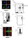
Figure 1.
Figure 1.
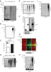
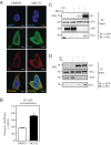
D2 is an ERAD pathway substrate. A, HEK-293 cells transiently expressing the double-tagged YFP/His D2 protein (D2HY) were fixed in 4% paraformaldehyde (PFA) and stained with a α-YFP for D2HY (red) and α-ERp72 for the ER marker ERp72 (green). Nucleus was stained with 4′,6-diamidino-2-phenylindole (DAPI). Scale bar, 7.5 μm. Lower panel, BiP protein levels in control or D2-expressing cells. B, Western blot analysis of HEK-293 cells transiently expressing D2HY or control YFP with an α-YFP antibody. D2HY migrates at an approximate molecular weight of 62 kDa and YFP migrates at 30 kDa. Overexposure of same blot reveals high-molecular weight D2HY bands (solid arrows). BiP (70 kDa; middle panel) and β-actin (50 kDa; lower panel) are shown. C, D2HY protein levels in HEK-293 transiently expressing D2HY and treated with either vehicle or 1 μM MG132 for 24 hours. In red, D2HY bands are observed with an α-His antibody. In green, D2HY bands are observed with an α-YFP antibody. The overlay of both images and high-molecular weight D2HY bands is seen on the right panel, with overlaying bands appearing in yellow. D, Western blot analysis with an α-YFP antibody of lysates of control or D2HY-expressing HEK-293 cell treated with vehicle, 1 μM MG132, or 10 μM EERI for 24 hours. E, Western blot analysis of α-YFP immunoprecipitates of HEK-293 cells expressing either control YFP or D2HY and treated with vehicle or 1 μM MG132 for 24 hours. Blot was probed with α-ubiquitin antibody. LC, IgG light chain. F, D2 activity of HEK-293 cells stably expressing the D2HY protein and treated with vehicle, 1 μM MG132, or 10 μM EERI for 24 hours. DMSO, dimethylsulfoxide.
Figure 2.
Figure 2.
UbD2 is composed of substrate-induced K48-ubiquitin chains. A, Isolated microsomes (300 ng) of vehicle-treated D2HY-expressing HEK-293 cells subject to a 4-hour in vitro ubiquitination reaction followed by IP and analyzed by Western blot with α-ubiquitin (Ub; left panel) and α-K48-linked chain antibodies (K48; right panel). B, α-K48-linked ubiquitin chain Western blot analysis of α-YFP immunoprecipitates of control YFP or D2HY-expressing HEK-293 cells treated with vehicle or 1 μM MG132 for 24 hours. C, D2 activity of HEK-293 cells transiently transfected with wild-type (WT) D2 and wild-type or K48R ubiquitin. Transfection efficiency was monitored by the cotransfection of secreted alkaline phosphatase (SEAP). D2 activity was expressed as fmol/min/mg/a.u serum alkaline phosphatase (SEAP). D, α-Ubiquitin and α-K48-linked ubiquitin chain (panel E) Western blot analysis of α-YFP immunoprecipitates of D2HY-expressing HEK-293 cells cultivated in stripped serum and supplemented with vehicle or 20 μM T4 in the presence of dimethylsulfoxide (DMSO), 1 μM MG132, or 10 μM EERI for 24 hours. F, α-YFP Western blot of lysates from HEK-293 cells transiently expressing either a YFP or a Cys133AlaD2 (AlaD2HY) isoform. AlaD2HY-specific bands are indicated by solid arrowheads. In lower panels, BiP and β-actin protein levels of same samples from upper panel. G, α-His (red) and α-YFP (green) Western blots of AlaD2HY-expressing HEK-293 cells treated with DMSO or 1 μM MG132 for 24 hours. AlaD2HY-specific bands are indicated by solid arrows. Overlay of α-His and α-YFP Western blots is shown on the panel on the right. On the lower panel, β-actin protein levels are shown. H, Western blot analysis with either an α-ubiquitin or K48-linkage (panel I) of α-YFP immunoprecipitates of HEK-293 cells expressing either control YFP or AlaD2HY and treated with vehicle or 1 μM MG132 for 24 hours. C, Data are shown as mean ± SD; n = 3; *, P < .001 by t test. LC, IgG light chain; WB, Western blot.
Figure 3.
Figure 3.
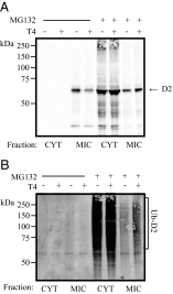
D2 is ubiquinated at the ER level. α-YFP (panel A) and α-ubiquitin (panel B) Western blot of cytosolic (CYT) and microsomal (MIC) cellular fractions of cells expressing D2HY protein, grown in CSS, and treated with either control or 1 μM MG132 and in combination with vehicle or 20 μM T4 for 24 hours.
Figure 4.
Figure 4.
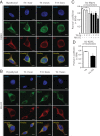
Upon ubiquitination D2 exits the ER. HEK-293 cells stably expressing D2HY were maintained with charcoal-stripped media and treated with dimethylsulfoxide (DMSO) (A) or 1 μM MG132 (B) in combination with 20 μM T4 for the indicated times. Following the treatment window, cells were methanol fixed and stained for D2HY (red) and the ER marker ERp72 (green) with α-YFP and α-Erp72 antibodies, respectively. Nucleus was stained with 4′,6-diamidino-2-phenylindole (DAPI). Scale bar, 7.5 μm. C, Quantification of Pearson's coefficient between D2HY and ERp72 colocalization from panels A and B and quantification of Pearson's coefficient between D2 and KDEL colocalization after treatment with 20 μM T4 for 3 hours (panel D). *, P < .01 vs DMSO-treated group at time 0; $, P < .01 vs. MG132-treated group at time 0.
Figure 5.
Figure 5.
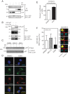
UbD2 interaction with the ATPase p97 blunts D2-USP33 binding. A, An aliquot of the same D2HY- or control-expressing samples treated with either vehicle or MG132 from Figure 1D were immunoprecipitated with an α-YFP and processed for Western blot analysis with an α-p97 antibody. B, HEK-293 cells transiently expressing a GFP-tagged p97 protein (GFP-p97) in combination with a FLAG-tagged D2 were subject to α-GFP immuprecipitation as in Figures 1 and 2 and analyzed by Western blot with α-S5a and α-FLAG antibodies. C, α-p97 Western blot of α-YFP immuprecipitates of HEK-293 cells expressing D2HY treated with vehicle or T4, and in combination with dimethylsulfoxide (DMSO), 1 μM MG132, or 10 μM EERI for 24 hours. D, HEK-293 cells stably expressing D2HY were treated with 10 μM EERI for 3 hours and methanol fixed and immunostained with α-ERp72 and α-YFP antibodies to detect the ER marker ERp72 (fluorescein isothiocyanate [FITC]) and D2 (Texas Red), respectively. Overlay is shown in yellow. Nuclei were stained with 4′,6-diamidino-2-phenylindole (DAPI). Scale bar, 13 μm. E, Pearson's coefficient of D2HY and ERp72 treated with DMSO or 10 μM EERI for 3 hours. Data are displayed as ±SEM. F, D2-USP-33 interaction in cells treated with either vehicle or 10 μM EERI for 24 hours as measured by FRET. Panel on the right shows representative scan images of YFP and CFP FRET signal of D2-USP-33 in cells treated with DMSO, EERI, or MG132. F, Data were acquired with at least 40 measurements and displayed as ±SD of % of control FRET signal, where * represents P < .05 by one-way ANOVA with Dunnett's post-test. IB, immunoblotting; WB, Western blot.
Figure 6.
Figure 6.
UbD2 complexes colocalize with the 20S proteasome after exiting the ER. A, HEK-293 cells stably expressing D2HY were maintained with charcoal-stripped media and treated with dimethylsulfoxide (DMSO) or 1 μM MG132 for 24 hours and were paraformaldehyde (PFA)-fixed and stained for D2HY (Texas Red), the α- and β-subunit of the 20S proteasome chamber (fluorescein isothiocyanate [FITC]) with an α-YFP and α-20S (with α/β-subunits) antibodies, respectively. Nucleus was stained with DAPI. Scale bar, 7.5 μm. B, Quantification of Pearson's coefficient of conditions from panels A and B, where * represents P < .01 vs DMSO-treated group by Student's t test. C, HEK-293 cells transiently expressing either D2HY or YFP were treated with vehicle or 1 μM MG132 for 24 hours, immunoprecipitated with α-YFP antibody, and analyzed by Western blot with an α-S5a antibody. D, D2HY- or YFP-expressing HEK-293 cells were grown in charcoal-stripped medium and supplemented with vehicle or 20 μM T4, treated with 1 μM MG132 for 24 hours and processed by α-YFP IP followed by Western blot analysis with an α-S5a antibody. IB, immunoblotting.

Similar articles

Cited by

References

    1. Ciehanover A, Hod Y, Hershko A. A heat-stable polypeptide component of an ATP-dependent proteolytic system from reticulocytes. Biochem Biophys Res Commun. 1978;81:1100–1105 - PubMed
    1. Hershko A, Ciechanover A, Heller H, Haas AL, Rose IA. Proposed role of ATP in protein breakdown: conjugation of protein with multiple chains of the polypeptide of ATP-dependent proteolysis. Proc Natl Acad Sci USA. 1980;77:1783–1786 - PMC - PubMed
    1. Komander D, Rape M. The ubiquitin code. Annu Rev Biochem. 2012;81:203–229 - PubMed
    1. Mukhopadhyay D, Riezman H. Proteasome-independent functions of ubiquitin in endocytosis and signaling. Science. 2007;315:201–205 - PubMed
    1. Chau V, Tobias JW, Bachmair A, et al. A multiubiquitin chain is confined to specific lysine in a targeted short-lived protein. Science. 1989;243:1576–1583 - PubMed

Publication types