Review
. 2013 Nov 5;14(11):21899-942.
doi: 10.3390/ijms141121899.

Solid lipid nanoparticle-based calix[n]arenes and calix-resorcinarenes as building blocks: synthesis, formulation and characterization

Affiliations
Review

Solid lipid nanoparticle-based calix[n]arenes and calix-resorcinarenes as building blocks: synthesis, formulation and characterization

Imed Montasser et al. Int J Mol Sci. .

Abstract

Solid lipid nanoparticles (SLNs) have attracted increasing attention during recent years. This paper presents an overview about the use of calix[n]arenes and calix-resorcinarenes in the formulation of SLNs. Because of their specific inclusion capability both in the intraparticle spaces and in the host cavities as well as their capacity for functionalization, these colloidal nanostructures represent excellent tools for the encapsulation of different active pharmaceutical ingredients (APIs) in the area of drug targeting, cosmetic additives, contrast agents, etc. Various synthetic routes to the supramolecular structures will be given. These various routes lead to the formulation of the corresponding SLNs. Characterization, properties, toxicological considerations as well as numerous corresponding experimental studies and analytical methods will be also exposed and discussed.


Figures

Figure 1
Figure 1
Proposed structure of solid lipid nanoparticles (SLNs), the interior structure varies from amorphous to crystalline.
Figure 2
Figure 2
Synthetic route to different amphiphilic structures based on calix[4]arene. Alk = CH3(CH2)n, n = 4 (a), 6 (b), 8 (c), 10 (d).
Figure 3
Figure 3
Non-contact mode atomic force microscopy (AFM) images of 2d-based SLNs reconstituted after freeze-drying in a solution of glucose (2%) at 5 mm scan range.
Figure 4
Figure 4
Formulae of 2d, 5d and 6d.
Figure 5
Figure 5
General formulae of the amphiphilic calix-arenes used in the haemolysis experiment.
Figure 6
Figure 6
Chemical structures of amphiphilic calixarenes.
Figure 7
Figure 7
(a) Static 129Xe NMR spectra recorded under continuous flow of hyperpolarized xenon at an effective Xe pressure of 7 Torr and T 293 K; (b) Plot of the chemical shift of Xe in the host cavity versus the chain length of the amphiphilic calixarenes [68].
Figure 8
Figure 8
In situ transformation of 2a SLNs (bottom) after a pulse of methylene chloride (next to bottom) as monitored by continuous flow hyperpolarized 129Xe MAS NMR. Consecutive spectra were recorded every 4.5 min at room temperature. Sharp signals at ±40 ppm are spinning sidebands [68].
Figure 9
Figure 9
Differential scanning calorimetry traces for para-hexanoyl calix-[4]arene 2a[68].
Figure 10
Figure 10
Top view to the cavity of para-hexanoylcalix[4]arene 2a (left) and para-hexylcalix[4]arene (right) [70].
Figure 11
Figure 11
Structures of para-Hexanoyl Calix[4]arene (2a) and t-EHMC.
Figure 12
Figure 12
Capsular structure of inclusion complex of para-hexanoyl calix[4]-arene with t-EHMC (2*2a. t-EHMC). Disorder and H-atoms are omitted for clarity [71].
Figure 13
Figure 13
Powder XRD patterns of 2a SLNs: unloaded SLN (a); SLNEHMC-1.2 (b); and crystalline inclusion complex 2*2a.1t-EHMC (c) [71].
Figure 14
Figure 14
Structures of para-hexanoyl calix[4]arene (2a) and MT.
Figure 15
Figure 15
Electron spin resonance (ESR) spectra of SLN of 2a loaded with MT upon treatment by the solution of sodium ascorbate at room temperature in buffer solution of pH 7.4. Insets: integrated spectra. The asterisk denotes the ascorbate anion-radical [75].
Figure 16
Figure 16
Synthetic route to the amphiphilic para-acyl-calix[9]arenes, 9ae.
Figure 17
Figure 17
Synthetic route to the compounds 14 and 15.
Figure 18
Figure 18
Noncontact mode AFM image of the solid lipid nanoparticles formed by 14 on mica at 5 × 5 μm scan size, z axis is 94 nm. A 20 μL portion of a suspension of the SLNs was deposited, and imaging was performed after drying for 24 h at 25 °C [86].
Figure 19
Figure 19
Non-contact mode AFM image of the solid lipid nanoparticles formed by 15 on mica at 5 × 5 μm scan size, z axis is 140 nm. A 20 μL portion of a suspension of the SLNs was deposited, and imaging was performed after drying for 24 h at 25 °C [86].
Figure 20
Figure 20
Variation of the diameter of solid lipid nanoparticles formed by 14 in the presence of varying concentrations of Na+ (♦); K+ (•); Mg2+ (▪); and Ca2+ (▴) [86].
Figure 21
Figure 21
Variation of the diameter of solid lipid nanoparticles formed by 15 in the presence of varying concentrations of Na+ (♦); K+ (•); Mg2+ (▪); and Ca2+ (▴) [86].
Figure 22
Figure 22
Noncontact mode AFM image of the solid lipid nanoparticles of 15 in the presence of 3 mM CaCl2 on mica at10 μm scan size [86].
Figure 23
Figure 23
Synthetic route to 17.
Figure 24
Figure 24
Atomic force micrsocope image of 17-based SLNs spread on mica and imaged in air in noncontact mode [92].
Figure 25
Figure 25
Agarose gel electrophoresis of 17-based SLNs incubated with increasing concentrations of plasmid DNA (values are expressed in mg mL−1) [98].
Figure 26
Figure 26
ζ-potential values for 17-based SLNs incubated with an increasing amount of DNA [98].
Figure 27
Figure 27
ζ-potential values during the LbL formation; +D: DNA addition; +C: chitosan addition [98].
Figure 28
Figure 28
Transfection rates of MDCK cells, normalized with the values obtained for lipofection experiments, using SLNs coated with DNA (a); DNA + chitosan: DC (b); D–C–D (c); D–C–D–C (d); D–C–D–C–D (e); D–C–D–C–D–C (f); D–C–D–C–D–C–D (g); and D–C–D–C–D–C–D–C (h); black bars represent the SLNs having the chitosan as the last layer [98].
Figure 29
Figure 29
Confocal micrograph of a transfected cell that has expressed green fluorescent protein (GFP). The red color representing the labeled-chitosan appeared to be in the cytoplasmic compartment of the cell [98].
Figure 30
Figure 30
Chemical formula of 5,11,17,23-tetracarboxy-25,26,27,28-tetradodecyloxycalix[4]arene.
Figure 31
Figure 31
AFM images of a suspension of 18 spread on a mica surface (top) at scan ranges of 15 (left) and 2 mm (right) and SEM images of 18-based SLNs [104].
Figure 32
Figure 32
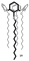
Molecular formula of para-H-tetra-O-dodecyl-calix[4]arene 19.
Figure 33
Figure 33
Non-contact mode AFM image of 19-based nanoparticles ([107]).
Figure 34
Figure 34
Formula of c-2, c-8, c-14, c-20-tetraundecylclix[4]resorcinarene 20.
Figure 35
Figure 35
Non-contact mode AFM images of 20-based SLNs on glass (a) and on mica (b) at 10 μm scan ranges [109].
Figure 36
Figure 36
Non-contact mode AFM images of SLNs based on mixtures of 20 and pluronic® F68 acid (10%) at 10 × 10 μm scan range [109].
Figure 37
Figure 37
Synthetic route to l-RA-Pro 21.
Figure 38
Figure 38
Scanning electron miroscopy image of l-RA-Pro-based SLNs spread on a glass surface (scale bar 200 nm) [120].
Figure 39
Figure 39
Non-contact mode AFM image of l-RA-Pro-based SLNs spread on a freshly cleaned glass slide [120].
Figure 40
Figure 40
Schematic representation of the proposed interaction mechanism. The lateral chain of Phe is represented by a dashed line for the l-enantiomer (For clarity, only the prolyl moiety of l-RA-Pro is represented) [121].
Figure 41
Figure 41
Synthetic route to proteo-SLNs 23[120].
Figure 42
Figure 42
Non-contact mode AFM of proteo-SLNs immobilized on a gold surface via antibody-antigen interactions and schematic representation of the strategy used to attach the SLNs on the surface (1: activated ester; 2: deactivated amide functions, 3: antibody anti-BSA) [120].
Figure 43
Figure 43
Scanning electron microscopy image of proteo-SLNs spread on a glass substrate (scale bars 1 μm and 200 nm (inset)) [120].
Figure 44
Figure 44
Structure of the resorcinarene bis-crowns CNBC5 24, R = CnH2n+1 where n = 2, 3, 4, 5, 7, 9, 10, 11 with selected crystallographic numbering. cw and ccw enantiomers are shown; the A/C plane runs through the upright aryl rings (A and C) and the B/D plane through parallel aryl rings (B and D) respective to the methine plane C7–C14–C21–C28 [124].
Figure 45
Figure 45
SEM image of C11BC5 SLN on SiOx; spherical particles with a mean diameter of 300 nm are shown [124].
Figure 46
Figure 46
SLN diameters for all CNBC5 24 (mean of the size distribution from DLS showing standard deviation). SLNs with a constant 0.1 mM concentration (black) and with a constant mass 100 mg L−1 (grey) show increasing diameter for longer alkyl chains [124].

Similar articles

Cited by

References

    1. Wissing S.A., Kayser O., Muller R.H. Solid lipid nanoparticles for parenteral drug delivery. Adv. Drug Deliv. Rev. 2004;56:1257–1272. - PubMed
    1. Luks S., Muller R. EP0605497 A1. Medication vehicles made of solid lipid particles. 1992 Sep 16;
    1. Gasco M.R. U.S. Patent US5250236 A. Method for producing solid lipid microspheres having a narrow size distribution. 1993 Oct 5;
    1. Freitas C., Muller R.H. Effect of light and temperature on zeta potential and physical stability in solid lipid nanoparticles. Int. J. Pharm. 1998;168:221–229.
    1. Muller R.H., Mehnert W., Lucks J.S., Schwarz C., Zur Muhlen A., Meyhers H., Freitas C., Ruhl D. Solid Lipid Nanoparticles (SLN)-An alternative colloidal carrier system for controlled drug delivery. Eur. J. Pharm. Biopharm. 1995;41:62–65.

Publication types

LinkOut - more resources