. 2013 Nov 6;33(45):17656-66.
doi: 10.1523/JNEUROSCI.1090-13.2013.

A local source of FGF initiates development of the unmyelinated lineage of sensory neurons

Affiliations

A local source of FGF initiates development of the unmyelinated lineage of sensory neurons

Saïda Hadjab et al. J Neurosci. .

Abstract

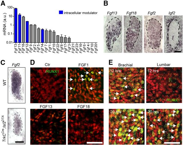
The principle by which unmyelinated primary sensory neurons transducing thermal, itch and pain perception are specified in early development is unknown. These classes of sensory neurons diversify from a common population of late-born neurons, which initiate expression of Runt homology domain transcription factor RUNX1 and the nerve growth factor receptor TrkA. Here, we report that signals emanating from within the mouse dorsal root ganglion mediated partly by early-born neurons destined to a myelinated phenotype participate in fating late-born RUNX1(+)/TrkA(+) neurons. Inductive factors include FGFs via activation of FGF receptor 1 (FGFR1). Consistently, FGF2 is sufficient to induce expression of RUNX1, and Fgfr1 conditional mutant mice display deficits in fating of the common population of late-born RUNX1(+)/TrkA(+) neurons that develop into unmyelinated neurons. Thus, the distinct lineages of sensory neurons are acquired in response to increasing FGF levels provided by a rising number of born neurons.


Figures

Figure 1.
Figure 1.
RUNX1 induction in chicken and mouse DRG. A, B, qPCR analysis of Runx1 mRNA relative expression in embryonic chicken (A) and mouse (B) brachial DRG shows the onset of expression at E5 in chicken and between E11.5 and E12.5 in mouse. Data are presented as mean ± SEM. C, Triple immunostaining for ISL1, TrkA, and RUNX1 on brachial DRG sections from mouse embryo showing RUNX1 protein first detected at E12.5 in cells that already express TrkA and at E13.5 in a large majority of TrkA-positive cells. D, DRG cultures from E11.5 mouse recapitulate the dynamics of RUNX1 induction seen in vivo. Note expression of RUNX1 in neurons (arrows) after 24 h, which is not affected by AraC. a.u., Arbitrary units. Scale bars: C, D, 50 μm.
Figure 2.
Figure 2.
Cre activity in DRG from TrkCCre;R26YFP mice. A, Triple immunostaining for YFP/TrkC/ISL1, YFP/RET/NF200, YFP/TrkA/NF200, and YFP/RET/TrkB on P0 brachial DRG sections from TrkCCre;R26YFP mice. Reporter expression in 100% of TrkC-expressing cells and in the majority of myelinated (NF200+) neurons. B, Pie chart summarizing reporter expression in the major types of DRG neurons. YFP expression in ∼88% and 17% of the myelinated and unmyelinated neurons, respectively. In the quantitative experiments, the different types of myelinated neurons have been classified according to their expression of TrkA, TrkB, TrkC, RET, and NF200. There is a small number of YFP+/TrkA+/NF200 cells in B. Quantification was performed in two different animals (five DRG sections/animal). Scale bar: A, 100 μm.
Figure 3.
Figure 3.
Early-born sensory neurons contribute to the development of later-born TrkA/RUNX1 neurons. A, Immunostaining for ISL1 and TrkC, RET, TrkB, TrkA, or RUNX1 (E12.5) and for TrkA/RUNX1 (P0) on brachial TrkCCre;Isl2DTA DRG sections. At E12.5, a complete absence of TrkC+ cells, very few if any RET+ and TrkB+ cells, and a decrease of TrkA+ and RUNX1+ cells. There is low expression of RUNX1 in the mutant animals (insets). B, C, Quantification of A at E12.5 (B) and P0 (C). Number of cells are in the left and intensity of RUNX1 fluorescence in the two right-most bars of the graphs. D, Whole-mount TrkA and NF165 (insets) immunostaining of E14.5 TrkCCre;Isl2DTA and WT embryos reveals severe impairments of forelimb innervation in the absence of early-born DRG neurons. Fiber extension and arborization throughout the limb are limited in the mutant embryos, with the lateral aspect of both the anterior (A) and posterior (P) regions of the limb and the second, third, and fourth digits lacking innervation. E, Immunostaining of brachial ventral neural tube shows similar pattern of ISL1+ motor neurons (MNs) in E12.5 TrkCCre;Isl2DTA and WT embryos. Data are presented as mean ± SEM. n = 2–4 animals per genotype. *p < 0.05 (two-tailed, unpaired t test, statistical comparison with WT). **p < 0.01 (two-tailed, unpaired t test, statistical comparison with WT). ***p < 0.001 (two-tailed, unpaired t test, statistical comparison with WT). a.u., Arbitrary units. Scale bars: A, D, E, 100 μm.
Figure 4.
Figure 4.
FGF signaling induces RUNX1 in DRG cultures. A, Blocking of RUNX1 induction in E11.5 DRG cultures with inhibitor of MAPK/ERK and PI3K/Akt pathways (PD98059 and LY294002) indicates the involvement of a signaling factor for RUNX1 induction. B, FGF and IGF signaling increases RUNX1 mRNA in cultured E5 chicken DRG as measured by qPCR. ER81 upregulation by NT3 serves as a positive control. C, FGF and IGF signaling induces Runx1 mRNA expression also in mouse E11.5 brachial DRG cultures. D, Induction of RUNX1 protein in E11.5 brachial DRG 12 h after addition of FGF2 or IGF1. There is absence of RUNX1 expression in control condition (3 h in vitro). E, Inhibitors of FGF receptors (PD166866) and IGF receptors (PPP) block an endogenous induction of RUNX1 in 24 h cultures of E11.5 brachial DRG. F, FGF-induced expression of RUNX1 in E11.5 brachial DRG is reduced by PD98059 and inhibited by LY294002. Data are presented as mean ± SEM. **p < 0.005 (one-way ANOVA with Tukey's post hoc test, statistical comparison with control). ***p < 0.001 (one-way ANOVA with Tukey's post hoc test, statistical comparison with control). a.u., Arbitrary units; Ctr, control. Scale bars: A, D, E, and F, 50 μm.
Figure 5.
Figure 5.
FGFs and IGFs are expressed in the developing DRG. A, qPCR analysis for mRNA relative expression of FGF ligands in E11.5 brachial DRG (a.u., Arbitrary units, normalized to Fgf2). Blue represents FGF homologous factor subfamily members (FGF11-FGF14). Experiment performed in duplicate, each sample representing 80 brachial DRG from different embryos. B, In situ hybridization for Fgf and Igfligands in E11.5 brachial mouse DRG shows expression of Fgf13, Fgf18, and Fgf2 within the DRG. Igf2 is expressed in the DRG periphery and surrounding mesenchyme. C, Fgf2 expression is largely decreased in TrkCCre;Isl2DTA brachial DRG at E12.5 (n = 2 animals per genotype; experiment done in duplicate for each animal). D, A total of 20 ng/ml FGF1, but not 200 ng/ml FGF13 or FGF18, induces RUNX1 expression in neurons from DRG explants after 12 h (arrows). E, A time-dependent induction of RUNX1 by FGF2. At E11.5, brachial but not lumbar DRGs are competent to respond to FGF2 treatment within 12 h (arrows indicate positive neurons). However, lumbar DRG neurons upregulate RUNX1 within 20 h in vitro with FGF2. Scale bars: B, 50 μm; C, 100 μm; D, 20 μm; E, 50 μm.
Figure 6.
Figure 6.
Expression and function of FGF receptors in developing DRG. A, In situ hybridization on E12.5 brachial DRG sections shows that Fgfr1, but not Fgfr2, Fgfr3, or Fgfr5, is expressed in the early DRG. B, Fgfr1 mRNA expression in brachial DRG appears at E11.5 and reaches high levels by E12.5. C, Immunostaining for TrkA and ISL1 after in situ hybridization for Fgfr1 on E12.5 brachial DRG section shows expression of Fgfr1 in TrkA+ neurons. D, Immunostaining for βIII-tubulin and Tomato fluorescence on brachial DRG section from E12.5 Wnt1cre;Tomato (cross between the Wnt1cre transgenic line and the R26Tomato reporter mouse line). There is recombination in all neurons. E, In situ hybridization for Fgfr1 on E14.5 brachial DRG sections from Fgfr1lox/lox and Fgfr1cKO shows a nearly complete loss of Fgfr1 in Fgfr1cKO DRG. F, EdU staining (in green, 2 h after injection) on brachial DRG sections from E10.5 or E12 Fgfr1lox/lox and Fgfr1cKO embryos immunostained for SOX10 and ISL1. G, Quantification done within the DRGs depicted by dashed lines in F (n = 2 animals per genotype). H, Immunostaining for RUNX1, TrkA/ISL1, and TrkC/ISL1 on brachial DRG sections from E12.5 control Fgfr1lox/lox (n = 4) and Wnt1Cre;FGFR1lox/lox (FGFR1cKO) (n = 4) mice shows a decrease of RUNX1+ and TrkA+ cells at E12.5. There is lower intensity of RUNX1 in the mutant DRG (insets). I, Quantification of H. Data are presented as mean ± SEM. *p < 0.05 (two-tailed, unpaired t test, statistical comparison with Fgfr1lox/lox). **p < 0.01 (two-tailed, unpaired t test, statistical comparison with Fgfr1lox/lox). a.u., Arbitrary units. J, Whole-mount TrkA immunostaining of E14.5 Fgfr1lox/lox and Fgfr1cKO embryos reveals similar innervation pattern of the forelimb. K, Schematic illustration of the mechanism resulting in the birth of unmyelinated sensory neurons in the coalescing DRG (1) and intracellular signaling pathways participating (2). Scale bars: A, B, D–F, 50 μm; C, 10 μm; H, J, 100 μm.

References

    1. Abdel Samad O, Liu Y, Yang FC, Kramer I, Arber S, Ma Q. Characterization of two RUNX1-dependent nociceptor differentiation programs necessary for inflammatory versus neuropathic pain. Mol Pain. 2010;6:45. doi: 10.1186/1744-8069-6-45. - DOI - PMC - PubMed
    1. Angevine JB, Jr, Sidman RL. Autoradiographic study of cell migration during histogenesis of cerebral cortex in the mouse. Nature. 1961;192:766–768. doi: 10.1038/192766b0. - DOI - PubMed
    1. Bachy I, Franck MC, Li L, Abdo H, Pattyn A, Ernfors P. The transcription factor Cux2 marks development of an A-δ sublineage of TrkA sensory neurons. Dev Biol. 2011;360:77–86. doi: 10.1016/j.ydbio.2011.09.007. - DOI - PubMed
    1. Bertrand N, Castro DS, Guillemot F. Proneural genes and the specification of neural cell types. Nat Rev Neurosci. 2002;3:517–530. doi: 10.1038/nrn874. - DOI - PubMed
    1. Brent AE, Tabin CJ. FGF acts directly on the somitic tendon progenitors through the Ets transcription factors Pea3 and Erm to regulate scleraxis expression. Development. 2004;131:3885–3896. doi: 10.1242/dev.01275. - DOI - PubMed

Publication types

MeSH terms

Substances