. 2013 Nov 6;33(45):17691-709.
doi: 10.1523/JNEUROSCI.3429-13.2013.

Modality-based organization of ascending somatosensory axons in the direct dorsal column pathway

Affiliations

Modality-based organization of ascending somatosensory axons in the direct dorsal column pathway

Jingwen Niu et al. J Neurosci. .

Abstract

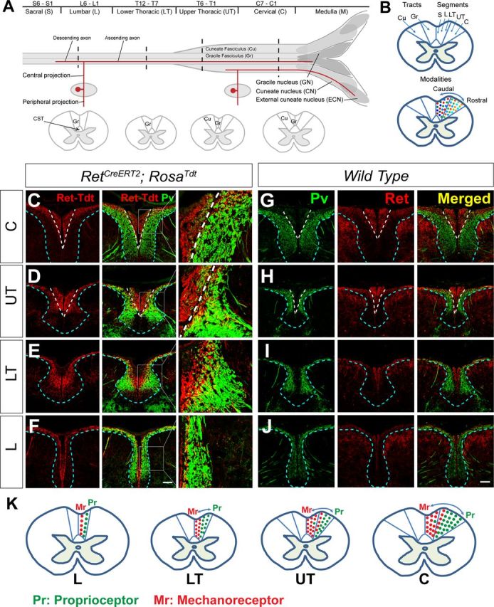
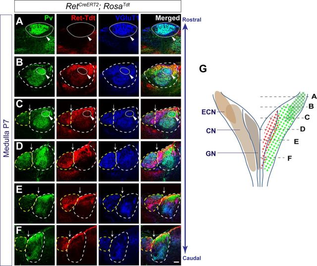
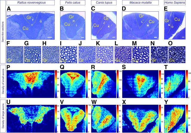
The long-standing doctrine regarding the functional organization of the direct dorsal column (DDC) pathway is the "somatotopic map" model, which suggests that somatosensory afferents are primarily organized by receptive field instead of modality. Using modality-specific genetic tracing, here we show that ascending mechanosensory and proprioceptive axons, two main types of the DDC afferents, are largely segregated into a medial-lateral pattern in the mouse dorsal column and medulla. In addition, we found that this modality-based organization is likely to be conserved in other mammalian species, including human. Furthermore, we identified key morphological differences between these two types of afferents, which explains how modality segregation is formed and why a rough "somatotopic map" was previously detected. Collectively, our results establish a new functional organization model for the mammalian direct dorsal column pathway and provide insight into how somatotopic and modality-based organization coexist in the central somatosensory pathway.


Figures

Figure 1.
Figure 1.
The DDC pathway and modality segregation of ascending axons of RA mechanoreceptors and proprioceptors in the P7 mouse dorsal column. A, Schematic illustration shows the rodent dorsal column in both longitudinal and cross section views. The dorsal column is composed of three tracts: (1) the medial gracile fasciculus (Gr) through the entire dorsal column; (2) the lateral cuneate fasciculus (Cu) that formed above thoracic segment 6 (T6), which innervate the GN, CN, and ECN in the medulla; and (3) the corticospinal tract (CST) at the most ventral area of the dorsal column. Primary somatosensory afferents from S6–T7 and T6–C1 DRGs travel in the Gr and Cu, respectively. S, Sacral; L, lumbar; LT, lower thoracic; UT, upper thoracic; C, cervical. B, Illustration of the “somatotopic map” model for the cervical mammalian dorsal column. Different colors represent different types of somatosensory afferents, and the dark to light gradient of color represents caudal to rostral segments. C–F, Immunostaining of P7 RetCreERT2; Rosa26Tdt spinal cord sections with Pv antibody. RA mechanoreceptors are specifically labeled by Tdt, and proprioceptors are stained by Pv. Ascending axons of mechanoreceptors and proprioceptors are segregated from each other in a medial–lateral pattern at different levels of the spinal cord. G–J, Immunostaining of Pv and Ret antibodies of P7 WT mouse spinal cord sections. Ret antibody stains RA mechanosensory axons in the dorsal column, whereas Pv antibody stains proprioceptive axons. Ascending axons of RA mechanoreceptors and proprioceptors are segregated from each other in a medial–lateral pattern at different levels of the spinal cord. n = 3 mice. Scale bar, 50 μm. K, Illustration of spinal cord transverse views to depict the modality-based segregation of ascending axons of RA mechanoreceptor (Mr) (red) and proprioceptors (Pr) axons (green) at L, LT, UT, and C levels.
Figure 2.
Figure 2.
Modality segregation of RA mechanoreceptors and proprioceptors in the P7 mouse DCNs. A–F, Representative images from serial sections of a P7 RetCreERT2; Rosa26Tdt mouse medulla, which were stained with antibodies against Pv and VGluT1. VGluT1 stains synapses of primary mechanosensory and proprioceptive afferents and thus was used as a marker together with the dorsal sulcus (white arrows) to define the DCNs. Dashed yellow circle represents GN; dashed white circle represents CN; dotted white circle and white arrowheads represent ECN. At caudal levels, Tdt+ RA mechanoreceptors mainly innervate the lateral part of GN, whereas proprioceptors innervate CN (E, F). At more rostral levels, RA mechanoreceptors start to innervate the dorsal medial part of CN, but their termination domain is still segregated from that of proprioceptors (C, D). At the most rostral levels where GN ends, proprioceptors highly innervate the ECN (A, B). There are some Pv+ interneurons present in GN and CN. Scale bar, 100 μm. G, Schematic view of the DCNs shows the section levels and innervation pattern of RA mechanoreceptors (red dots) and proprioceptors (light and dark green dots indicate innervation domains in CN and ECN, respectively).
Figure 3.
Figure 3.
Characterization of PvCre(Arbr); TaumGFP mice. A–C, Immunostaining of P7 PvCre(Arbr); TaumGFP DRG sections with GFP and Pv antibodies. GFP+/Pv neurons, indicated by white arrows, were found in the limb-level DRGs, such as C6 (A) and L5 (C), but not in non–limb-level DRGs, such as T6 (B). Scale bar, 100 μm. D, Low magnification of a P7 PvCre(Arbr); TaumGFP spinal cord, double-stained with antibodies against GFP and Pv. GFP+/Pv+ fibers innervate the intermediate Clarke's nucleus and ventral horn of the spinal cord. E, High magnification of double-stained PvCre(Arbr);TaumGFP dorsal spinal cord shows boxed area in D. GFP+ fibers also innervate layer III to IV of the dorsal horn, circled with dashed white lines, which are Pv. This staining pattern is consistent with previous report that Pv is a proprioceptor-specific marker at P7. F–H, Double staining of GFP and S100 in P21 PvCre(Arbr);TaumGFP mice showing that GFP+ fibers innervate mechanosensory end organs in the periphery, including Meissner corpuscles (white arrow) in the dermal papillae of hindpaw footpads (F), Pacinian corpuscles (white arrow) in the interosseous membrane of fibula (G), and longitudinal lanceolate endings (circled by a dashed white ellipse) associated with follicles on back hairy skin (H). I, Triple staining of Pv, Ret, and GFP of E14.5 PvCre(Arbr); TaumGFP DRG sections shows that Cre-mediated recombination begins at this stage, as some Pv+ neurons have now initiated the expression of GFP. Yellow arrows indicate Pv+/Ret+ cells, and white arrows indicate GFP+/Pv+/Ret+ neurons. Scale bar, 50 μm. n = 3 for each age.
Figure 4.
Figure 4.
Dynamic expression of Pv in Ret+ neurons during development. A, Whole-mount staining of Pv and Ret of P0 WT DRGs shows the expression of Pv in Ret+ DRG neurons at limb levels. White arrows indicate Pv+/Ret+ cells, which can be found in C5–T2 DRGs at forelimb and L3–L6 DRGs at hindlimb levels. Scale bar, 50 μm. B, Quantification of the percentage of Pv+/Ret+ DRG neurons over the total number of Pv+ neurons in different DRGs at E14.5, P0, and P7. Error bars indicate SEM. n = 4 mice for each developmental stage. The expression of Pv is mainly found in limb-level Ret+ DRG neurons, which decreases postnatally.
Figure 5.
Figure 5.
Specific labeling of proprioceptors using PvCreERT2; Rosa26iAP mice. A, Whole-mount Pv staining of a P7 L4 DRG, combined with Fast red-AP color reaction. Scale bar, 50 μm. B, Quantification of AP+ and Pv+/AP+ DRG neurons over the total number of AP+ DRG neurons in PvCreERT2; Rosa26iAP mice (n = 4). 97.6% of all AP+ neurons are Pv+ proprioceptors at P7. C–F, Fast red-AP color reaction and VGluT1 staining of adult PvCreERT2; Rosa26iAP spinal cord sections at cervical (C), upper thoracic (D), lower thoracic (E), and lumbar levels (F). AP+ fibers do not innervate the dorsal horn (dashed white lines), indicating that mechanoreceptors are not labeled in PvCreERT2; Rosa26iAP mice. Scale bar, 100 μm. G–J, BCIP/NBT AP color reaction of the PvCreERT2; Rosa26iAP spinal cord sections revealed the specific innervation pattern of proprioceptors at different levels. Dashed blue lines indicate the dorsal horn. Few Pv+ spinal cord interneurons are also labeled. Scale bar, 100 μm. n = 3.
Figure 6.
Figure 6.
Complementary innervation pattern of RA mechanoreceptors and proprioceptors in the adult mouse dorsal column and DCNs. A–D, Immunostaining of adult RetCreERT2; TaumGFP spinal cord sections with GFP antibody. GFP+ ascending axons of RA mechanoreceptors are enriched in the middle of the adult dorsal column. E–H, Fluorescent AP color reaction of adult PvCreERT2; Rosa26iAP spinal cord sections. AP+ ascending axons of proprioceptors are only found in the lateral region of the dorsal column. Different levels of the RetCreERT2; TaumGFP and PvCreERT2; Rosa26iAP spinal cord are matched and compared. Dashed yellow lines indicate the segregation between AP+ proprioceptive axons and GFP+ RA mechanosensory axons, and dashed white lines indicate the boundary between the dorsal column and the dorsal horn. Scale bar, 100 μm. I–M, Adult medulla innervation pattern of RA mechanoreceptors, which were genetically labeled by GFP in RetCreERT2; TaumGFP mice. N–R, Adult medulla innervation pattern of proprioceptors, which were genetically labeled by AP in PvCreERT2; Rosa26iAP mice. Serial sections were cut through the entire medulla oblongata and costained with VGluT1. Representative images from different levels (similar to those of P7) are shown. RA mechanoreceptors and proprioceptors innervate distinct domains of DCNs, showing a complementary pattern. Dashed yellow lines, dashed white lines, and dotted white lines circle GN, CN, and ECN, respectively, as defined by VGluT1 staining and the dorsal sulcus (white arrows). The ECN is also indicated by white arrowheads. Scale bar, 100 μm. n = 3 for both genotypes.
Figure 7.
Figure 7.
Modality-specific correlation with axonal size in the mouse dorsal column. A–D, Semithin sections of adult RetCreERT2; Rosa26iAP (A, B) and PvCreERT2; Rosa26iAP (C, D) cervical dorsal columns. High magnifications of the boxed areas are shown in B and D. Most RA mechanoreceptors are small-diameter axons (red arrows), whereas few of them are large-diameter axons (magenta arrows). These AP+ large-diameter axons are not proprioceptive axons as indicated by their spinal cord innervation pattern (J). On the other hand, AP+ proprioceptive axons (green arrows) are large-diameter axons found in the lateral area of the dorsal column (D). Scale bars: A, C, 100 μm; B, D, 10 μm. E, Comparison of the transverse areas of AP+ axons of RA mechanoreceptors and proprioceptors. Error bars indicate SEM; 2840 RA mechanosensory axons and 150 proprioceptive axons were measured. n = 3 mice for each genotype. F, G, Distribution of transverse areas of labeled RA mechanosensory axons in RetCreERT2; Rosa26iAP mice. G, Inset, Distribution of large-diameter RA mechanosensory axons, which make up <1% of the totally labeled ones. H, I, Distribution of transverse areas of labeled proprioceptive axons in PvCreERT2; Rosa26iAP mice. Both absolute count (F, H) and percentage are shown (G, I). J, Low- and high-magnification of whole-mount AP color reaction of 6- to 8-week-old RetCreERT2; Rosa26iAP spinal cord sections. AP+ fibers highly innervate layers III-V of the spinal cord at different levels, indicating that AP specifically label RA mechanoreceptors. AP also labels motor neurons at the ventral spinal cord, which are also Ret+ at E11.5 and E12.5. Scale bar, 100 μm.
Figure 8.
Figure 8.
Method of generating heatmaps to represent the density of small- and large-diameter dorsal column fibers. A–E, A computer program was written and run in MATLAB to automatically calculate the axonal size and density in the dorsal column. A, An example raw image cropped from a mouse sample (current size, 500 × 300 pixels). B, Binary image showing areas identified as axons (white). C, Image showing categorization of axons as large (red) and small (cyan) with the 88% as the category threshold. D, Heatmap of large axon density in sliding windows. E, Heatmap of small axon density in sliding windows. F, Probability density functions for AP labeled mechanosensory axons (red trace) and proprioceptive axons (green) and cumulative distribution function for all dorsal column axons (black). Arrows indicate axon area values corresponding to 1st and 5th percentile of proprioceptor axons, which correspond to 80th and 88th percentile of all axons, respectively. G, H, Cumulative distribution functions for all MATLAB-extracted axons of cervical (G) and lumbar (H) spinal cord sections of different mammalian species.
Figure 9.
Figure 9.
Modality segregation revealed by distribution of small- and large-diameter axons in the mouse dorsal column. A–G, Semithin sections of the cervical (A–D) and lumbar dorsal column (E–G) of a 3 month WT mouse. Dashed white lines indicate the boundary between Gr and Cu; dashed cyan lines indicate the edge between the dorsal column and the dorsal horn. A–C, Overview of the cervical dorsal column (A), and higher magnified images of the boxed area in gracile (B), cuneatefasciculus (C), and the boundary areas of Gr and Cu (D). E–G, Overview of the lumbar dorsal column (E) and higher-magnification images in the medial (F) and lateral (G) part of the Gr. Scale bars: A, E, 100 μm; B–D, F, G, 10 μm. H–K, Heatmaps showing the density of small- and large-diameter axons. H, I, In the cervical dorsal column, small-diameter axons are enriched in the medial gracile fasciculus and the medial marginal zone of the cuneate fasciculus, whereas large-diameter axons are mainly in the lateral cuneate fasciculus. J, K, At lumbar level, small-diameter axons are mainly in the medial gracile fasciculus whereas large-diameter axons are more laterally located.
Figure 10.
Figure 10.
Modality-based organization in the cervical dorsal column of multiple mammalian species. A–E, The semithin sections of rat (A), feline (B), canine (C), monkey (D), and human (E) cervical dorsal column. F–O, Higher-magnification images of boxed areas in A–E. M–P, Heatmaps showing the density of small (P–T) and large (U–Y) axons of the cervical dorsal column of each species. Dashed yellow lines indicate the boundary between gracile and cuneate fasciculi; dashed cyan lines indicate the edge between the dorsal column and the dorsal horn. Scale bars: A, B, 200 μm; C–E, 300 μm; F–O, 10 μm.
Figure 11.
Figure 11.
Axon distribution reveals modality-based organization in the lumbar dorsal column of multiple mammalian species. A–C, The semithin sections of rat (A), canine (B), and monkey (C) lumbar dorsal column (stained with toluidine blue). D–I, Higher-magnification images of boxed areas in A–C, showing the axonal size difference between medial and lateral gracile fasciculus of each species. J–O, Heatmaps show the density of small (J–L) and large (M–O) axons of the lumbar dorsal column of each species. Scale bars: A, 100 μm; B, 200 μm; C, 300 μm; D–I, 10 μm.
Figure 12.
Figure 12.
Background recombination of RetCreERT2; Rosa26iAP mice. A–C, Whole-mount AP color reaction of the P21 RetCreERT2; Rosa26iAP spinal cord without 4HT treatment showing that two types of morphologically different neurons are labeled by AP. One type of neurons grows long projections (B), which bifurcate at the dorsal root entry zone and send long ascending and descending axons in the dorsal column, and the other ones are locally projecting neurons (C). D–I, Immunostaining of CGRP or VGluT1 of adult RetCreERT2; Rosa26iAP spinal cord sections combined with Fast-red AP color reaction. AP+ fibers innervate different dorsal horn layers. D–F, Dashed white lines indicate the boundary between layers I and II defined by CGRP staining. G–I, Dashed white lines indicate the boundary between layers II and III, defined by VGluT1 staining. E, H, White arrows point to the AP+ fibers that innervate superficial layers; yellow arrows indicate the AP+ fibers that innervate deep layers of the dorsal horn. J, Quantification of the percentages of long versus locally projecting neurons of P0 and P21 RetCreERT2; Rosa26iAP mice without 4HT treatment, and that of P21 RetCreERT2; Rosa26iAP mice with 0.25 mg 4HT treatment at E12.5. K–P, Whole-mount DRG immunostaining of anti-NFH and Fast-red AP color reaction. M, An example of NFH+/AP+ DRG neuron. P, An example of NFH/AP+ DRG neuron. Scale bar, 100 μm. Q, Quantification of the percentages of NFH+/AP+ and NFH/AP+ DRG neurons of P0 and P21 RetCreERT2; Rosa26iAP mice without 4HT treatment, and that of P21 RetCreERT2; Rosa26iAP mice with 0.25 mg 4HT treatment at E12.5. Error bars indicate SEM. n = 6–8 mice. *p < 0.001.
Figure 13.
Figure 13.
Sparse labeling of RA mechanoreceptors and proprioceptors uncovers key morphological differences. A, B, AP color reaction of whole-mount RetCreERT2; Rosa26iAP and PvCreERT2; Rosa26iAP DRGs, showing one labeled RA mechanoreceptor (A) and proprioceptor (B), respectively. C, D, Dorsal view of the whole-mount spinal cord shows the central projection of RA mechanoreceptors and proprioceptors. After bifurcation, one lumbar RA mechanoreceptor travels 3–4 segments before joining the dorsal column pathway (C), whereas proprioceptors enter the dorsal column within the same segment (D). E, F, AP+ ascending axons of RA mechanoreceptors (E) and proprioceptors (F) travel in the dorsal column. F, Red arrow indicates the endpoint of a proprioceptive axon. C–F, Images are oriented as caudal (left) and rostral (right). G, H, Dorsal views of the medulla show the different termination domains of RA mechanoreceptors (G) and proprioceptors (H) in the DCNs. Dashed yellow lines outline the DCN areas. Scale bar, 100 μm. I, J, Summary of ascending and descending axons of 100 RA mechanoreceptors from 14 mice (I) and 135 proprioceptors from 13 mice (J). Black dots indicate where the cell bodies are located; blue and red lines indicate the ascending and descending fibers respectively. Almost all RA mechanoreceptors reach medulla regardless of their cell body locations, whereas only proprioceptors from DRGs above T6 reach medulla.
Figure 14.
Figure 14.
3D reconstruction of genetically traced RA mechanoreceptors and proprioceptors. Blue lines indicate the first- and second-order central projections; magenta lines indicate the interstitial collaterals innervating the spinal cord or DCNs. A–E, A representative example of 3D reconstructed lumbar RA mechanoreceptors and of P21 RetCreERT2; Rosa26iAP mice. A, Overview of the L2 RA mechanoreceptor, with an ascending axon ending at medulla and a descending axon stopping at S2. The ascending axon sends several collaterals to the dorsal spinal cord before it enters the dorsal column at the T10 level but grows no collaterals afterward (represented by dashed blue line) until it reaches medulla. The descending axon also grows a few collaterals. Enlarged view of the bifurcation area and collaterals near the dorsal root entry zone (B), collaterals near the descending endpoint (C), the nerve endings in the medulla (D), and the collaterals arising from the ascending axon (E) are also shown. F–N, Representative examples of 3D reconstructed lumbar group Ia (F–J) and Ib (K–N) proprioceptors of P21 PvCreERT2; Rosa26iAP mice. F, Overview of an L5 group Ia proprioceptor, with an ascending fiber ending at T12 and a descending fiber stopping at S1. Enlarged view of different areas are also shown, including the bifurcation area (G), collaterals innervating the intermediate area (red circle) and those innervating ventral horn (green ellipse) of the spinal cord (H), descending endpoint at S1 (I), and the ascending endpoint at T12 (J). K, Overview of an L2 group Ib proprioceptor, which has an ascending fiber ending at T5 and a descending fiber stopping at L3. Enlarged view of the bifurcation area and descending endpoint (L), the collaterals arising from the main afferent to show the innervation of the intermediate area in the spinal cord (M), and the ascending endpoint at T5 (N) are also shown. Both group Ia and Ib fibers from the lumbar levels drop out of the ascending pathway with a terminal innervating interstitially in the spinal cord. O–W, Representatives of 3D reconstructed cervical RA mechanoreceptor (O–R) and proprioceptor (S–W). Both have their ascending fibers ending at medulla.
Figure 15.
Figure 15.
Central root rhizotomy suggests that lumbar proprioceptors drop out of the ascending pathway. A–D, Semithin sections of L2 (A), T6 (B), and C3 (C) mouse spinal cord with L4–L5 central root rhizotomy. Dashed magenta lines indicate degenerating axons; dashed yellow lines indicate the boundary between gracile and cuneate fasciculi; dashed cyan lines indicate the edges between the dorsal column and the dorsal horn. Scale bar, 50 μm. D, Quantification of degenerating axons at L2, T6, and C3 with L4–L5 central root rhizotomy. The number of degenerating axons decreased significantly at C3 (283 ± 7) and T6 (390 ± 18) compared with L2 (832 ± 14). n = 3 mice. *p < 0.001. E, Illustration of the model that lumbar proprioceptors drop out the ascending pathway. Red represents RA mechanoreceptors; green represents proprioceptors. Dark color represents fibers from posterior DRGs, and lighter color represents fibers from anterior DRGs.
Figure 16.
Figure 16.
Model of the functional organization of the mammalian dorsal column. A, Relative positions of labeled ascending axons of RA mechanoreceptors from different DRGs are replotted as the transverse view at C3, suggesting that RA mechanosensory axons are organized in a somatotopic manner in the DDC pathway. B, Coexisting model of the functional organization of the mammalian dorsal column. Schematic view of different morphologies of caudal and rostral RA mechanoreceptors and proprioceptors, which lead to modality segregation throughout the dorsal column and dorsal column nuclei. Caudal proprioceptors send collaterals to innervate the Clarke's column, which transmit the caudal proprioceptive information via the indirect pathway to the medulla. Most other collaterals of proprioceptors and RA mechanoreceptors are omitted for simplicity. C, Simulation to demonstrate how a modality-based organization can generate a rough somatotopic map in the caudal spinal cord dorsal column. Red lines indicate axons of RA mechanoreceptors; green lines indicate axons of proprioceptors; the dark to light gradient of color indicates caudal to rostral segments. In addition, dashed red lines indicate RA mechanosensory axons after bifurcation and before they enter the dorsal column. For this simulation, ascending axons of RA mechanoreceptors project ∼3 segments rostrally on average before joining the dorsal column, whereas ascending axons of lumbar proprioceptors terminate after traveling ∼6.5 segments. These numbers are derived from the experimental observations (Fig. 13J).

References

    1. Badea TC, Nathans J. Quantitative analysis of neuronal morphologies in the mouse retina visualized by using a genetically directed reporter. J Comp Neurol. 2004;480:331–351. doi: 10.1002/cne.20304. - DOI - PubMed
    1. Badea TC, Hua ZL, Smallwood PM, Williams J, Rotolo T, Ye X, Nathans J. New mouse lines for the analysis of neuronal morphology using CreER(T)/loxP-directed sparse labeling. PLoS One. 2009;4:e7859. doi: 10.1371/journal.pone.0007859. - DOI - PMC - PubMed
    1. Boshes B, Padberg F. Studies on the cervical spinal cord of man: sensory pattern after interruption of the posterior columns. Neurology. 1953;3:90–101. doi: 10.1212/WNL.3.2.90. - DOI - PubMed
    1. Brown AG, editor. Organization in the spinal cord. New York: Springer; 1981.
    1. Burgess PR, Clark FJ. Characteristics of knee joint receptors in the cat. J Physiol. 1969;203:317–335. - PMC - PubMed

Publication types

LinkOut - more resources