. 2013 Nov 6;33(45):17897-907.
doi: 10.1523/JNEUROSCI.0511-13.2013.

Axonal sprouting and formation of terminals in the adult cerebellum during associative motor learning

Affiliations

Axonal sprouting and formation of terminals in the adult cerebellum during associative motor learning

Henk-Jan Boele et al. J Neurosci. .

Abstract

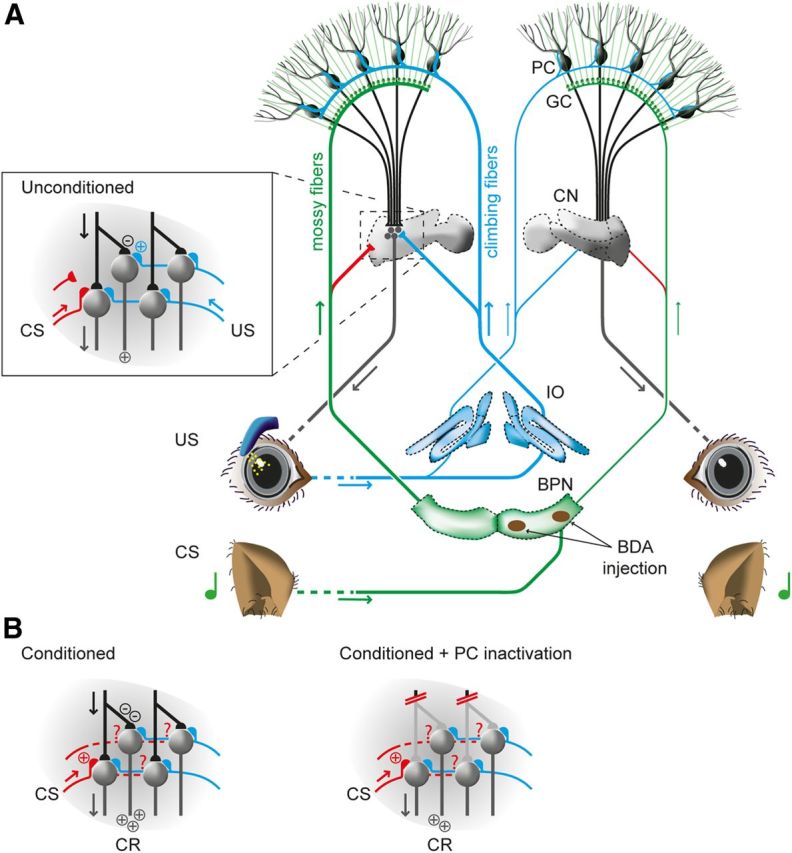
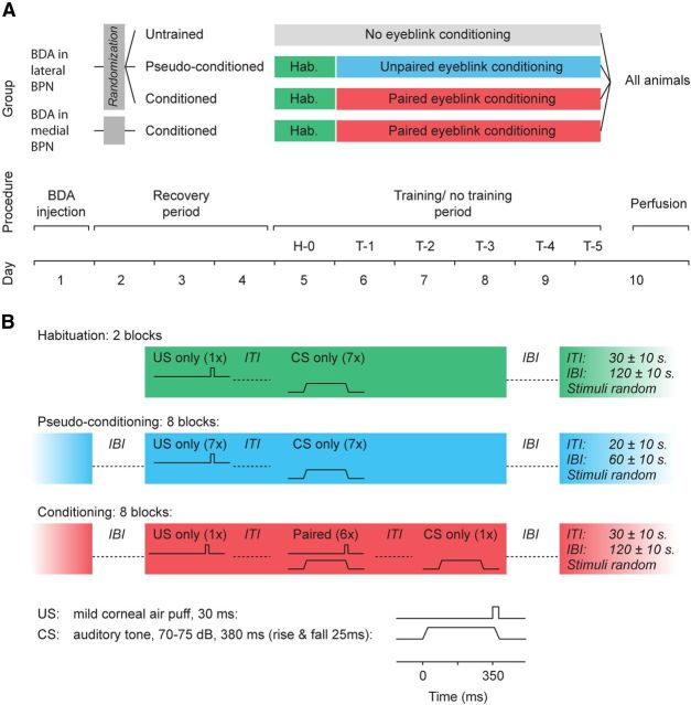
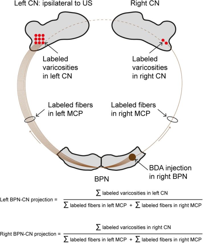
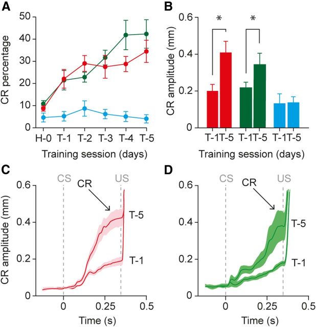
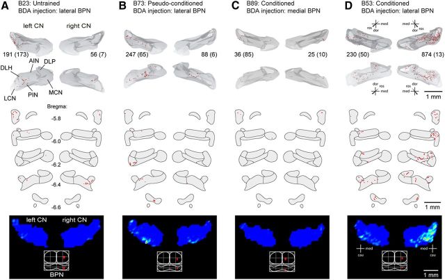
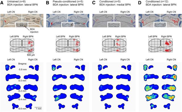
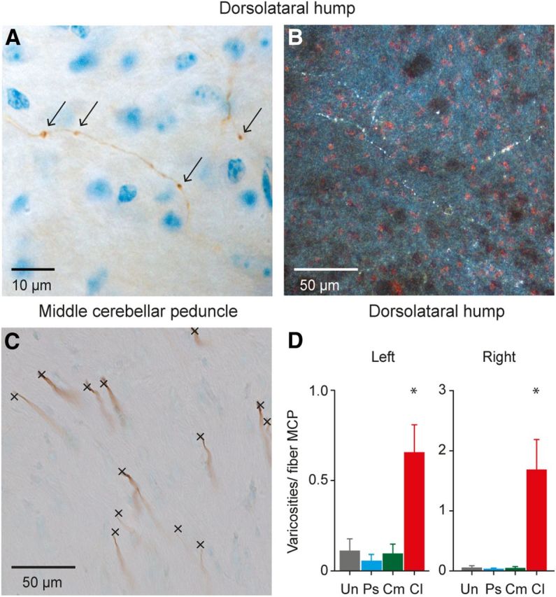
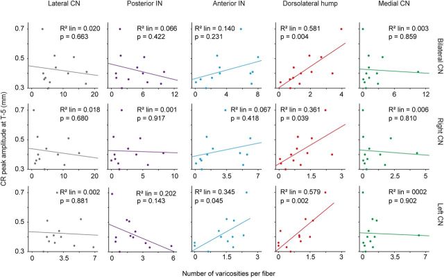
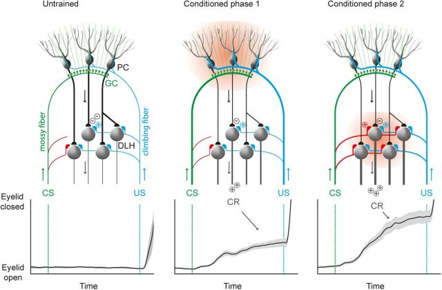
Plastic changes in the efficacy of synapses are widely regarded to represent mechanisms underlying memory formation. So far, evidence for learning-dependent, new neuronal wiring is limited. In this study, we demonstrate that pavlovian eyeblink conditioning in adult mice can induce robust axonal growth and synapse formation in the cerebellar nuclei. This de novo wiring is both condition specific and region specific because it does not occur in pseudoconditioned animals and is particularly observed in those parts of the cerebellar nuclei that have been implicated to be involved in this form of motor learning. Moreover, the number of new mossy fiber varicosities in these parts of the cerebellar nuclei is positively correlated with the amplitude of conditioned eyelid responses. These results indicate that outgrowth of axons and concomitant occurrence of new terminals may, in addition to plasticity of synaptic efficacy, contribute to the formation of memory.


Figures

Figure 1.
Figure 1.
Neurocircuits engaged during pavlovian eyeblink conditioning. A, The CS pathway (shown in green) is formed by mossy fibers, which transmit the auditory tone from the lateral BPN bilaterally to PCs in eyeblink-controlling zones in the cerebellar cortex. From the pontine region with the BDA injection, there are bilateral projections to the cerebellar cortex, with a preference for the contralateral site from the injection (represented by the thick green line). The US pathway (shown in blue) is formed by climbing fibers, which transmit the eye-puff signal from the inferior olive (IO) bilaterally to both the cerebellar cortex and the CN, with a preference for the ipsilateral site from the stimulated eye (represented by the thick blue line). In untrained animals, the projections from the BPN to the CN are very sparse (set in). B, Left, In trained animals, the presentation of the CS will result in a perfectly timed inhibition of the PC input to the CN. Right, Inactivation of the PC input to the CN cannot completely abolish the CRs, suggesting cerebellar nuclear plasticity. However, in untrained animals, there is no convergence of the CS and US at the level of the CN. GC, Granule cell.
Figure 2.
Figure 2.
Scheme of experimental procedures. A, At day 1 in all groups, BDA was injected iontophoretically in the lateral or medial right BPN. After a recovery period of 3 d, mice with BDA injected in the lateral right BPN were randomly divided in a conditioned group (n = 12), a pseudoconditioned group (n = 5), and an untrained group (n = 8). Mice with the BDA injection in the medial part of the right BPN (n = 5) were all conditioned. Both conditioned groups were subjected to one habituation session (H-0), followed by five training sessions (T-1–T-5). The pseudoconditioned group received essentially the same treatments as the conditioned group, except that these mice were exposed to an explicitly unpaired presentation of the CS and the US. The untrained group was not subjected to any additional training after surgery. At day 10, all animals were transcardially perfused. B, Overview of the stimuli that were delivered during the habituation, pseudoconditioning, and conditioning procedures. IBI, interblock interval; ITI, intertrial interval.
Figure 3.
Figure 3.
Quantification of BDA-labeled mossy fibers from the right BPN to the CN. BDA was injected in the right BPN. From here, mossy fibers project via the MCP to both the left and the right CN but with a clear preference for the left CN. Because we use an air-puff US on the left eye and because the cerebellum is assumed to represent mainly ipsilateral movements, we made the BDA injection in the right BPN. To allow direct comparison of the BPN–CN projection between animals, we correct for the size of the BDA injection, which is reflected the number of labeled MCP fibers, by dividing the number of labeled varicosities in the CN by the total number of labeled MCP fibers.
Figure 4.
Figure 4.
Eyeblink conditioning after paired and pseudopaired presentation of CS and US. A, The mean percentage of CRs per daily training session (T-1–T-5) gradually increased for animals that were trained with a paired presentation of the CS and US (red, conditioned with BDA injection in lateral right BPN; green, conditioned with BDA injection in medial right BPN), whereas pseudoconditioning with explicitly unpaired presentations of the CS and US did not result in CRs (blue). Before training, all animals were habituated (H-0) to the experimental setup. B, Animals that were subjected to a paired presentation of the CS and US also showed a significant increase of the amplitude of their eyelid CRs. *p = 0.027, conditioned with BDA in lateral right BPN; *p = 0.04, conditioned with BDA in medial right BPN. C, Average ± SEM raw MDMT eyeblink traces of T1 and T5 for conditioned animals with BDA injected in the lateral part of the right BPN. Maximum eyelid closure during the CR was reached just before the onset of the US at 350 ms after CS onset. For comparison, a full eyelid closure (the unconditioned response) has an amplitude of ∼1 mm. D, Same as C but now for conditioned animals with BDA injected in the medial part of the right BPN.
Figure 5.
Figure 5.
Differences in collateralization of pontine mossy fibers after conditioning. Four examples of each experimental group are shown. Top, Three-dimensional reconstruction based on every other section through the CN. The bottom pair of reconstructions represent caudal view of the left and right hand nuclei; the top pair represents an inverted rostral view. Numbers between reconstruction pairs indicate number of plotted varicosities and number of labeled fibers within the MCP (between brackets). Middle, Labeled varicosities in the bilateral CN plotted in standardized sections going from bregma −5.8 to −6.6 mm. Bottom, Dorsal view of the CN showing the density profile determined from counting the number of plotted varicosities in mediolateral bins of 80 μm wide per section and visualized with MATLAB routines. Bottom shows a dorsal and caudal view of the BPN with a representation of the injection site. Note that, in all groups, the number of labeled fibers is highest in the contralateral MCP. The number of plotted varicosities/labeled fibers is highest ipsilaterally of the conditioned eye. Scale bars, 1 mm. DLP, Dorsolateral protuberance; MCN, medial cerebellar nucleus.
Figure 6.
Figure 6.
Expansion of mossy fiber collaterals in CN induced by eyeblink conditioning. Top, BDA tracer injections in the BPN and the labeling in the CN for an untrained, a pseudoconditioned, a conditioned animal with medial BDA injection in the BPN, and a conditioned animal with lateral BDA injection in the BPN. Each red dot represents a labeled varicosity. Middle, Dorsal (top panel) and caudal view (bottom panel) of the BPN with a representation of the BDA injection site. Bottom, Mean color-coded density plots for the number of varicosities per mossy fiber in the MCP for all animals per group from bregma −5.8 to −6.6 corrected for group size and the number of labeled varicosities in the MCP. MCN, Medial cerebellar nucleus.
Figure 7.
Figure 7.
Labeled mossy fiber collaterals and varicosities in the DLH of conditioned animals with BDA injected in the lateral part of the right BPN. A, Light microscopic image of a BDA-labeled mossy fiber collateral with varicosities in the DLH region of a conditioned animal. Scale bar, 10 μm. Arrows indicate labeled varicosities. B, Dark-field microscopic image of BDA-labeled mossy fiber collaterals with varicosities in the DLH region of a conditioned animal. Scale bar, 50 μm. C, BDA-labeled mossy fibers in the MCP, which were used to correct for the size of the BDA injection, by dividing the number of labeled varicosities in the CN by the number of labeled fibers in the MCP. Scale bar, 50 μm. D, Total number of synapses in the DLH per labeled MCP fiber for all four groups. Conditioned animals with BDA injection in the lateral right BPN show a significant increase in their number of varicosities per fiber in the MCP. Cl, Conditioned with BDA injection in the lateral right BPN; Cm, conditioned with BDA injection in the medial right BPN; Ps, pseudoconditioned with BDA injected in the lateral right BPN; Un, untrained with BDA injected in the lateral right BPN. *p value left DLH 0.003; p value right DLH <0.001.
Figure 8.
Figure 8.
Mean number of labeled varicosities per labeled mossy fiber in the MCP plotted against the mean CR amplitude (millimeters) at the last training session for conditioned animals with BDA injected in the lateral BPN. Top panels are for the bilateral CN, middle panels for the right CN, and bottom panels for the left CN. In conditioned animals, the number of labeled varicosities in the DLH per MCP fiber correlates positively with the amplitude of the eyelid CRs in the last training sessions. This correlation was found for bilateral, left, and right DLH region. IN, Interposed nucleus.
Figure 9.
Figure 9.
Proposed model showing how the cerebellar cortex and CN may both contribute to the learning process during pavlovian eyeblink conditioning. DLH neurons receive inhibitory PC input (black), excitatory climbing fiber input as the US (blue), and excitatory mossy fiber input as the CS (red) and innervate the eyelid muscles via the red nucleus and facial nucleus. In naive animals, the CS will not elicit a blink response. During the first phase of acquisition during eyeblink conditioning, plasticity is induced in the cerebellar cortex, resulting in a well timed PC firing pause (indicated by 2 minus symbols), which behaviorally is reflected with a small-amplitude CR. Prolonged training will induce growth of mossy fiber collateral to eyeblink-controlling neurons in the CN, which behaviorally is reflected with a larger-amplitude CR. GC, Granule cell.

References

    1. Boele HJ, Koekkoek SK, De Zeeuw CI. Cerebellar and extracerebellar involvement in mouse eyeblink conditioning: the ACDC model. Front Cell Neurosci. 2010;3:19. doi: 10.3389/neuro.03.019.2009. - DOI - PMC - PubMed
    1. Brodal P, Dietrichs E, Walberg F. Do pontocerebellar mossy fibres give off collaterals to the cerebellar nuclei? An experimental study in the cat with implantation of crystalline HRP-WGA. Neurosci Res. 1986;4:12–24. doi: 10.1016/0168-0102(86)90013-1. - DOI - PubMed
    1. Campolattaro MM, Freeman HJ. Examination of bilateral eyeblink conditioning in rats. Behav Neurosci. 2009;6:1346–1352. - PMC - PubMed
    1. Cicirata F, Zappalà A, Serapide MF, Parenti R, Pantò MR, Paz C. Different pontine projections to the two sides of the cerebellum. Brain Res Brain Res Rev. 2005;49:280–294. doi: 10.1016/j.brainresrev.2005.02.002. - DOI - PubMed
    1. Dancause N, Barbay S, Frost SB, Plautz EJ, Chen D, Zoubina EV, Stowe AM, Nudo RJ. Extensive cortical rewiring after brain injury. J Neurosci. 2005;25:10167–10179. doi: 10.1523/JNEUROSCI.3256-05.2005. - DOI - PMC - PubMed

Publication types

LinkOut - more resources