. 2013 Nov 6;33(45):17908-20.
doi: 10.1523/JNEUROSCI.1312-13.2013.

Human immunodeficiency virus-1 Tat protein increases the number of inhibitory synapses between hippocampal neurons in culture

Affiliations

Human immunodeficiency virus-1 Tat protein increases the number of inhibitory synapses between hippocampal neurons in culture

Nicholas J Hargus et al. J Neurosci. .

Abstract

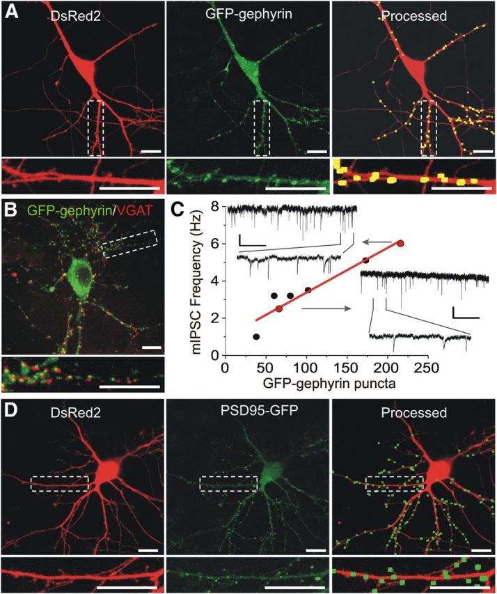
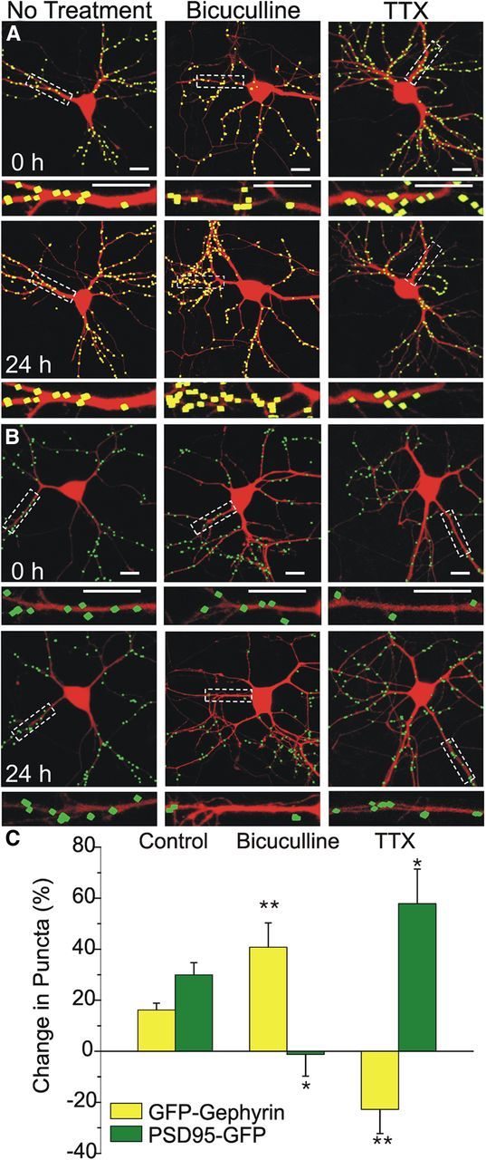
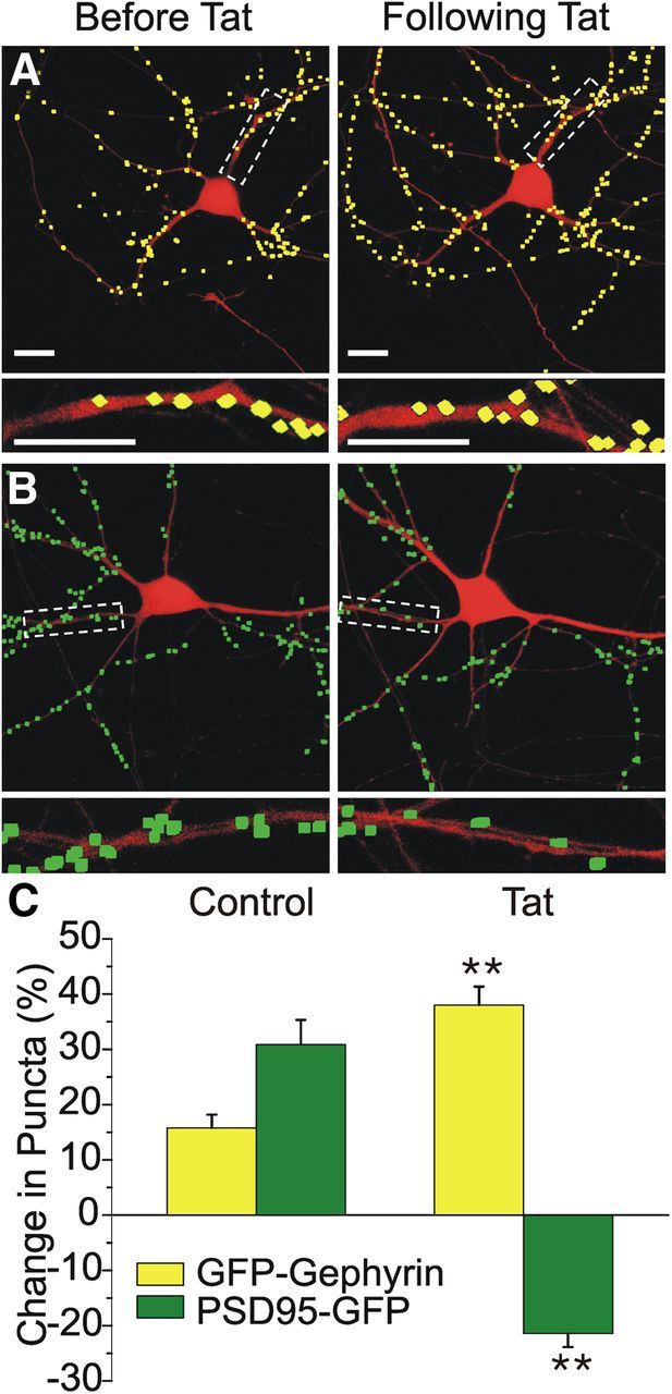
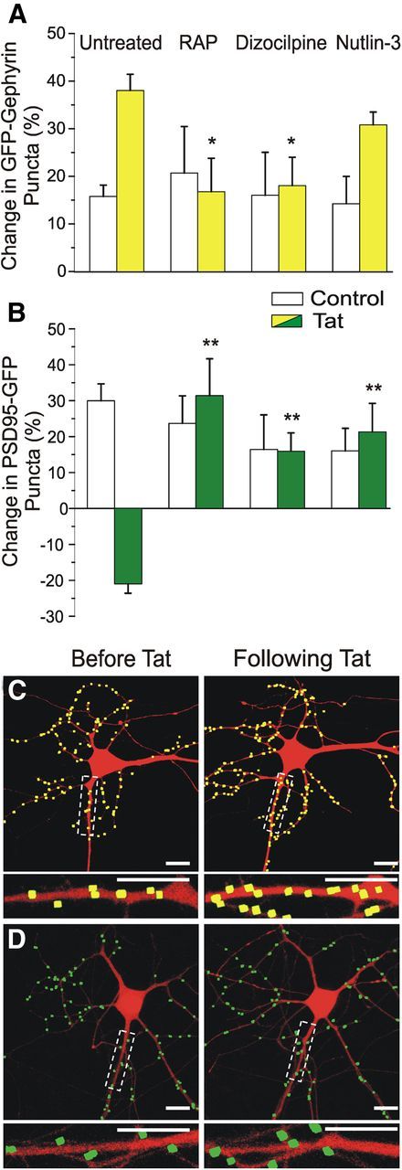
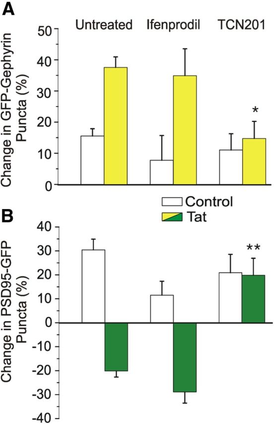
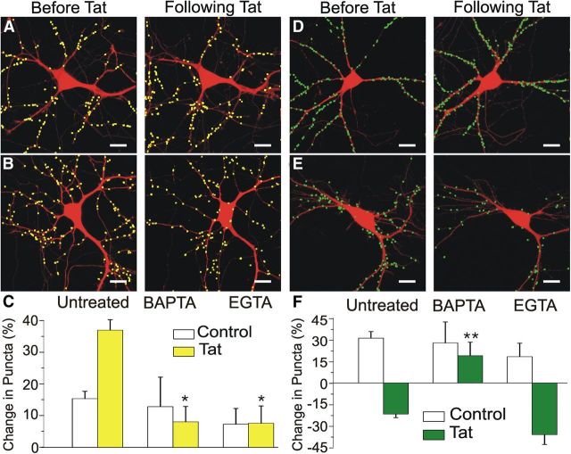
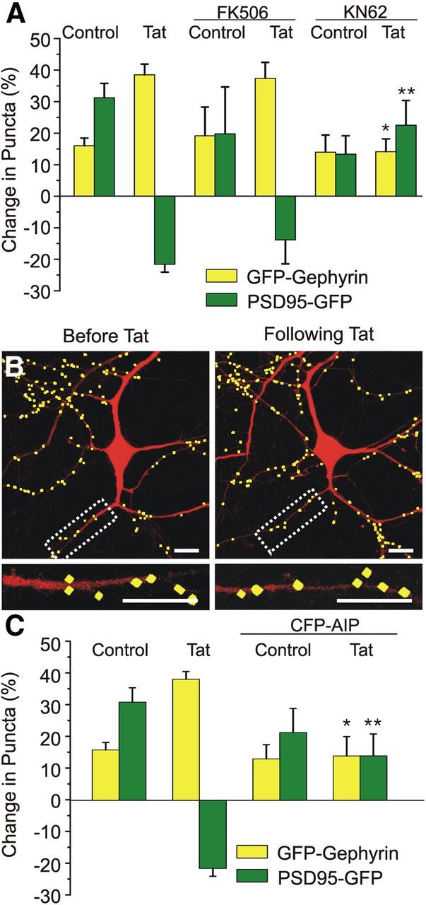
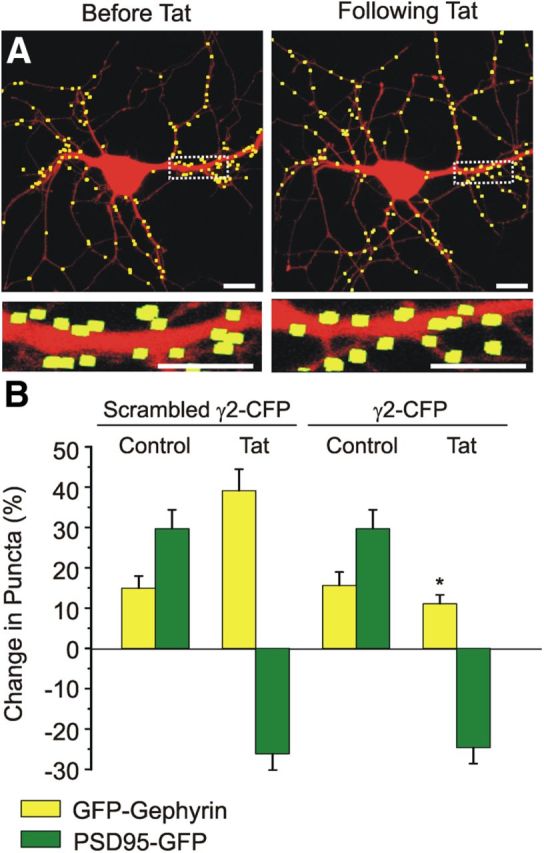
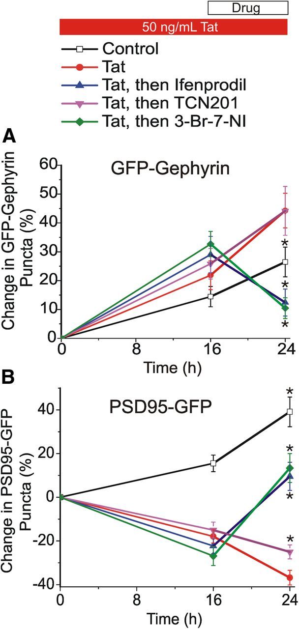
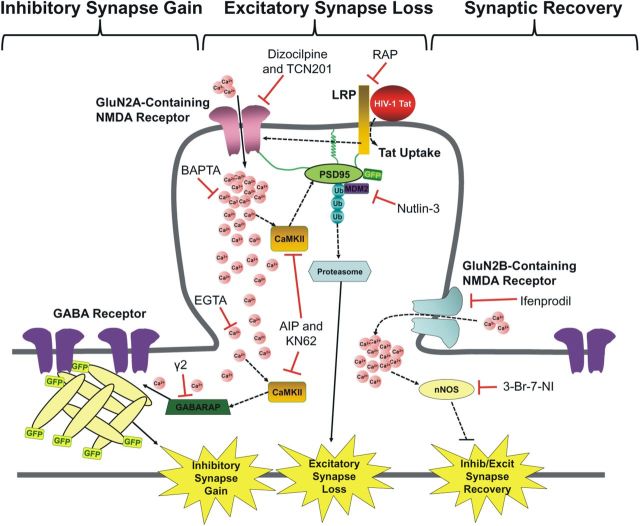
Synaptodendritic damage correlates with cognitive decline in many neurodegenerative diseases, including human immunodeficiency virus-1 (HIV-1)-associated neurocognitive disorders (HAND). Because HIV-1 does not infect neurons, viral-mediated toxicity is indirect, resulting from released neurotoxins such as the HIV-1 protein transactivator of transcription (Tat). We compared the effects of Tat on inhibitory and excitatory synaptic connections between rat hippocampal neurons using an imaging-based assay that quantified clusters of the scaffolding proteins gephyrin or PSD95 fused to GFP. Tat (24 h) increased the number of GFP-gephyrin puncta and decreased the number of PSD95-GFP puncta. The effects of Tat on inhibitory and excitatory synapse number were mediated via the low-density lipoprotein receptor-related protein and subsequent Ca(2+) influx through GluN2A-containing NMDA receptors (NMDARs). The effects of Tat on synapse number required cell-autonomous activation of Ca(2+)/calmodulin-dependent protein kinase II (CaMKII). Ca(2+) buffering experiments suggested that loss of excitatory synapses required activation of CaMKII in close apposition to the NMDAR, whereas the increase in inhibitory synapses required Ca(2+) diffusion to a more distal site. The increase in inhibitory synapses was prevented by inhibiting the insertion of GABAA receptors into the membrane. Synaptic changes induced by Tat (16 h) were reversed by blocking either GluN2B-containing NMDARs or neuronal nitric oxide synthase, indicating changing roles for pathways activated by NMDAR subtypes during the neurotoxic process. Compensatory changes in the number of inhibitory and excitatory synapses may serve as a novel mechanism to reduce network excitability in the presence of HIV-1 neurotoxins; these changes may inform the development of treatments for HAND.


Figures

Figure 1.
Figure 1.
GFP–gephyrin labels functional inhibitory postsynaptic sites. Laser scanning confocal microscopy was used to image a cultured hippocampal neuron expressing DsRed2 (A, D, left) and either GFP–gephyrin (A, middle) or PSD95–GFP (D, middle). Maximum z-projections from eight 1 μm steps were created from DsRed2 z-series and GFP–gephyrin or PSD95–GFP z-series. After enhancing contrast, puncta reaching appropriate size and intensity criteria and in contact with a DsRed2 mask were enlarged and superimposed on the DsRed2 image (A, D, Processed). A more detailed description of image processing is described in Materials and Methods. The insets are enlarged images of the boxed region. Scale bars, 10 μm. B, Neurons expressing GFP–gephyrin (green) were fixed and labeled with an antibody for VGAT (red, 1:500) as described in Materials and Methods. The inset displays an enlarged image of the boxed region. Scale bars, 10 μm. C, mIPSCs were recorded from neurons expressing GFP–gephyrin as described previously, and their mIPSC frequency was plotted as a function of GFP–gephyrin puncta number. The red line is a linear regression of points demonstrating correlation between number of puncta and the frequency of inhibitory synaptic activity (r2 = 0.91, n = 7, p < 0.001). Insets show examples of mIPSC recordings from the cells indicated (red circles), demonstrating an increase in frequency between low and high puncta counts. Calibration: 25 pA, 1 or 0.1 s (for expanded trace).
Figure 2.
Figure 2.
Pharmacological modulation of synaptic transmission alters GFP–gephyrin and PSD95–GFP puncta counts. A, B, Processed confocal images of neurons expressing DsRed2 and either GFP–gephyrin (A, yellow) or PSD95–GFP (B, green) before (0 h) and 24 h after treatment with 10 μm bicuculline or 1 μm TTX as indicated. Insets are enlarged images of the boxed regions. Scale bars, 10 μm. C, Bar graph summarizes the effects of no treatment or 24 h treatment with bicuculline or TTX on changes in GFP–gephyrin (yellow) or PSD95–GFP (green) puncta counts. Data are mean ± SEM. *p < 0.05, **p < 0.01 relative to corresponding control group (ANOVA with Bonferroni's post hoc test).
Figure 3.
Figure 3.
HIV-1 Tat induced concurrent upregulation of GFP–gephyrin puncta and loss of PSD95–GFP puncta. A, B, Processed confocal images of neurons expressing DsRed2 and either GFP–gephyrin (A, yellow) or PSD95–GFP (B, green) before and 24 h after Tat treatment (50 ng/ml). Insets are enlarged images of the boxed regions. Scale bars, 10 μm. C, Bar graph summarizes the effects of 24 h treatment with HIV-1 Tat or no treatment (control) on changes in GFP–gephyrin (yellow bars) or PSD95–GFP (green bars) expression. Data are mean ± SEM. **p < 0.01 relative to corresponding control group (Student's t test).
Figure 4.
Figure 4.
Tat-induced upregulation of GFP–gephyrin puncta is dependent on NMDA and LRP but not MDM2. Bar graphs summarize the changes in GFP–gephyrin (A) or PSD95–GFP (B) puncta in the absence (open bars) or presence (filled bars) of 50 ng/ml HIV-1 Tat for 24 h. Cells were untreated or pretreated (30 min) with 50 nm RAP, 10 μm dizocilpine, or 1 μm nutlin-3 as indicated. Data are mean ± SEM. *p < 0.05, **p < 0.01 relative to corresponding untreated + Tat groups (ANOVA with Bonferroni's post hoc test). C, D, Processed confocal images of neurons expressing DsRed2 and either GFP–gephyrin (C) or PSD95–GFP (D) before (left) and 24 h after (right) Tat (50 ng/ml) application. Cells were pretreated for 30 min with 1 μm nutlin-3. Note that nutlin-3 failed to affect the Tat-induced increase in GFP–gephyrin puncta count, whereas it blocked the effects of Tat on the number of PSD95–GFP puncta. Insets are enlarged images of the boxed regions. Scale bars, 10 μm.
Figure 5.
Figure 5.
Tat-induced upregulation of GFP–gephyrin puncta is dependent on GluN2A-containing NMDARs. A, B, Bar graphs summarize the changes in GFP–gephyrin (A) or PSD95–GFP (B) puncta in the absence (control, open bars) or presence (filled bars) of 50 ng/ml HIV-1 Tat for 24 h. Cells were untreated or pretreated for 30 min with 10 μm ifenprodil or 10 μm TCN201, as indicated. Data are mean ± SEM. *p < 0.05, **p < 0.01 relative to corresponding untreated + Tat groups (ANOVA with Bonferroni's post hoc test).
Figure 6.
Figure 6.
Chelating intracellular calcium prevents HIV-1 Tat-induced increases in inhibitory synapses. A, B, Processed confocal images of neurons expressing DsRed2 and GFP–gephyrin before (left) and 24 h after (right) Tat application (50 ng/ml). Cells were pretreated for 30 min with 10 μm BAPTA-AM (A) or 10 μm EGTA-AM (B). Scale bars, 10 μm. C, Bar graph summarizes the changes in GFP–gephyrin puncta in the absence (control, open bars) or presence (filled bars) of 50 ng/ml Tat for 24 h. Cells were untreated or pretreated for 30 min with 10 μm BAPTA-AM or 10 μm EGTA-AM, as indicated. D, E, Processed confocal images of neurons expressing DsRed2 and PSD95–GFP before (left) and 24 h after (right) Tat application (50 ng/ml). Cells were pretreated for 30 min with 10 μm BAPTA-AM (D) or 10 μm EGTA-AM (E). Scale bars, 10 μm. F, Bar graph summarizes the changes in PSD95–GFP puncta in the absence (control, open bars) or presence (filled bars) of 50 ng/ml Tat for 24 h. Cells were untreated or pretreated for 30 min with 10 μm BAPTA-AM or 10 μm EGTA-AM, as indicated. Data are mean ± SEM. *p < 0.05, **p < 0.01 relative to corresponding untreated + Tat (ANOVA with Bonferroni's post hoc test).
Figure 7.
Figure 7.
CaMKII activity is required for Tat-induced increases in inhibitory synapses. A, Bar graph summarizes the changes in GFP–gephyrin (yellow bars) or PSD95–GFP (green bars) puncta after the indicated treatments. Cells were untreated (control) or treated with Tat (50 ng/ml; 24 h), FK506 (10 μm), or KN-62 (10 μm) as indicated. Drugs were applied 30 min before addition of Tat. B, Processed representative confocal images of neurons expressing DsRed2, GFP–gephyrin, and CFP–AIP before (left) and 24 h after Tat (50 ng/ml; right). Insets are enlarged images of the boxed region. Scale bars, 10 μm. C, Bar graph summarizes the changes in GFP–gephyrin (yellow bars) or PSD95–GFP (green bars) puncta in the absence or presence of expressed CFP–AIP. Cells were either untreated (control) or treated with Tat (50 ng/ml, 24 h), as indicated. Data are mean ± SEM. *p < 0.05, **p < 0.01 relative to corresponding Tat groups (ANOVA with Bonferroni's post hoc test).
Figure 8.
Figure 8.
Inhibiting gephyrin-mediated insertion of GABAARs into the membrane prevents Tat-induced increases in inhibitory synapses. A, Processed representative confocal images of neurons expressing DsRed2, GFP–gephyrin, and γ2–CFP before (left) and 24 h after treatment with Tat (50 ng/ml, right). Insets are enlarged images of the boxed region. Scale bars, 10 μm. B, Bar graph summarizes the changes in GFP–gephyrin (yellow bars) or PSD95–GFP (green bars) puncta when cotransfected with scrambled γ2–CFP or active γ2–CFP. Cells were either untreated (control) or treated with Tat (50 ng/ml, 24 h), as indicated. Data are mean ± SEM. *p < 0.05 relative to corresponding Tat + scrambled γ2–CFP group (ANOVA with Bonferroni's post hoc test).
Figure 9.
Figure 9.
Inhibiting GluN2B-containing NMDARs or nNOS restores the balance of inhibitory synapses after Tat. Graph summarizes changes in GFP–gephyrin (A) or PSD95–GFP (B) puncta in the absence (control, open squares) or presence (24 h, filled symbols) of 50 ng/ml Tat. After 16 h exposure to Tat, cells were left untreated (red circles) or treated with 10 μm ifenprodil (blue triangles), 10 μm TCN201 (purple inverted triangles), or 10 μm 3-Br-7-NI (green diamonds). Data are expressed at mean ± SEM. *p < 0.05 relative to Tat (ANOVA with Bonferroni's post hoc test).
Figure 10.
Figure 10.
Hypothetical mechanism for HIV-1 Tat-induced increase in inhibitory synapses and recovery.

References

    1. Akram A, Schmeidler J, Katsel P, Hof PR, Haroutunian V. Association of ApoE and LRP mRNA levels with dementia and AD neuropathology. Neurobiol Aging. 2012;33:628.e1–628.e14. doi: 10.1016/j.neurobiolaging.2011.04.010. - DOI - PMC - PubMed
    1. Albensi BC. The NMDA receptor/ion channel complex: a drug target for modulating synaptic plasticity and excitotoxicity. Curr Pharm Des. 2007;13:3185–3194. doi: 10.2174/138161207782341321. - DOI - PubMed
    1. Arnold DB, Clapham DE. Molecular determinants for subcellular localization of PSD-95 with an interacting K+ channel. Neuron. 1999;23:149–157. doi: 10.1016/S0896-6273(00)80761-8. - DOI - PubMed
    1. Avenet P, Léonardon J, Besnard F, Graham D, Frost J, Depoortere H, Langer SZ, Scatton B. Antagonist properties of the sterioisomers of ifenprodil at NR1A/NR2A and NR1A/NR2B subtypes of the NMDA receptor expressed in Xenopus oocytes. Eur J Pharmacol. 1996;296:209–213. doi: 10.1016/0014-2999(95)00700-8. - DOI - PubMed
    1. Bacsksai BJ, Xia MQ, Strickland DK, Rebeck GW, Hyman BT. The endocytic receptor protein LRP also mediates neuronal calcium signaling via N-methyl-d-aspartate receptors. Proc Natl Acad Sci U S A. 2000;97:11551–11556. doi: 10.1073/pnas.200238297. - DOI - PMC - PubMed

Publication types

MeSH terms

Substances

LinkOut - more resources