

New World simian foamy virus infections in vivo and in vitro
- PMID: 24198412
- PMCID: PMC3911628
- DOI: 10.1128/JVI.03154-13
New World simian foamy virus infections in vivo and in vitro
Abstract
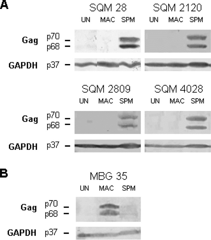
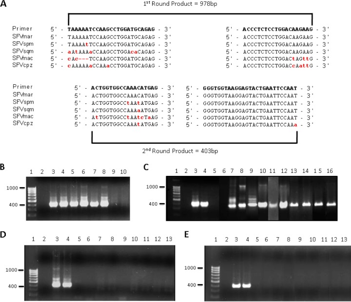
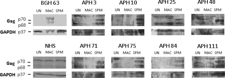
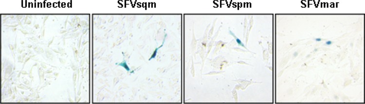
Foamy viruses (FV) are complex retroviruses that naturally infect all nonhuman primates (NHP) studied to date. Zoonotic transmission of Old World NHP simian foamy viruses (SFV) has been documented, leading to nonpathogenic persistent infections. To date, there have been no reports concerning zoonotic transmission of New World monkey (NWM) SFV to humans and resulting infection. In this study, we developed a Western blot assay to detect antibodies to NWM SFV, a nested PCR assay to detect NWM SFV DNA, and a β-galactosidase-containing indicator cell line to assay replication of NWM SFV. Using these tools, we analyzed the plasma and blood of 116 primatologists, of whom 69 had reported exposures to NWM. While 8 of the primatologists tested were seropositive for SFV from a NWM, the spider monkey, none had detectable levels of viral DNA in their blood. We found that SFV isolated from three different species of NWM replicated in some, but not all, human cell lines. From our data, we conclude that while humans exposed to NWM SFV produce antibodies, there is no evidence for long-term viral persistence.
Figures
Similar articles
-
Simian Foamy Viruses in Central and South America: A New World of Discovery.Viruses. 2019 Oct 20;11(10):967. doi: 10.3390/v11100967. Viruses. 2019. PMID: 31635161 Free PMC article. Review.
-
Wide distribution and ancient evolutionary history of simian foamy viruses in New World primates.Retrovirology. 2015 Oct 29;12:89. doi: 10.1186/s12977-015-0214-0. Retrovirology. 2015. PMID: 26514626 Free PMC article.
-
An expanded search for simian foamy viruses (SFV) in Brazilian New World primates identifies novel SFV lineages and host age-related infections.Retrovirology. 2015 Nov 14;12:94. doi: 10.1186/s12977-015-0217-x. Retrovirology. 2015. PMID: 26576961 Free PMC article.
-
Identification and characterization of highly divergent simian foamy viruses in a wide range of new world primates from Brazil.PLoS One. 2013 Jul 3;8(7):e67568. doi: 10.1371/journal.pone.0067568. Print 2013. PLoS One. 2013. PMID: 23844033 Free PMC article.
-
Origin, evolution and innate immune control of simian foamy viruses in humans.Curr Opin Virol. 2015 Feb;10:47-55. doi: 10.1016/j.coviro.2014.12.003. Epub 2015 Feb 17. Curr Opin Virol. 2015. PMID: 25698621 Free PMC article. Review.
Cited by
-
Endemic Viruses of Squirrel Monkeys (Saimiri spp.).Comp Med. 2015 Jun;65(3):232-40. Comp Med. 2015. PMID: 26141448 Free PMC article. Review.
-
An Immunodominant and Conserved B-Cell Epitope in the Envelope of Simian Foamy Virus Recognized by Humans Infected with Zoonotic Strains from Apes.J Virol. 2019 May 15;93(11):e00068-19. doi: 10.1128/JVI.00068-19. Print 2019 Jun 1. J Virol. 2019. PMID: 30894477 Free PMC article.
-
Simian Foamy Viruses in Central and South America: A New World of Discovery.Viruses. 2019 Oct 20;11(10):967. doi: 10.3390/v11100967. Viruses. 2019. PMID: 31635161 Free PMC article. Review.
-
Simian Foamy Virus Co-Infections.Viruses. 2019 Sep 27;11(10):902. doi: 10.3390/v11100902. Viruses. 2019. PMID: 31569704 Free PMC article. Review.
-
Wide distribution and ancient evolutionary history of simian foamy viruses in New World primates.Retrovirology. 2015 Oct 29;12:89. doi: 10.1186/s12977-015-0214-0. Retrovirology. 2015. PMID: 26514626 Free PMC article.
References
-
- Linial ML. 2007. Foamy viruses, p 2245–2263 In Knipe DM, Howley PM. (ed), Fields virology, 5th ed. Lippincott Williams & Wilkins, Philadelphia, PA
Publication types
MeSH terms
Grants and funding
LinkOut - more resources
Full Text Sources
Other Literature Sources
