. 2010 Nov 23:3:165-73.
doi: 10.2147/SCCAA.S14388. eCollection 2010.

Involvement of Notch1 inhibition in serum-stimulated glia and oligodendrocyte differentiation from human mesenchymal stem cells

Affiliations

Involvement of Notch1 inhibition in serum-stimulated glia and oligodendrocyte differentiation from human mesenchymal stem cells

Yi-Jang Lee et al. Stem Cells Cloning. .

Abstract

The use of in vitro oligodendrocyte differentiation for transplantation of stem cells to treat demyelinating diseases is an important consideration. In this study, we investigated the effects of serum on glia and oligodendrocyte differentiation from human mesenchymal stem cells (KP-hMSCs). We found that serum deprivation resulted in a reversible downregulation of glial- and oligodendrocyte-specific markers. Serum stimulated expression of oligodendrocyte markers, such as galactocerebroside, as well as Notch1 and JAK1 transcripts. Inhibition of Notch1 activation by the Notch inhibitor, MG132, led to enhanced expression of a serum-stimulated oligodendrocyte marker. This marker was undetectable in serum-deprived KP-hMSCs treated with MG132, suggesting that inhibition of Notch1 function is additive to serum-stimulated oligodendrocyte differentiation. Furthermore, a dominant-negative mutant RBP-J protein also inhibited Notch1 function and led to upregulation of oligodendrocyte-specific markers. Our results demonstrate that serum-stimulated oligodendrocyte differentiation is enhanced by the inhibition of Notch1-associated functions.

Keywords: Notch1 signaling; glia and oligodendrocyte differentiation; mesenchymal stem cells; serum deprivation.


Figures

Figure 1
Figure 1
Serum induces the upregulation of markers for glial differentiation. A) Western blot analysis for comparison of glial and neuronal markers in human mesenchymal stem cells cultured with or without 10% FBS. B) Densitometric measurement of Western blot protein bands (*P < 0.05). C) Immunofluorescence staining of Gal-C and GFAP expression in human mesenchymal stem cells cultured in different concentration of FBS. D) effects of serum deprivation on C6 rat glial cells expressing GFAP and Gal-C proteins. Abbreviations: SF, serum-free medium; FBS, fetal bovine serum; HS, 5% horse serum; GFAP, glial fibrillary acidic protein; Gal-C, galactocerebroside.
Figure 2
Figure 2
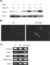
Recovery of glial markers by serum supplementation in serum-deprived human mesenchymal stem cells. A) Western blot analysis detecting the expression of Gal-C in human mesenchymal stem cells that were cultured in serum-free medium for two days and then supplemented with FBS. B) Immunofluorescence staining of glial fibrillary acidic protein expression in human mesenchymal stem cells cultured in either SF for two days or thereafter supplemented with 10% FBS for an additional two days (SF + FBS). C) Semiquantitative real-time polymerase chain reaction detecting mRNA levels of glial and oligodendrocyte markers using the above experimental conditions. C is a control that was continuously cultured in FBS-supplemented medium. Abbreviations: FBS, fetal bovine serum; SF, serum-free medium; Gal-C, galactocerebroside; MBP, myelin basic protein.
Figure 3
Figure 3
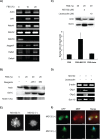
Effects of Notch signaling on serum-induced oligodendrocyte differentiation. A) Semiquantitative real-time polymerase chain reaction for detecting mRNA levels of JAK1 and Notch-associated markers in human mesenchymal stem cells cultured in different concentration of FBS. B) Western blotting for detecting the Gal-C and Notch1 cleaved internal domain in human mesenchymal stem cells treated with JAK1 inhibitor and Notch inhibitor MG132 for 24 hours. C) Comparison of effects of MG132 and lactacystin on expression of Gal-C. Upper panel: Western blot analysis. Lower panel: densitometric measurement of protein bands (*P < 0.05 compared with FBS control). D) Comparison of mRNA levels of oligodendrocyte-specific transcription factors (Olig-1, Olig-2, and SOX-10) in human mesenchymal stem cells treated with MG132 or lactacystin. E) Morphologic changes in GFP-labeled human mesenchymal stem cells. F) Comparison of the expression of MBP-1 in GFP-labeled human mesenchymal stem cells before (−) and after (+) MG132 treatment for 24 hours. Human mesenchymal stem cells were maintained in FBS during the treatment. The nuclei were stained by 4′,6-diamidino-2-phenylindole (DAPI). The sizes of scale bars for E and F were 20 and 40 μm, respectively. Abbreviations: FBS, fetal bovine serum; GFP, green fluorescence protein; MBP, myelin basic protein; Gal-C, galactocerebroside.
Figure 4
Figure 4
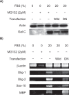
Inhibition of Notch activity by mutant RBP-J protein enhances the serum-stimulated expression of oligodendrocyte markers. A) Western blot analysis for comparison of Gal-C expression in human mesenchymal stem cells transfected with wild-type or the DN mutant form of RBP-J cDNA. B) Semiquantitative real-time polymerase chain reaction detecting the mRNA levels of other oligodendrocyte markers. MG132 (24 hours of treatment) was unable to induce these markers under serum-free conditions. Abbreviations: Gal-C, galactocerebroside; DN, dominate negative; FBS, fetal bovine serum; MBP, myelin basic protein.

Similar articles

References

    1. Bruck W. The pathology of multiple sclerosis is the result of focal inflammatory demyelination with axonal damage. J Neurol. 2005;252(Suppl 5):v3–v9. - PubMed
    1. Lubetzki C, Williams A, Stankoff B. Promoting repair in multiple sclerosis: Problems and prospects. Curr Opin Neurol. 2005;18:237–244. - PubMed
    1. Lublin F. History of modern multiple sclerosis therapy. J Neurol. 2005;252(Suppl 3):iii3–iii9. - PubMed
    1. Keirstead HS. Stem cells for the treatment of myelin loss. Trends Neurosci. 2005;28:677–683. - PubMed
    1. Pittenger MF, Mackay AM, Beck SC, et al. Multilineage potential of adult human mesenchymal stem cells. Science. 1999;284:143–147. - PubMed