

A three-dimensional anatomy of the posterolateral compartment of the knee: the use of a new technology in the study of musculoskeletal anatomy
- PMID: 24198580
- PMCID: PMC3781892
- DOI: 10.2147/OAJSM.S28705
A three-dimensional anatomy of the posterolateral compartment of the knee: the use of a new technology in the study of musculoskeletal anatomy
Abstract
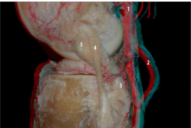
Background: Recently, an interest has developed in understanding the anatomy of the posterior and posterolateral knee. The posterolateral compartment of the knee corresponds to a complex arrangement of ligaments and myotendinous structures. Undiagnosed lesions in this compartment are the main reason for failure of the anterior and posterior cruciate ligament reconstructions. Understanding the anatomy of these structures is essential to assist in the diagnosis and treatment of these lesions. The aim of this study was to better understand the relationship between these structures of the knee using three-dimensional technology.
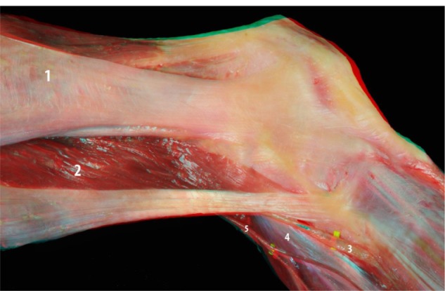
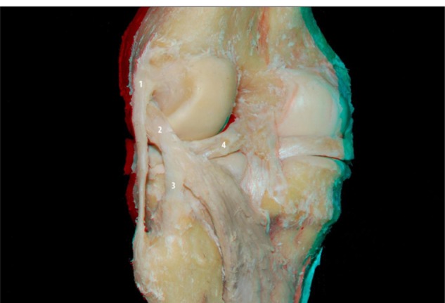
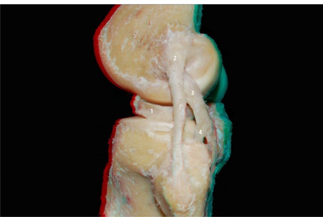
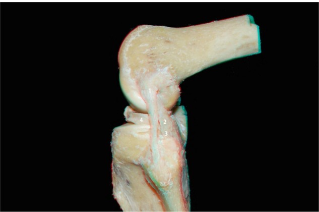
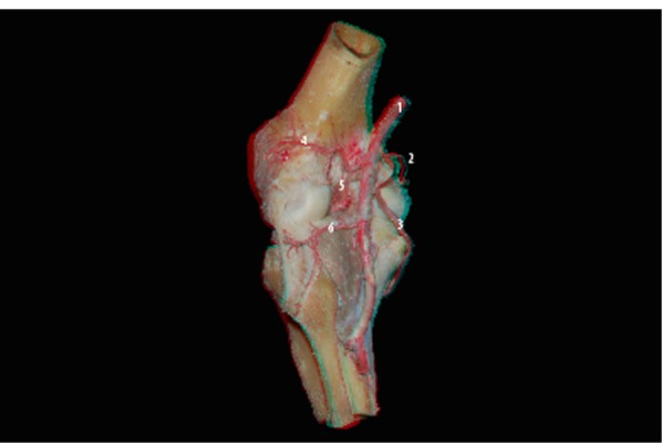
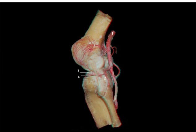
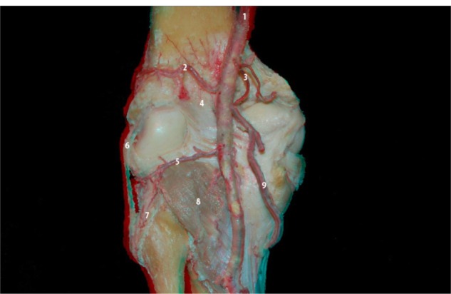
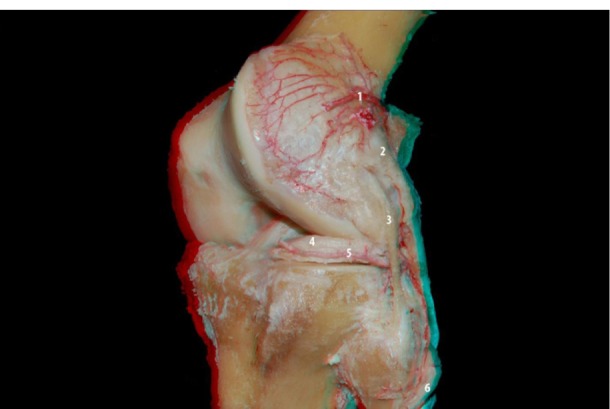
Methods: Ten knees were included from cadaver lower limbs of adult patients. The skin and subcutaneous tissue were removed leaving only the muscle groups and ligaments. The neurovascular bundles and their ramifications were preserved. Images were acquired from the dissections using a Nikon D40 camera with AF-S Nikkor 18-55 mm (1:3.5 5.6 GII ED) and Micro Nikkor 105 mm (1:2.8) lenses. The pair of images were processed using Callipyan 3D and AnaBuilder software, which transforms the two images into one anaglyphic image.
Results: During the dissection of the knees, twelve pictures were acquired and transformed into anaglyphic images.
Conclusion: The use of three-dimensional images in this study demonstrates that this technique is useful to improve the knowledge in anatomy of the knee as well as for knee reconstruction surgery.
Keywords: anatomy education; education; eyeglasses; humans; knee joint anatomy and histology; medical methods; photography methods.
Figures
References
-
- Scapinelli R. Vascular anatomy of the human cruciate ligaments and surrounding structures. Clin Anat. 1997;10(3):151–162. - PubMed
-
- Park SE, Stamos BD, DeFrate LE, Gill TJ, Li G. The effect of posterior knee capsulotomy on posterior tibial translation during posterior cruciate ligament tibial inlay reconstruction. Am J Sports Med. 2004;32(6):1514–1519. - PubMed
-
- Mariani PP, Margheritini F. Full arthroscopic inlay reconstruction of posterior cruciate ligament. Knee Surg Sports Traumatol Arthrosc. 2006;14(11):1038–1044. - PubMed
-
- Sanchez AR, 2nd, Sugalski MT, LaPrade RF. Anatomy and biomechanics of the lateral side of the knee. Sports Med Arthrosc. 2006;14(1):2–11. - PubMed
LinkOut - more resources
Full Text Sources
Research Materials
