Case Reports
. 2013 May 31;2(4):2047981613492532.
doi: 10.1177/2047981613492532. eCollection 2013.

Mazabraud's syndrome: case report and literature review

Affiliations
Case Reports

Mazabraud's syndrome: case report and literature review

Peter Svenssen Munksgaard et al. Acta Radiol Short Rep. .

Abstract

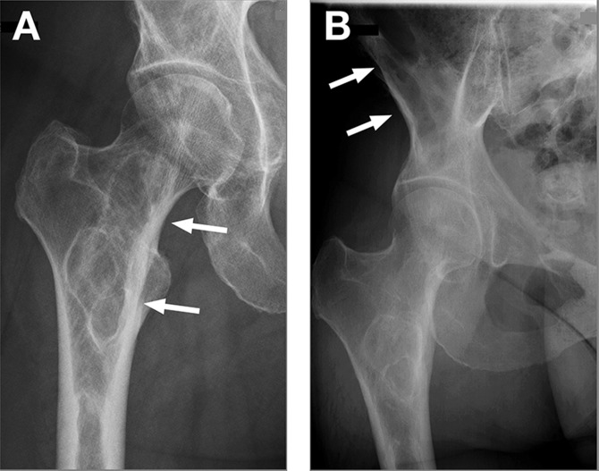
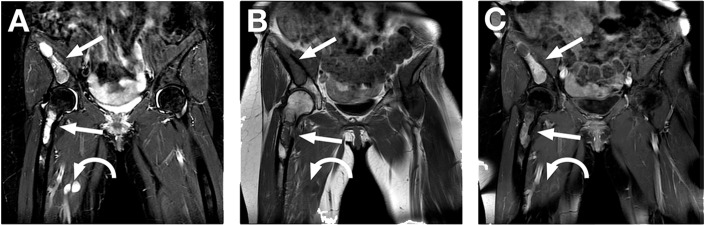
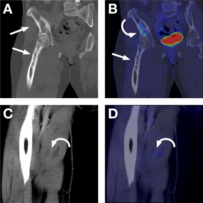
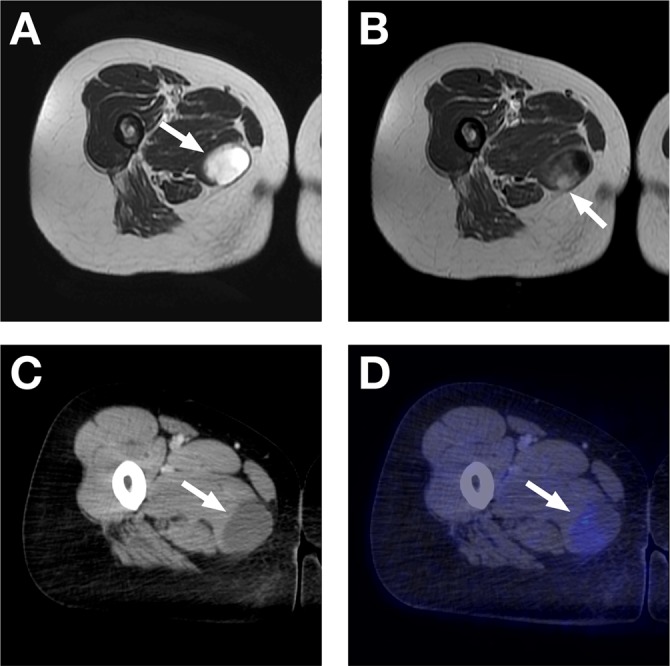
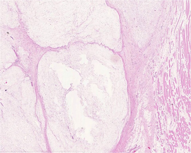
Mazabraud's syndrome is a rare disorder characterized by the association of single or multiple intramuscular myxomas with fibrous dysplasia. Here, we present the first case of Mazabraud's syndrome visualized on 18F-FDG PET/CT with histopathological confirmation of the myxoma. Our case demonstrates a slightly increased FDG uptake (SUVmax 2.1) within the myxomas and a moderately to highly increased tracer uptake (SUVmax 7.0) within the fibrous dysplastic lesions. The typical histological appearance of the intramuscular myxoma confirmed the radiological diagnosis. Further, we discuss the imaging findings and the histopathological features of this rare case with a review of the related literature.

Keywords: MRI; Mazabraud's syndrome; fibrous dysplasia; fluorodeoxyglucose F18 (18F-FDG PET/CT); myxoma.


Figures

Fig. 1
Fig. 1
(a, b) Anteroposterior radiographs of the proximal femur and right hip showing radiolucent cystic lesions with sclerotic margins and no periosteal reaction, findings in keeping with fibrous dysplasia (straight arrows)
Fig. 2
Fig. 2
(a) Coronal STIR, (b) T1, and (c) T1-weighted with fat-sat and gadolinium (Gd) contrast agent MR images. Bony lesions with high signal on STIR, low signal on T1, and heterogeneous contrast enhancement after Gd injection (straight arrows) can be observed. Soft tissue lesions with high signal on STIR, low signal on T1, and a mixture of rim and central contrast enhancement after Gd injection (curved arrows) can also be observed
Fig. 3
Fig. 3
18F-FDG PET/CT. (a) Coronal CT with a bone window showing multi-loculated expansile intramedullary fibrous dysplasia lesions in the ileum and proximal part of the femur (straight arrows). (b) Fused PET/CT with increased FDG uptake (SUVmax 7.0) only in the lower region of the fibrous dysplasia lesion in the right ileum (curved arrow). Notice that the region of the fibrous dysplasia taking up the FDG corresponds to the region on the coronal MR image in Fig. 2c that is the most enhanced by the Gd contrast agent. (c) Coronal CT with a soft tissue window showing the low-density myxomas in the medial thigh muscles (curved arrow). (d) Fused PET/CT showing slightly increased FDG uptake in the myxomas (curved arrow)
Fig. 4
Fig. 4
MRI and 18F-FDG PET/CT. (a) Axial T2-weighted MR image with high signal, partly cystic-appearing soft tissue lesion in the medial thigh muscle (straight arrow). (b) Axial T1-weighted MR image after intravenous Gd agent administration with contrast enhancement of the more solid-appearing region of the myxoma (straight arrow). (c) Axial CT with a soft tissue window showing the low-density, almost cystic-appearing lesion in the medial thigh muscle (straight arrow). (d) Fused PET/CT with slightly increased FDG uptake (SUV max 2.1) in the apparently more solid region of the myxoma. Notice that the region of the myxoma taking up the FDG corresponds to the Gd-enhanced region on the MRI (straight arrow)
Fig. 5
Fig. 5
Typical histological appearance of intramuscular myxoma: well-circumscribed, paucicellular lobulated myxoid tumour with skeletal muscle involvement

References

    1. van der Wal WA, Unal H, de Rooy JW, et al. Fibrous dysplasia of bone associated with soft-tissue myxomas as well as an intra-osseous myxoma in a woman with mazabraud's syndrome: A case report. J Med Case Reports 2011;5:239 - PMC - PubMed
    1. Henschen F Fall von ostitis fibrosa mit multiplen tumoren in der umgebenden muskulatur. Verh Dtsch Ges Pathol 1926;21:93–7
    1. Mazabraud A, Semat P, Roze R Apropos of the association of fibromyxomas of the soft tissues with fibrous dysplasia of the bones. Presse Med 1967;75:2223–8 - PubMed
    1. Gaumetou E, Tomeno B, Anract P Mazabraud's syndrome. A case with multiple myxomas. Orthop Traumatol Surg Res 2012;98:455–60 - PubMed
    1. Singnurkar A, Phancao JP, Chatha DS, et al. The appearance of mazabraud's syndrome on 18F-FDG PET/CT. Skeletal Radiol 2007;36:1085–9 - PubMed

Publication types

LinkOut - more resources