. 2013 Oct 7;6(1):77.
doi: 10.1186/1756-8722-6-77.

Inhibition of Hedgehog signaling sensitizes NSCLC cells to standard therapies through modulation of EMT-regulating miRNAs

Affiliations

Inhibition of Hedgehog signaling sensitizes NSCLC cells to standard therapies through modulation of EMT-regulating miRNAs

Aamir Ahmad et al. J Hematol Oncol. .

Abstract

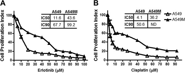
Background: Epidermal growth factor receptor- tyrosine kinase inhibitors (EGFR-TKIs) benefit Non-small cell lung cancer (NSCLC) patients, and an EGFR-TKIi erlotinib, is approved for patients with recurrent NSCLC. However, resistance to erlotinib is a major clinical problem. Earlier we have demonstrated the role of Hedgehog (Hh) signaling in Epithelial-to-Mesenchymal transition (EMT) of NSCLC cells, leading to increased proliferation and invasion. Here, we investigated the role of Hh signaling in erlotinib resistance of TGF-β1-induced NSCLC cells that are reminiscent of EMT cells.

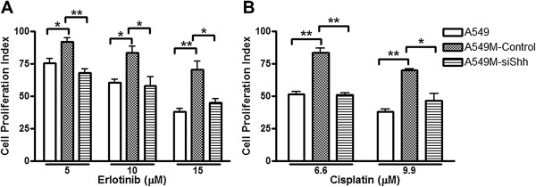
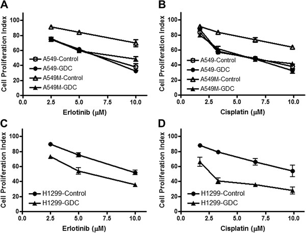
Methods: Hh signaling was inhibited by specific siRNA and by GDC-0449, a small molecule antagonist of G protein coupled receptor smoothened in the Hh pathway. Not all NSCLC patients are likely to benefit from EGFR-TKIs and, therefore, cisplatin was used to further demonstrate a role of inhibition of Hh signaling in sensitization of resistant EMT cells. Specific pre- and anti-miRNA preparations were used to study the mechanistic involvement of miRNAs in drug resistance mechanism.

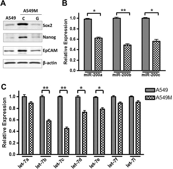
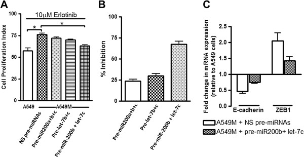
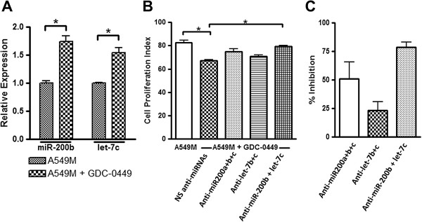
Results: siRNA-mediated inhibition as well as pharmacological inhibition of Hh signaling abrogated resistance of NSCLC cells to erlotinib and cisplatin. It also resulted in re-sensitization of TGF-β1-induced A549 (A549M) cells as well the mesenchymal phenotypic H1299 cells to erlotinib and cisplatin treatment with concomitant up-regulation of cancer stem cell (CSC) markers (Sox2, Nanog and EpCAM) and down-regulation of miR-200 and let-7 family miRNAs. Ectopic up-regulation of miRNAs, especially miR-200b and let-7c, significantly diminished the erlotinib resistance of A549M cells. Inhibition of Hh signaling by GDC-0449 in EMT cells resulted in the attenuation of CSC markers and up-regulation of miR-200b and let-7c, leading to sensitization of EMT cells to drug treatment, thus, confirming a connection between Hh signaling, miRNAs and drug resistance.

Conclusions: We demonstrate that Hh pathway, through EMT-induction, leads to reduced sensitivity to EGFR-TKIs in NSCLCs. Therefore, targeting Hh pathway may lead to the reversal of EMT phenotype and improve the therapeutic efficacy of EGFR-TKIs in NSCLC patients.


Figures

Figure 1
Figure 1
TGF-β1-induced EMT results in drug resistance phenotype. Dose–response curves shows that A549M cells exhibit increased cell viability, after treatment with erlotinib (A) and cisplatin (B), compared to A549 cells. Cells were treated with indicated concentrations of erlotinib/cisplatin for 72 hours and then subjected to MTT assay. The IC50 and IC90 values for different conditions are provided in the table within the individual figures. ND: IC90 could not be determined. *p<0.05.
Figure 2
Figure 2
Knock-down of Shh sensitizes A549M cells to standard therapies. Cell proliferation of A549M cells was significantly reduced after treatment with erlotinib (A) and cisplatin (B) following Shh knock-down. Cells were first treated with vehicle (A549M-control) or with specific si-RNA against Shh (A549M-siShh) for 48 hours and then with indicated concentrations of erlotinib/cisplatin for 24 hours. Parental A549 cells were included as a control to verify the induced resistance of A549M cells to erlotinib/cisplatin. All the plotted values are relative to vehicle-treated A549 cells. *p<0.05 and **p<0.01.
Figure 3
Figure 3
Hedgehog inhibitor, GDC-0449 (GDC) sensitizes A549M as well as H1299 cells to standard therapies. Pre-treatment with GDC-0449 (20nM) markedly reduced cell proliferation of A549M cells (A549M-GDC) (A-B) as well as H1299 cells (H1299-GDC) (C-D), compared to vehicle treated respective control cells, when they were exposed to erlotinib or cisplatin for 72 hours. Control A549 cells did not exhibit such sensitization (A-B). All the plotted values are relative to vehicle-treated cells.
Figure 4
Figure 4
Modulation of CSC markers and miRNAs accompanies EMT of NSCLC cells. (A) A549M cells exhibit increased expression of CSC markers Sox2, Nanog and EpCAM and GDC-0449 inhibited such TGF-β-induced expression of CSC markers. TGF-β1-induced EMT also involved changes in the expression levels of (B) miR-200 family and (C) let-7 family of miRNAs. RNU6B and RNU48 were used as miRNA controls against which the data was normalized. *p<0.05 and **p<0.01.
Figure 5
Figure 5
Mechanistic role of miRNAs in TGF-β1 induced drug resistance. (A) Re-expression of miR-200s and let-7s sensitized A549M cells to erlotinib treatment. (B) Data from Figure 5A was used to calculate the extent of sensitization by re-expression of miRNAs upon erlotinib treatment, as measured by % inhibition of A549M resistance compared to parental A549 cells. (C) Re-expression of miR-200b+let-7c reversed EMT. E-cadherin and ZEB1 mRNA levels were determined by real time RT-PCR using GAPDH as the internal control. All the plotted values in Figure 5A are relative to vehicle-treated A549 cells. RNU6B and RNU48 were used as miRNA controls against which the data was normalized. *p<0.05.
Figure 6
Figure 6
Mechanistic role of miRNAs in GDC-0449 effects on drug resistance. (A) GDC-0449 up-regulated the expression of miR-200b and let-7c, the two top regulated miRNAs in their respective families. (B) Down-regulation of miR-200 and let-7 family miRNAs abrogated the GDC-0449 mediated inhibition of erlotinib resistance in A549M cells. (C) Data from Figure 6B was used to calculate the abrogation of GDC-0449 effect through the use of anti-miRNAs, as measured by % inhibition of A549M sensitivity to GDC-0449, compared to control A549M cells. All the plotted values in Figure 6B are relative to vehicle-treated A549M cells and the first bar in the same figure represents A549M cells treated only with 10μM erlotinib. RNU6B and RNU48 were used as miRNA controls against which the data was normalized. *p<0.05.

References

    1. Siegel R, Naishadham D, Jemal A. Cancer statistics, 2013. CA Cancer J Clin. 2013;63:11–30. doi: 10.3322/caac.21166. - DOI - PubMed
    1. Ma J, Ward EM, Smith R, Jemal A. Annual number of lung cancer deaths potentially avertable by screening in the United States. Cancer. 2013;119:1381–1385. doi: 10.1002/cncr.27813. - DOI - PubMed
    1. Maitah MY, Ali S, Ahmad A, Gadgeel S, Sarkar FH. Up-regulation of sonic hedgehog contributes to TGF-beta1-induced epithelial to mesenchymal transition in NSCLC cells. PLoS One. 2011;6:e16068. doi: 10.1371/journal.pone.0016068. - DOI - PMC - PubMed
    1. Ahmad A, Aboukameel A, Kong D, Wang Z, Sethi S, Chen W. et al.Phosphoglucose isomerase/autocrine motility factor mediates epithelial-mesenchymal transition regulated by miR-200 in breast cancer cells. Cancer Res. 2011;71:3400–3409. doi: 10.1158/0008-5472.CAN-10-0965. - DOI - PMC - PubMed
    1. Gomes LR, Terra LF, Sogayar MC, Labriola L. Epithelial-Mesenchymal Transition: Implications in Cancer Progression and Metastasis. Curr Pharm Biotechnol. 2011;12(11):1881–1890. doi: 10.2174/138920111798377102. - DOI - PubMed

MeSH terms