

Advanced MR imaging techniques in the evaluation of nonenhancing gliomas: perfusion-weighted imaging compared with proton magnetic resonance spectroscopy and tumor grade
- PMID: 24199813
- PMCID: PMC4202829
- DOI: 10.1177/197140091302600506
Advanced MR imaging techniques in the evaluation of nonenhancing gliomas: perfusion-weighted imaging compared with proton magnetic resonance spectroscopy and tumor grade
Abstract
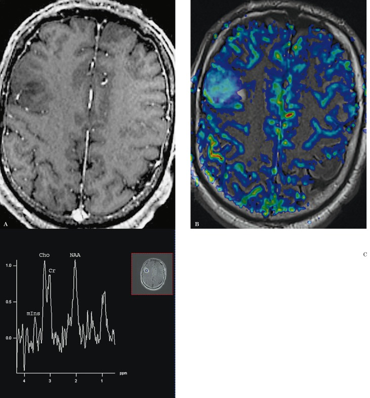
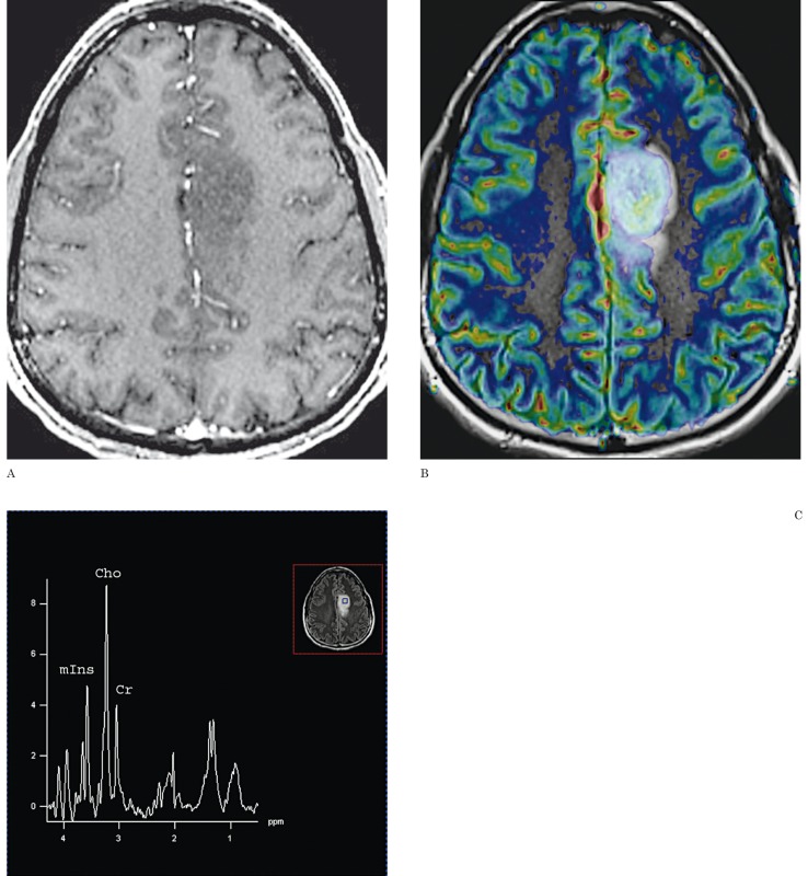
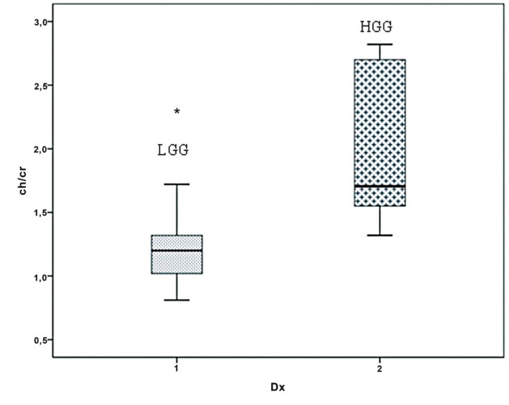
A significant number of nonenhancing (NE) gliomas are reported to be malignant. The purpose of this study was to compare the value of advanced MR imaging techniques, including T2*-dynamic susceptibility contrast PWI (DSC-PWI) and proton magnetic resonance spectroscopy ((1)HMRS) in the evaluation of NE gliomas. Twenty patients with NE gliomas underwent MRI including DSC-PWI and (1)HMRS. The relative CBV (rCBV) measurements were obtained from regions of maximum perfusion. The peak ratios of choline/creatine (Cho/Cr) and myo-inositol/creatine (mIns/Cr) were measured at a TE of 30 ms. Demographic features, tumor volumes, and PWI- and (1)HMRS-derived measures were compared between low-grade gliomas (LGGs) and high-grade gliomas (HGGs). In addition, the association of initial rCBV ratio with tumor progression was evaluated in LGGs. No significant difference was noted in age, sex or tumor size between LGGs and HGGs. Cho/Cr ratios were significantly higher in HGGs (1.7±0.63) than in LGGs (1.2±0.38). The receiver operating characteristic analysis demonstrated that a Cho/Cr ratio with a cutoff value of 1.3 could differentiate between LGG and HGG with a specificity of 100% and a sensitivity of 71.4%. There was no significant difference in the rCBV ratio and the mIns/Cr ratio between LGG and HGG. However, higher rCBV ratios were observed with more rapid progressions in LGGs. The results imply that Cho/Cr ratios are useful in distinguishing NE LGG from HGG and can be helpful in preoperative grading and biopsy guidance. On the other hand, rCBV ratios do not help in the distinction.
Keywords: MR spectroscopy; nonenhancing glioma; perfusion MRI.
Figures

Similar articles
-
Glioma grading: sensitivity, specificity, and predictive values of perfusion MR imaging and proton MR spectroscopic imaging compared with conventional MR imaging.AJNR Am J Neuroradiol. 2003 Nov-Dec;24(10):1989-98. AJNR Am J Neuroradiol. 2003. PMID: 14625221 Free PMC article.
-
MR diffusion tensor and perfusion-weighted imaging in preoperative grading of supratentorial nonenhancing gliomas.Neuro Oncol. 2011 Apr;13(4):447-55. doi: 10.1093/neuonc/noq197. Epub 2011 Feb 4. Neuro Oncol. 2011. PMID: 21297125 Free PMC article.
-
Measurements of diagnostic examination performance using quantitative apparent diffusion coefficient and proton MR spectroscopic imaging in the preoperative evaluation of tumor grade in cerebral gliomas.Eur J Radiol. 2011 Nov;80(2):462-70. doi: 10.1016/j.ejrad.2010.07.017. Epub 2010 Aug 13. Eur J Radiol. 2011. PMID: 20708868
-
Optimal differentiation of high- and low-grade glioma and metastasis: a meta-analysis of perfusion, diffusion, and spectroscopy metrics.Neuroradiology. 2016 Apr;58(4):339-50. doi: 10.1007/s00234-016-1642-9. Epub 2016 Jan 15. Neuroradiology. 2016. PMID: 26767528
-
The role of imaging in the management of adults with diffuse low grade glioma: A systematic review and evidence-based clinical practice guideline.J Neurooncol. 2015 Dec;125(3):457-79. doi: 10.1007/s11060-015-1908-9. Epub 2015 Nov 3. J Neurooncol. 2015. PMID: 26530262
Cited by
-
Hydrogen Proton Magnetic Resonance Spectroscopy (MRS) in Differential Diagnosis of Intracranial Tumors: A Systematic Review.Contrast Media Mol Imaging. 2022 May 18;2022:7242192. doi: 10.1155/2022/7242192. eCollection 2022. Contrast Media Mol Imaging. 2022. Retraction in: Contrast Media Mol Imaging. 2023 Sep 14;2023:9805474. doi: 10.1155/2023/9805474. PMID: 35655732 Free PMC article. Retracted.
-
Usefulness of quantitative peritumoural perfusion and proton spectroscopic magnetic resonance imaging evaluation in differentiating brain gliomas from solitary brain metastases.Neuroradiol J. 2016 Jun;29(3):160-7. doi: 10.1177/1971400916638358. Epub 2016 Mar 17. Neuroradiol J. 2016. PMID: 26988081 Free PMC article.
-
Improving the Grading Accuracy of Astrocytic Neoplasms Noninvasively by Combining Timing Information with Cerebral Blood Flow: A Multi-TI Arterial Spin-Labeling MR Imaging Study.AJNR Am J Neuroradiol. 2016 Dec;37(12):2209-2216. doi: 10.3174/ajnr.A4907. Epub 2016 Aug 25. AJNR Am J Neuroradiol. 2016. PMID: 27561831 Free PMC article.
-
Brainstem glioma: Prediction of histopathologic grade based on conventional MR imaging.Neuroradiol J. 2018 Feb;31(1):10-17. doi: 10.1177/1971400917743099. Epub 2017 Nov 17. Neuroradiol J. 2018. PMID: 29148317 Free PMC article.
-
Magnetic resonance perfusion for differentiating low-grade from high-grade gliomas at first presentation.Cochrane Database Syst Rev. 2018 Jan 22;1(1):CD011551. doi: 10.1002/14651858.CD011551.pub2. Cochrane Database Syst Rev. 2018. PMID: 29357120 Free PMC article.
References
-
- Kleihues P, Cavenee WK. Pathology and genetics of tumours of the nervous system. Lyon:: International Agency for Research on Cancer; 2000.
-
- Morita N, Wang S, Chawla S, et al. Dynamic susceptibility contrast perfusion weighted imaging in grading of nonenhancing astrocytomas. J Magn Reson Imaging. 2010;32(4):803–808. - PubMed
-
- Ginsberg LE, Fuller GN, Hashmi M, et al. The significance of lack of MR contrast enhancement of supratentorial brain tumors in adults: histopathological evaluation of a series. Surg Neurol. 1998;49(4):436–440. - PubMed
-
- Al-Okaili RN, Krejza J, Wang S, et al. Advanced MR imaging techniques in the diagnosis of intraaxial brain tumors in adults. Radiographics. 2006;26(Suppl 1):S173–189. - PubMed
Publication types
MeSH terms
LinkOut - more resources
Full Text Sources
Other Literature Sources
Medical
