Review
. 2013 Nov 4;23(21):R972-9.
doi: 10.1016/j.cub.2013.09.033.

ERK as a model for systems biology of enzyme kinetics in cells

Affiliations
Review

ERK as a model for systems biology of enzyme kinetics in cells

Alan S Futran et al. Curr Biol. .

Abstract

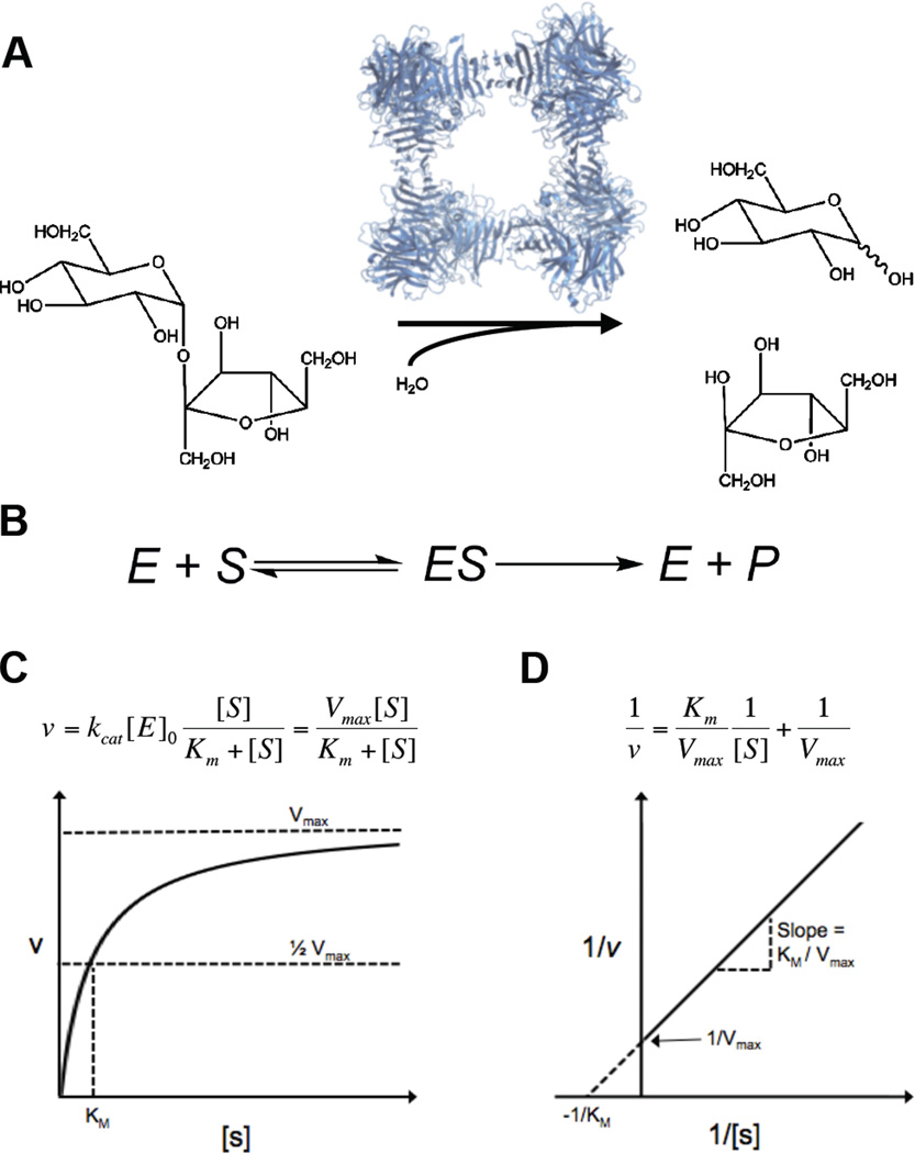
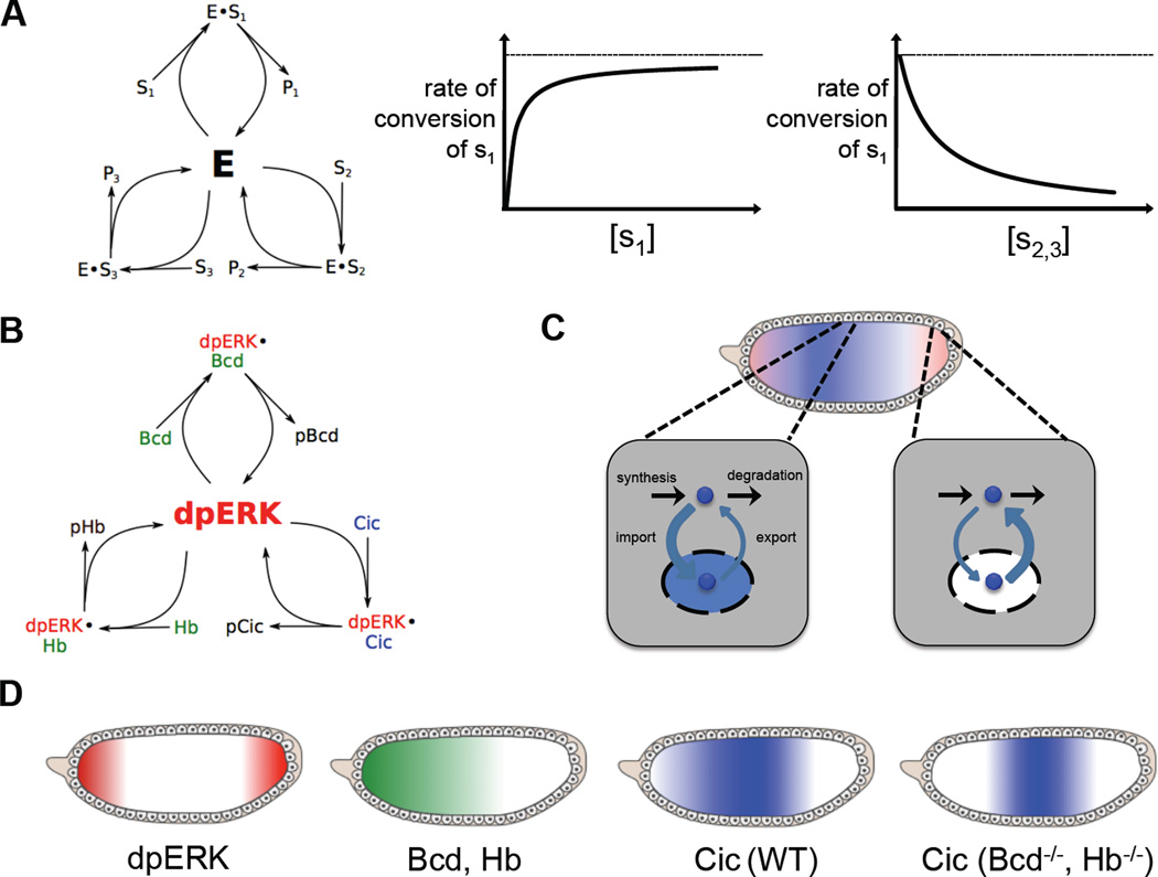
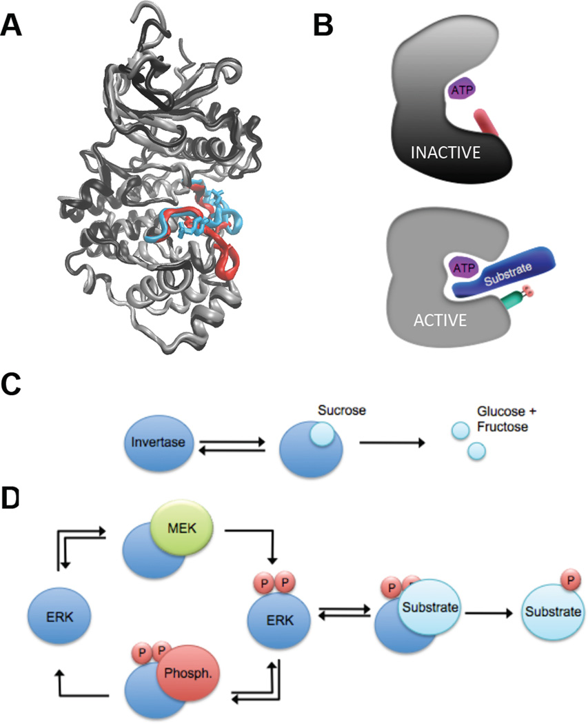
A key step towards a chemical picture of enzyme catalysis was taken in 1913, when Leonor Michaelis and Maud Menten published their studies of sucrose hydrolysis by invertase. Based on a novel experimental design and a mathematical model, their work offered a quantitative view of biochemical kinetics well before the protein nature of enzymes was established and complexes with substrates could be detected. Michaelis-Menten kinetics provides a solid framework for enzyme kinetics in vitro, but what about kinetics in cells, where enzymes can be highly regulated and participate in a multitude of interactions? We discuss this question using the Extracellular Signal Regulated Kinase (ERK), which controls a myriad functions in cells, as a model of an important enzyme for which we have crystal structures, quantitative in vitro assays, and a vast list of binding partners. Despite great progress, we still cannot quantitatively predict how the rates of ERK-dependent reactions respond to genetic and pharmacological perturbations. Achieving this goal, which is important from both fundamental and practical standpoints, requires measuring the rates of enzyme reactions in their native environment and interpreting these measurements using simple but realistic mathematical models--the two elements which served as the cornerstones for Michaelis' and Menten's seminal 1913 paper.


Figures

Figure 1
Figure 1
The Michaelis-Menten model of enzyme kinetics. (A) Yeast invertase and the hydrolysis of sucrose to glucose (top) and fructose (bottom). Structure of S. cerevisiae invertase drawn from PDB file 4EQV (81). (B) The model proposed by Michaelis and Menten, wherein an enzyme and a substrate bind reversibly to form a complex, which is converted to a product and the enzyme. (C) The Michaelis-Menten equation and its graphical representation. Plotting the initial rate vs. substrate concentration enables the determination of both Vmax and Km. (D) Lineweaver Burk representation of the Michaelis-Menten equation. Taking the reciprocal of both sides of the Michaelis-Menten equation yields a linear relationship. Plotting the reciprocal of the initial rate vs. the reciprocal of substrate concentration allows the determination of Km and Vmax from the y and×intercept and the slope of the line.
Figure 2
Figure 2
Patterns of ERK activation in organisms. (A) ERK activation requires its phosphorylation by MEK. Active ERK controls cellular processes by phosphorylating multiple substrates. (B) Active ERK (red) at three different time points in Drosophila embryos. Active ERK is first detected at the embryonic poles, where it specifies the nonsegmented terminal regions of the future larva. Within the next 30 minutes, ERK is activated in a lateral domain corresponding to the presumptive neurogenic ectoderm. After gastrulation, ERK is active along the ventral midline and in tracheal pits.
Figure 3
Figure 3
ERK2 docking domains and ERK2 substrate docking sites. (A) ERK2 structure with docking domains – DRS domain (left) and FRS domain (right) – and active site (middle) in space filling representation. Structure drawn from PDB file 1ERK (33) (B) Schematic representation of ERK2 substrate sequence containing a docking site and a (S/T)P phosphorylation site. (C) Alignment of F-site and D-Site sequences from multiple ERK2 interacting substrates and regulators. (D) Schematic representation of ERK2 interactions with substrate docking site and (S/T)P phosphorylation motif. (E) Effects of mutations on substrate docking site and phosphorylation site and insights into molecular mechanisms of ERK2 catalysis. (i) Wild type substrate and associated Michaelis-Menten parameters. (ii) Substrate with docking site mutations and associated Michaelis-Menten parameters. Mutating the docking site significantly increases the Km but has little effect on kcat. (iii) Substrate with phosphorylation motif mutations and associated Michaelis-Menten parameters. Mutating the phosphorylation motif significantly decreases the kcat but has little effect on Km.
Figure 4
Figure 4
Identification of ERK substrates in C. elegans. (A) Dissected C. elegans hermaphrodite germ line from wild type animals oriented from left to right, stained for dpERK (red) and DNA (white). Wild type germ lines reveal two zones of ERK activation, zone 1 and 2, with brief down regulation of active ERK in the loop region. (B) Schematic representation of ERK substrates identified in C. elegans by searching the genome for ERK docking site motifs and screening putative substrates in vivo.
Figure 5
Figure 5
Substrate Competition in the ERK pathway. (A) Substrate competition and its effects on enzyme kinetics. When multiple substrates compete for the activity of a single enzyme, the rate of conversion of a single substrate decreases as the concentration of competing substrates increases (B) ERK substrate competition in the Drosophila embryo. Three substrates – Bicoid (Bcd), Capicua (Cic) and Hunchback (Hb) – compete for ERK activity. (C) ERK-mediated downregulation of Cic. At the embryonic poles, where ERK is active, Cic is exported out of the nucleus into the cytoplasm, where it is degraded. (D) Distribution of ERK and its substrates in the Drosophila embryo in the presence and absence of substrate competition. ERK is doubly phosphorylated and activated at the poles of the embryo. Two substrates – Bcd and Hb – are present only at the anterior pole of the embryo. In wild type embryos, Cic is downregulated at the poles unevenly, with a higher concentration at the anterior pole. However, when the anteriorly located ERK substrates Bcd and Hb are removed, Cic is downregulated more evenly, indicating that ERK’s ability to phosphorylate and downregulate Cic is inhibited by the presence of these other ERK substrates at the anterior pole.
Figure 6
Figure 6
Regulation of ERK activity. (A) Superimposition of inactive (black and red) and active (grey and cyan) ERK2 highlighting the activation loop and phosphorylated residues. Structures of inactive and active ERK2 drawn from PDB files 1ERK and 2ERK, respectively (33, 34). (B) Schematic representation of structural changes upon ERK activation and the effects on ATP/Substrate binding and orientation. Upon activation, the N terminal and C terminal lobes of ERK rotate toward one another and residues is the active site are repositioned, allowing proper binding and orientation of substrates for catalysis. (C) A classical single enzyme/substrate network, exemplified by the system used by Michaelis and Menten – invertase-mediated hydrolysis of sucrose. (D) A simplified ERK network involving multiple individual enzyme/substrate interactions – ERK activation by MEK, ERK inactivation by a phosphatase, and ERK-mediated phosphorylation of one substrate.

Similar articles

Cited by

References

    1. Johnson KA, Goody RS. The Original Michaelis Constant: Translation of the 1913 Michaelis–Menten Paper. Biochemistry. 2011;50(39):8264–8269. - PMC - PubMed
    1. Michaelis L, Menten ML. Die kinetik der invertinwirkung. Biochem. Z. 1913;49:333–369.
    1. Fruton JS. Proteins, Enzymes, Genes: The Interplay of Chemistry and Biology. Yale University Press; 1999.
    1. Cornish-Bowden A. The origins of enzyme kinetics. FEBS letters. 2013;587(17):2725–2730. - PubMed
    1. Johnson KA. A century of enzyme kinetic analysis, 1913 to 2013. FEBS letters. 2013;587(17):2753–2766. - PMC - PubMed

Publication types

Substances

LinkOut - more resources