. 2014 Apr;10(3):491-501.
doi: 10.1016/j.nano.2013.10.010. Epub 2013 Nov 4.

Photoactivated rose bengal functionalized chitosan nanoparticles produce antibacterial/biofilm activity and stabilize dentin-collagen

Affiliations

Photoactivated rose bengal functionalized chitosan nanoparticles produce antibacterial/biofilm activity and stabilize dentin-collagen

Annie Shrestha et al. Nanomedicine. 2014 Apr.

Abstract

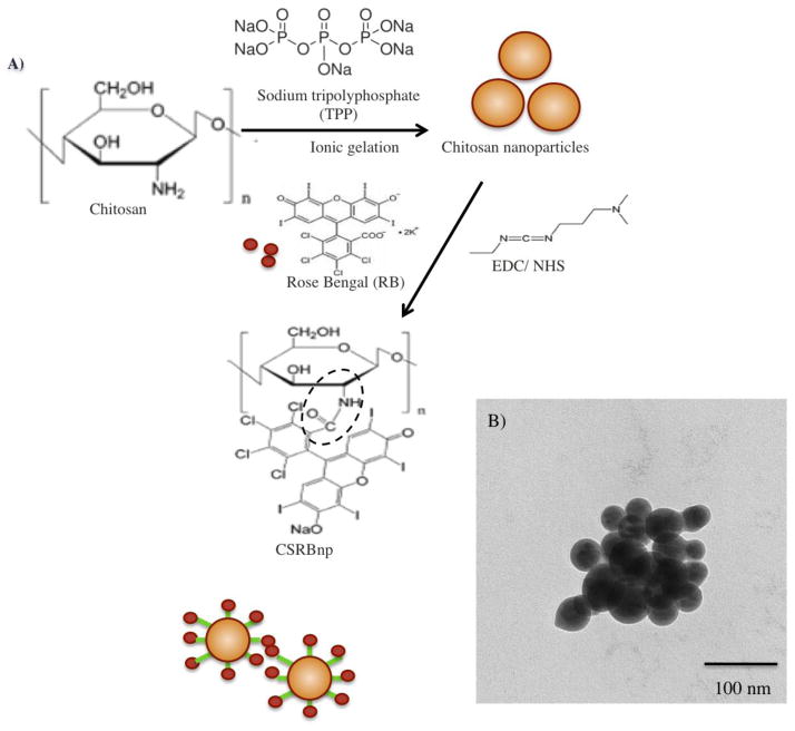
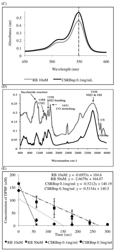
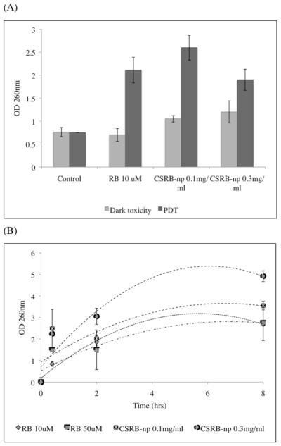
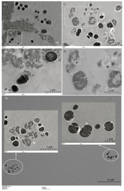
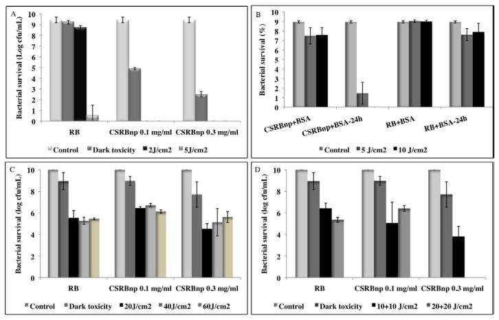
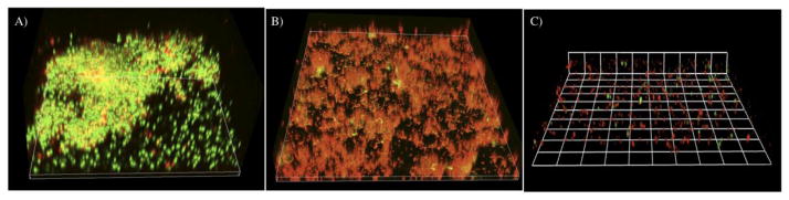
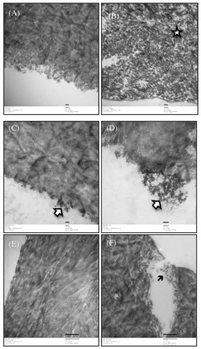
Treatment of infected teeth presents two major challenges: persistence of the bacterial-biofilm within root canals after treatment and compromised structural integrity of the dentin hard-tissue. In this study bioactive polymeric chitosan nanoparticles functionalized with rose-bengal, CSRBnp were developed to produce antibiofilm effects as well as stabilize structural-integrity by photocrosslinking dentin-collagen. CSRBnp were less toxic to fibroblasts and had significant antibacterial activity even in the presence of bovine serum albumin. CSRBnp exerted antibacterial mechanism by adhering to bacterial cell surface, permeabilizing the membrane and lysing the cells subsequent to photodynamic treatment. Photoactivated CSRBnp resulted in reduced viability of Enterococcus faecalis biofilms and disruption of biofilm structure. Incorporation of CSRBnp and photocrosslinking significantly improved resistance to degradation and mechanical strength of dentin-collagen (P<0.05). The functionalized chitosan nanoparticles provided a single-step treatment of infected root dentin by combining the properties of chitosan and that of photosensitizer to eliminate bacterial-biofilms and stabilize dentin-matrix.

From the clinical editor: In this study, bioactive polymeric chitosan nanoparticles functionalized with rose-bengal (a photosensitizer), CSRBnp were developed to produce antibiofilm effects as well as stabilize structural-integrity of dental root dentin by photocrosslinking dentin-collagen, leading to efficient elimination of bacterial-biofilms and stabilization of dentin-matrix.

Keywords: Biofilms; Chitosan; Collagen; Dentin; Functionalized nanoparticles; Photodynamic therapy.


Figures

Figure 1
Figure 1
Chemical reaction during conjugation of CS nanoparticles with rose bengal (RB) in the presence of EDC (N-ethyl-N′-(3-dimethyl aminopropyl) carbodiimide) and NHS (N-Hydroxysuccinimide) (A). Transmission electron microscopy (TEM) image of CSRBnp (scale bar= 100nm). The CSRBnp were of 60±20 nm in size (B). Absorption spectra of RB and CSRBnp with peak maxima at 550 nm (C). FTIR spectra of chitosan and CSRBnp (400 to 4000 cm−1 wave number) (D). The singlet oxygen yield monitored photometrically using oxidation of 1,3-diphenylisobenzofuran (DPBF). The rate of singlet oxygen yield in case of CSRBnp was slower than that of RB (E).
Figure 1
Figure 1
Chemical reaction during conjugation of CS nanoparticles with rose bengal (RB) in the presence of EDC (N-ethyl-N′-(3-dimethyl aminopropyl) carbodiimide) and NHS (N-Hydroxysuccinimide) (A). Transmission electron microscopy (TEM) image of CSRBnp (scale bar= 100nm). The CSRBnp were of 60±20 nm in size (B). Absorption spectra of RB and CSRBnp with peak maxima at 550 nm (C). FTIR spectra of chitosan and CSRBnp (400 to 4000 cm−1 wave number) (D). The singlet oxygen yield monitored photometrically using oxidation of 1,3-diphenylisobenzofuran (DPBF). The rate of singlet oxygen yield in case of CSRBnp was slower than that of RB (E).
Figure 2
Figure 2
Graph showing release of cell constituents (absorbance at 260 nm) following treatment with RB and CSRBnp with and without PDT (A). Time dependent release of cell constituents following treatment with RB and CSRBnp (B). With increase in interaction time, CSRBnp showed highest cell membrane damage.
Figure 3
Figure 3
Transmission electron microscopy images for planktonic E. faecalis after treatment with CSRBnp for 15 min (A and B). Aggregates of CSRBnp could be seen surrounding the bacterial cell. Nanoparticles were found attached to the bacterial cell surface and forming an envelope (⏎) (B). The cells did not show any disruption of morphology. Following PDT of the sensitized bacteria, various stages of membrane damage as well as release of cell constituents were evident (C and D). Most of the bacteria showed some kind of cell membrane disruption (□), and release of cell constituents (✉) at higher magnification (D). However, the bacterial cells after PDT with RB as the photosensitizer showed both live (¤) and dead (⌘) cells (E).
Figure 4
Figure 4
Bacterial survival of planktonic E. faecalis after PDT using RB and CSRBnp (A). Bacterial survival of planktonic E. faecalis in the presence of bovine serum albumin (BSA) after treatment with different nanoparticles with and without PDT (B). CSRBnp after PDT and incubation for 24 hrs showed the best results as compared to CSRBnp and RB with/without PDT. Antibiofilm effect of CSRBnp and RB on 21 days old E. faecalis biofilm (C and D). Complete elimination was obtained only after fractionation of PDT dose in case of CSRBnp at the higher concentration (D) in contrast to both the photosensitizers even after PDT dosage of 60 J/cm2 (C). Error bars show the standard deviation from average value.
Figure 5
Figure 5
The three-dimensional confocal laser scanning microscopy reconstruction of the biofilms subjected to PDT using RB and CSRBnp. (A) The biofilm receiving no treatment showed a multilayered three dimensional structure with both live (green) and dead (red) cells. (B) The biofilms subjected to sensitization with RB and PDT (40J/cm2) showed significantly higher number of dead cells. The mat like biofilm structure was not disturbed. (C) In case of CSRBnp the biofilm structure was completely disrupted with only few live and dead cells remaining on the substrate.
Figure 6
Figure 6
Transmission electron micrographs of dentin-collagen without any treatment (A and B) and following photocrosslinking treatment with CSRBnp (C–F). The scale bars in AD represent 100 nm and in EF are of 500 nm.

Similar articles

Cited by

References

    1. Lewis K. Riddle of biofilm resistance. Antimicrob Agents Chemother. 2001;45:999–1007. - PMC - PubMed
    1. Gilbert GH, Tilashalski KR, Litaker MS, McNeal SF, Boykin MJ, Kessler AW. Outcomes of root canal treatment in Dental Practice-Based Research Network practices. Gen Dent. 2010;58:28–36. - PMC - PubMed
    1. Lumley PJ, Lucarotti PS, Burke FJ. Ten-year outcome of root fillings in the General Dental Services in England and Wales. Int Endod J. 2008;41:577–85. - PubMed
    1. Nair PN. Pathogenesis of apical periodontitis and the causes of endodontic failures. Crit Rev Oral Biol Med. 2004;15:348–81. - PubMed
    1. Vier FV, Figueiredo JA. Internal apical resorption and its correlation with the type of apical lesion. Int Endod J. 2004;37:730–7. - PubMed

Publication types

MeSH terms