. 2014;13(2):279-92.
doi: 10.4161/cc.27053. Epub 2013 Nov 18.

Effects of cyclic AMP response element binding protein-Zhangfei (CREBZF) on the unfolded protein response and cell growth are exerted through the tumor suppressor p53

Affiliations

Effects of cyclic AMP response element binding protein-Zhangfei (CREBZF) on the unfolded protein response and cell growth are exerted through the tumor suppressor p53

Rui Zhang et al. Cell Cycle. 2014.

Abstract

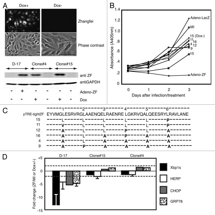
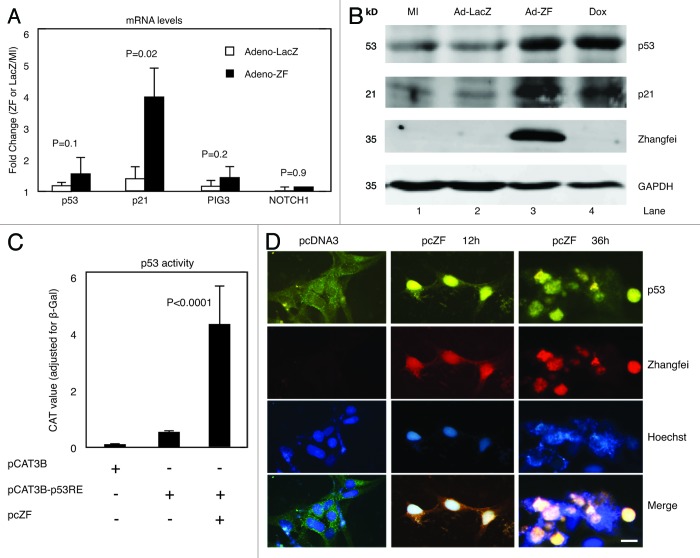
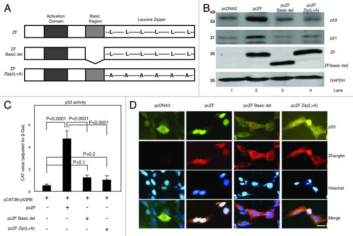
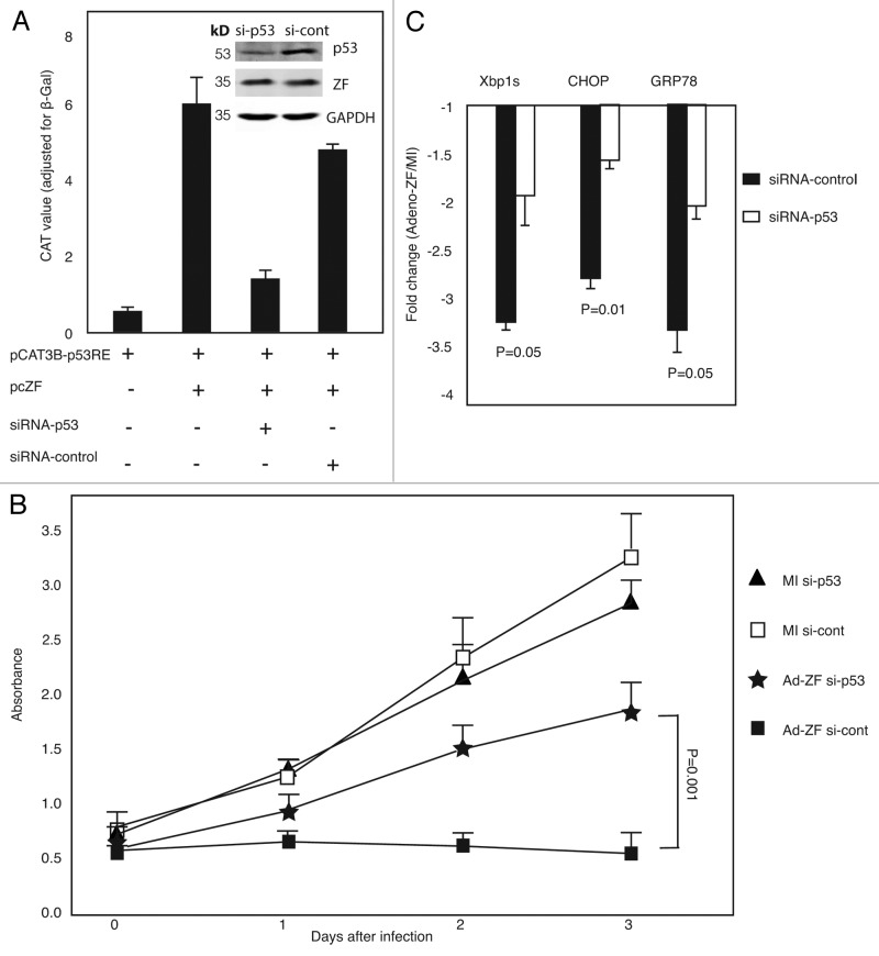
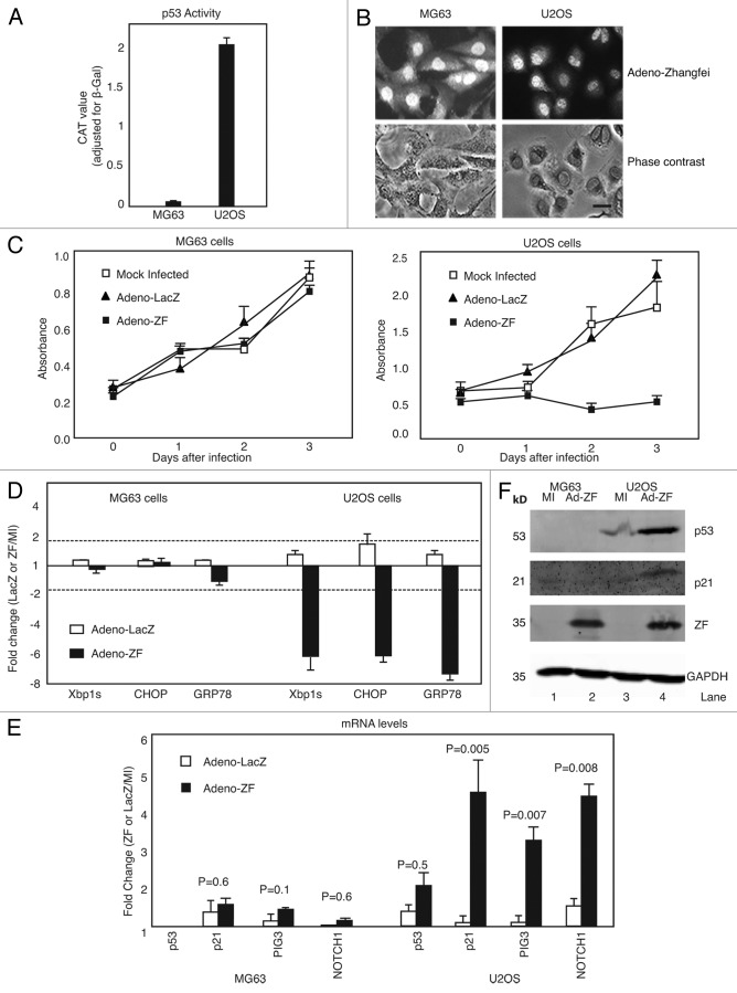
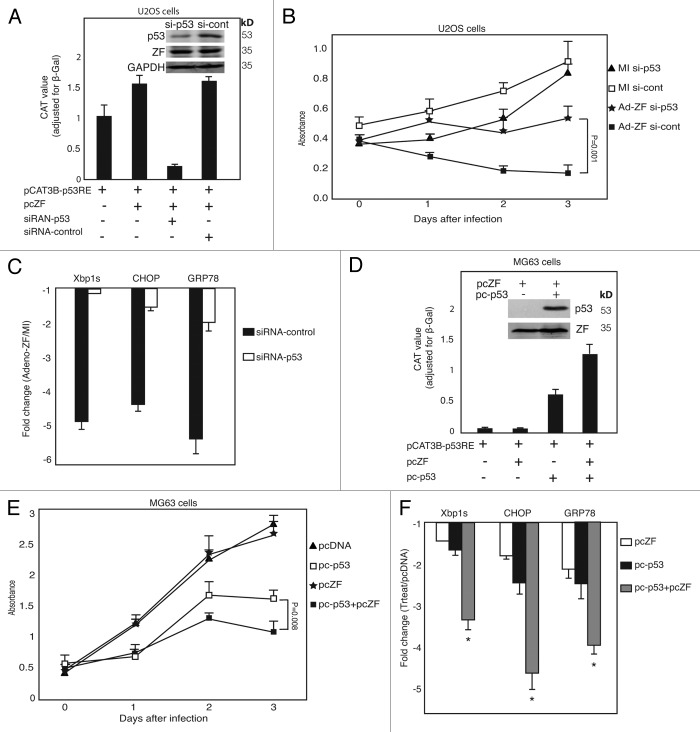
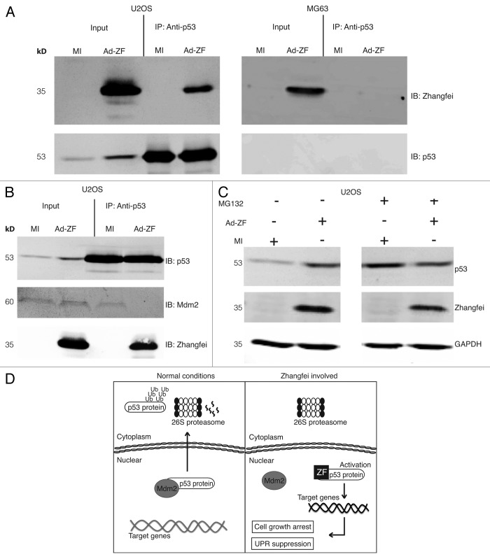
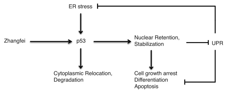
Zhangfei/CREBZF, a basic region-leucine zipper (bLZip) transcription factor, is a potent suppressor of growth and the unfolded protein response (UPR) in some cancer cell lines, including the canine osteosarcoma cell line, D-17. However, the effects of Zhangfei are not universal, and it has no obvious effects on untransformed cells and some cancer cell lines, suggesting that Zhangfei may act through an intermediary that is either not induced or is defective in cells that it does not affect. Here we identify the tumor suppressor protein p53 as this intermediary. We show the following: in cells ectopically expressing Zhangfei, the protein stabilizes p53 and co-localizes with it in cellular nuclei; the bLZip domain of Zhangfei is required for its profound effects on cell growth and interaction with p53. Suppression of p53 by siRNA at least partially inhibits the effects of Zhangfei on the UPR and cell growth. The effects of Zhangfei on D-17 cells is mirrored by its effects on the p53-expressing human osteosarcoma cell line U2OS, while Zhangfei has no effect on the p53-null osteosarcoma cell line MG63. In U2OS cells, Zhangfei displaces the E3 ubiquitin ligase mouse double minute homolog 2 (Mdm2) from its association with p53, suggesting a mechanism for the effects of Zhangfei on p53.

Keywords: Mdm2; Zhangfei/CREBZF; basic-leucine zipper domain; cell cycle; osteosarcoma; p53; protein domains; protein translocation; unfolded protein response.


Figures

None
Figure 1. Spontaneous mutation of leucine residues in the bLZip domain of Zhangfei in D-17 cells stably expressing the protein in the presence of tetracycline. (A) Doxycycline induces the expression of Zhangfei protein in D-17 cell clones. Twenty-four hours after induction with 1 µg/ml doxycycline, Zhangfei protein in D-17 clones was detected by immunofluorescence and immunoblotting. (B) Growth of D-17 cells after induction of Zhangfei. Six D-17 clones were treated with 1µg/ml doxycycline to induce Zhangfei expression. Cell growth characteristics were monitored at different time points using WST-1 assay. Growth characteristics of the original D-17 cells, mock-infected (MI) or infected with adenovirus vectors expressing either ZF (Adeno-ZF) or β-galactosidase (Adeno-LacZ) are shown for comparison. (C) Mutations in the leucine zipper of Zhangfei-expressing D-17 clones. Amino acid sequence of the LZip region for Zhangfei recovered by PCR from the vector used to develop the clones and the doxycycline-inducible Zhangfei expressing clones. Dashes (−) indicate no changes. Leucine residues (L) comprising the zipper are shown and mutations are in bold. (D) Induction of mutant Zhangfei has no effect on the UPR. Clones expressing ZF with mutations in the leucine zipper did not inhibit the UPR. Original D-17 cells and clones 4 and 15 were treated with 1 µg/ml doxycycline for 24 h, followed by 4 h of treatment of thapsigargin. Then cells were harvested and UPR transcripts were estimated by qRT-PCR. The original D-17 cells, mock-infected or infected with Adeno-ZF were also analyzed for comparison. The values represent fold changes in levels of transcripts between Adeno-ZF infected cells with mock-infected cells (in original D-17 cells) or between doxycycline treated with untreated cells (in Tet-on clones). Error bars indicated standard deviations from means of 3 individual experiments. Dashed lines indicate fold changes of less or more than 2-fold and changes exceeding these limits were arbitrarily regarded as significant. Bar = 100 µm.
None
Figure 2. Zhangfei regulates p53 in a post-translational level and induces p53 nuclear localization. (A) Zhangfei enhances the expression of p21, a p53-dependant gene. D-17 cells were mock-infected or infected with either Adeno-ZF or Adeno-LacZ, 24 h after infection, transcripts for p53, p21, PIG3, and NOTCH1 were determined by qRT-PCR. The values represented fold changes of transcripts between Adeno-LacZ (white bar) or Adeno-ZF (black bar) infected cells with mock-infected cells. Error bars indicated standard deviations from means. (B) Zhangfei stabilizes p53 and p21 proteins. p53, p21, Zhangfei, and GAPDH were detected by immunoblotting in D-17 cells either mock-infected (MI) or infected with either Adeno-LacZ or Adeno-ZF. As a positive control, cells were treated with 0.5 µM doxorubicin for 6 h. (C) Zhangfei activates p53-dependent transactivation. D-17 cells were transfected with a reporter plasmid containing the coding sequence for CAT linked to a promoter with 2 copies of p53-responsive element (pCAT3B-p53RE, 0.5 µg) in the presence or absence of a plasmid expressing Zhangfei (pcZF, 1 µg). The promoter-less parental reporter plasmid, pCAT3B was included as a control to show basal CAT activity. All samples also contained, as a control, a plasmid expressing β-galactosidase. 24 h after transfection, the CAT activity was determined. Values represented the relative CAT activity (normalized to the internal control, β-galactosidase) of different treatments. Standard deviations from means of three individual experiments are and the significant P values from ANOVA tests were noted above the bars. (D) Zhangfei alters the subcellular localization of p53. D-17 cells were transfected with 1 µg of pcZF or a control (pcDNA3), and 12 h and 36 h after transfection, endogenous p53 as well as Zhangfei were visualized by immunofluorescence with anti-p53 and anti-ZF antibody. The nucleus was detected by Hoechst staining (bar = 10 µm). The means and standard deviations of representative experiments (n = 3) were shown. P < 0.05 were considered to be significant.
None
Figure 3. The basic-region leucine zipper domain (bLZip) of Zhangfei is required for its effect on p53. (A) Schematic representation of the structures of Zhangfei (ZF) and Zhangfei mutants: ZF Basic del, basic region was deleted; ZF Zip (L > A), all leucines in the leucine-zipper domain were replaced with alanines. (B) The bLZip domain of Zhangfei is required for stabilization of p53 and p21 proteins. D-17 cells were transfected with 1 µg of plasmid expressing Zhangfei (pcZF) or mutants (pcZF Zip(L > A) or pcZF Basic del). Twenty-four h after transfection endogenous p53 and p21 proteins were detected by immunoblotting. (C) The bLZip domain of Zhangfei is required for p53-dependent transactivation. D-17 cells were transfected with 0.5 µg of p53 response element containing reporter plasmid pCAT3B-p53RE and 1 µg of pcZF or mutants (pcZF Zip [L > A] or pcZF Basic del). Twenty four hours after transfection, the CAT activity was determined. The means and standard deviations of experiments (n = 3) were shown. P < 0.05 were considered to be significant. (D) The bLZip domain of Zhangfei is required for p53 nuclear retention. D-17 cells were treated as described in (A), and endogenous p53 as well as Zhangfei were visualized by immunofluorescence. The nucleus was detected by Hoechst staining (bar = 10 µm).
None
Figure 4. Zhangfei regulates p53-mediated cell growth and UPR. (A) Zhangfei-induced transcription from a promoter with p53 response elements is mediated by p53. D-17 cells were transfected with 0.5 µg of p53 response element containing promoter (pCAT3B-p53), 1 µg of plasmid expressing Zhangfei (pcZF) and 100 pM of si-RNA against p53 (si-p53) or non-targeting siRNA control (si-cont), 48 h after transfection, cells were analyzed for CAT activity or p53, Zhangfei and GAPDH proteins (inset). (B) Zhangfei-mediated suppression of cell growth is mediated through p53. D-17 cells were transfected with 100 pM of si-RNA against p53 (siRNA-p53) or non-targeting siRNA control (siRNA-cont). Eight hours after transfection, cells were either mock-infected or infected with Adeno-ZF, and growth characteristics were measured using WST-1 assay at 0 h, 24 h, 48 h, and 72 h after infection. (C) Zhangfei-mediated suppression of UPR genes is mediated by p53. D-17 cells were transfected 100 pM of siRNA-p53 or non-targeting siRNA-control. Forty-eight hours after transfection, cells were treated with thapsigargin for 4 h and harvested. Differences in levels of transcripts (Xbp1s, CHOP, and GRP78) were determined by qRT-PCR. The results were expressed as fold changes relative to mock-infected cells. The means and standard deviations of experiments (n = 3) are shown. P < 0.05 were considered to be significant.
None
Figure 5. Zhangfei suppresses cell growth and UPR in wild-type p53-expressing U2OS cells, but not in p53-null MG63 cells. (A) p53-dependent transactivation in U2OS and MG63 cells. Cells were transfected with 0.5 µg of p53 reporter (pCAT3B-p53RE) for 24 h, then p53 transcriptional activity was determined by CAT ELISA assay. (B) U2OS and MG63 cells express Zhangfei when infected with adenovirus vector expressing the protein. Cells were infected with Adeno-ZF, and 24 h later Zhangfei protein was visualized by immunofluorescence (bar = 10 µm). (C) Ectopic expression of ZF suppresses cell growth in p53-wild type U2OS cells, but has no effect in p53-null MG63 cells. U2OS and MG63 human osteosarcoma cells were mock-infected or infected with Adeno-ZF or Adeno-LacZ and their growth rates were assessed at different time points after infection as absorbance at 405 nm after treatment with WST-1. (D) Zhangfei negatively regulates the UPR in U2OS cells but not in MG63 cells. U2OS and MG63 human cells were either mock-infected or infected with Adeno-ZF or Adeno-LacZ. Twenty-four hours after infection, cells were treated with thapsigargin for 4 h, and then harvested. The differences in levels of transcripts for UPR genes (Xbp1s, CHOP, and GRP78) were determined by qRT-PCR. (E) Zhangfei enhances p53-dependent transcripts in U2OS cells but not in MG63 cells. Transcripts for p53, p21, PIG3, and NOTCH1 in U2OS and MG63 cells either mock-infected or infected with Adeno-LacZ or Adeno-ZF were measured by pRT-PCR. (F) Zhangfei increases p53 and p21 proteins in U2OS cells but not in MG63 cells. p53 and p21 proteins were detected by immunoblotting in U2OS and MG63 cells either mock-infected or infected with Adeno-ZF. The values in (D and E) represented fold changes of transcripts between Adeno-LacZ (white bar) or Adeno-ZF (black bar) infected cells with mock-infected cells. The means and standard deviations of experiments (n = 3) were shown. P < 0.05 were considered to be significant. Error bars indicate standard deviations from means. Horizontal dotted line indicated a 2-fold change. A change of more than 2-fold was arbitrarily regarded as significant.
None
Figure 6. p53 mediates the suppressive effects of Zhangfei on cell growth and UPR in human osteosarcoma cells. (A) Zhangfei-mediated transcription from a p53 response element-containing promoter is mediated by p53 in U2OS cells. U2OS cells were transfected with 0.5 µg of p53 reporter plasmid (pCAT3B-p53RE) and 1 µg of plasmids expressing Zhangfei (pcZF) in the presence of 100 pM of siRNA-p53 or non-targeting siRNA-control. Forty eight hours after transfection CAT reporter activity and p53 protein were detected by ELISA and immunoblotting (inset). (B) Suppression of p53 by siRNA in U2OS cells restored Zhangfei-induced suppression of cell growth. U2OS were transfected with 100 pM of siRNA-p53 or non-targeting siRNA-control. Eight hours after transfection, cells were either mock-infected or infected with Adeno-ZF and measured growth rates as absorbance at 405 nm after treatment with WST-1. (C) Suppression of p53 prevents Zhangfei-induced inhibition of UPR genes. U2OS cells were transfected with 100 pM of siRNA-p53 or non-targeting siRNA-control. Forty eight hours after transfection, cells were treated with thapsigargin for 4h and harvested. Differences in levels of transcripts for UPR genes (Xbp1s, CHOP, and GRP78) were determined by qRT-PCR. The values represented fold changes of transcripts between Adeno-LacZ (white bar) or Adeno-ZF (black bar) infected cells with mock infected cells. (D) Transactivation activity and expression of p53 protein in p53-null MG63 cells. p53-null MG63 cells were transfected with 0.5 µg of p53 reporter plasmid and 1 µg of plasmids expressing Zhangfei (pcZF) in the presence of 1µg of plasmids expressing wild-type p53 (pc-p53, from U2OS cells) for 24 h, and then CAT reporter activity and p53 protein were detected by ELISA and immunoblotting. (E) Overexpression of p53 in p53-null MG63 cells suppressed cell growth and activated growth arrest induced by Zhangfei. MG63 cells were transfected with 3 µg of pcZF in the absence or presence of 3 µg of pc-p53 and measured growth rates as absorbance at 405 nm after treatment with WST-1. (F) Overexpression of p53 in p53-null MG63 cells activated the UPR suppression induced by Zhangfei. MG63 cells were transfected with 3 µg of pcZF in the absence or presence of 3 µg of pc-p53. Twenty-four hours after transfection, cells were treated with thapsigargin for 4 h and harvested. Differences in levels of transcripts for UPR genes (Xbp1s, CHOP, and GRP78) were determined by qRT-PCR. The values represented fold changes of transcripts between pcZF (white bar), pc-p53 (black bar), or pcZF plus pc-p53 (gray bar) transfected cells with pcDNA3 transfected cells. The total amount of DNA in each transfection above was made up to 5 μg with pcDNA3. The means and standard deviations of experiments (n = 3) were shown. *P < 0.05 were considered to be significant.
None
Figure 7. In vitro interaction of Zhangfei and p53. (A) Zhangfei associates with p53 in U2OS cells. Cell lysate from U2OS and MG63 human cells previously infected with adenovirus vector expressing Zhangfei (Ad-ZF) or mock infected (MI) were incubated with 2 µg of mouse anti-p53 antibody for 12 h at 4 °C, followed by an additional 4 h incubation with 100 µl of Protein A/G agarose beads (IP). The associated proteins were detected by immunoblotting (IB) using rabbit anti-Zhangfei antibody and mouse anti-p53 antibody. Input represented 1/10 of lysate used for immunoprecipitation. (B) Zhangfei displaces Mdm2 from p53 in U2OS cells. Cell lysates as in (A) were immunoprecipitated with anti-p53 antibody followed by immunoblotting with either anti-Mdm2 or anti-Zhangfei antibody. (C) Inhibition of proteasomal degradation reverses degradation of p53 in the absence of Zhangfei. Cells infected as in (A) were either treated with the proteasomal inhibitor MG132 (5 µM) or left untreated. Proteins in the cell lysates were separated by SDS-PAGE, and p53, Zhangfei and GAPDH (loading control) were detected by immunoblotting. (D) Schematic diagram of a model for the proposed mechanism by which Zhangfei inhibits cell growth and UPR. Under normal conditions, Mdm2 translocates the p53 protein out of the nucleus for degradation via the ubiquitin-dependent pathway. In Zhangfei-expressing cells, Zhangfei interacts with p53 and prevents it from binding to Mdm2. This results in p53 stabilization and nuclear accumulation, which, in turn, activates its target genes and suppresses tumor cell growth and the UPR. These results illustrate how Zhangfei controls the activity of p53 toward the cell growth and the UPR, and offer an explanation to why p53 is upregulated by Zhangfei in tumor cells.
None
Figure 8. Zhangfei and ER stress have opposing effects on p53. In Zhangfei-expressing cells, p53 is targeted for nuclear localization and stabilization, and further to prevent cell growth, induce cell apoptosis, as well as inhibit the UPR in response to ER stress. In contrast, ER stress promotes the cytoplasmic localization and degradation of p53.

References

    1. Lu R, Misra V. Zhangfei: a second cellular protein interacts with herpes simplex virus accessory factor HCF in a manner similar to Luman and VP16. Nucleic Acids Res. 2000;28:2446–54. doi: 10.1093/nar/28.12.2446. - DOI - PMC - PubMed
    1. López-Mateo I, Villaronga MA, Llanos S, Belandia B. The transcription factor CREBZF is a novel positive regulator of p53. Cell Cycle. 2012;11:3887–95. doi: 10.4161/cc.22133. - DOI - PMC - PubMed
    1. Hogan MR, Cockram GP, Lu R. Cooperative interaction of Zhangfei and ATF4 in transactivation of the cyclic AMP response element. FEBS Lett. 2006;580:58–62. doi: 10.1016/j.febslet.2005.11.046. - DOI - PubMed
    1. Xie YB, Lee OH, Nedumaran B, Seong HA, Lee KM, Ha H, Lee IK, Yun Y, Choi HS. SMILE, a new orphan nuclear receptor SHP-interacting protein, regulates SHP-repressed estrogen receptor transactivation. Biochem J. 2008;416:463–73. doi: 10.1042/BJ20080782. - DOI - PubMed
    1. Xie YB, Nedumaran B, Choi HS. Molecular characterization of SMILE as a novel corepressor of nuclear receptors. Nucleic Acids Res. 2009;37:4100–15. doi: 10.1093/nar/gkp333. - DOI - PMC - PubMed

Publication types

Substances