Case Reports
. 2014;54(3):219-26.
doi: 10.2176/nmc.cr2012-0433. Epub 2013 Nov 8.

Discrepancy between preoperative imaging and postoperative pathological finding of ruptured intracranial dissecting aneurysm, and its surgical treatment: case report

Affiliations
Case Reports

Discrepancy between preoperative imaging and postoperative pathological finding of ruptured intracranial dissecting aneurysm, and its surgical treatment: case report

Nakao Ota et al. Neurol Med Chir (Tokyo). 2014.

Abstract

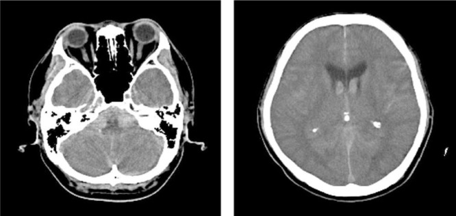
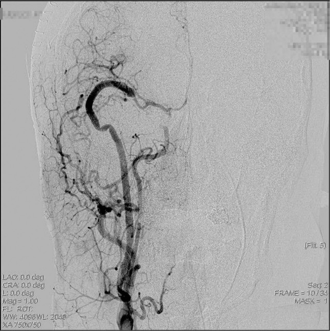
The choice of therapeutic strategy for intracranial dissecting aneurysm is often based on radiographic features, including characteristic geometry (e.g., irregular stenosis, segmental stenosis, aneurysm formation [pearl-and-string sign]), irregular fusiform or aneurysmal dilation, double lumen, and tapering occlusion. However, there is often a discrepancy between preoperative radiographic data and actual dissecting length. The present report describes three cases in which there was a discrepancy between preoperative radiographic data and actual dissecting length in patients undergoing direct trapping with or without revascularization. All three cases experienced good outcomes, but these cases underscore the fact that open surgery is a good option for management of ruptured intracranial dissecting aneurysms for determination of the rupture point, dissecting length, and the relationship between dissecting area and small arteries arising from the associated vessel.


Conflict of interest statement

Conflicts of Interest Disclosure

I have already submitted the self-reported potential COI disclosure statement of the Japan Neurosurgical Society. And as the corresponding author, I take all responsibility that all coauthors have disclosed all potential COIs concerning this manuscript, in accordance with the policy of the Japan Neurosurgical Society.

Figures

Fig. 1
Fig. 1
Computed tomography reveals subarachnoid hemorrhage with thick subarachnoid clot in the cerebellomedullary cistern. Back streaming of the hemorrhage in the lateral ventricle is also seen.
Fig. 2
Fig. 2
Computed tomography angiography (CTA) revealing a pearl-and-string sign in the V4 portion of the right vertebral artery (left). Slices a, b, and c correspond to the pathological specimens (right). Photomicrograph showing serial axial slices of an entry-only dissecting aneurysm. Elastica van Gieson stain, original magnification ×40. Panel a shows a slice taken from the narrowed lesion seen on CTA. The media is disrupted, and hematoma is seen just below the adventitia. The intravascular space is narrowed in this slice. Panel b shows a slice taken from the dilated lesion seen on CTA. The internal elastic lamina is disrupted, and the wall adjacent to the rupture site is only composed of fibrin and thin collagen, which forms a pseudoaneurysm. Panel c is a slice taken from the normal-appearing site seen on CTA. The internal elastic lamina is also disrupted in this specimen, while the intravascular space is relatively normal; this means that the angiographically “normal” lesion also includes the entry point.
Fig. 3
Fig. 3
Operating view of the right-sided lateral suboccipital transcondylar approach. Panels A and B show the distal end of the dissecting aneurysm. A: Perforating branches of vertebral artery are seen just distal to the dissecting aneurysm. B: Vertebral artery trapping was performed while securing this perforating branch. C: The proximal end of the dissecting aneurysm. Trapping was performed and involved the entire length of dissecting aneurysm.
Fig. 4
Fig. 4
Computed tomography reveals a Fisher Group three subarachnoid hemorrhage. The hematoma of the right side is dominant.
Fig. 5
Fig. 5
Preoperative computed tomography angiography reveals only an elevated margin (arrow) proximal to the origin of the posterior communicating artery (arrow head).
Fig. 6
Fig. 6
Operative view of the right anterior temporal approach. Dissecting aneurysm of the internal carotid artery is found in C2 to C1. The rupture point is found in the C2 portion (left, arrow head). The dissecting aneurysm extends to the distal portion of the C1 (right, arrow).
Fig. 7
Fig. 7
Photomicrograph showing the disrupted internal elastic lamina and hematoma below the adventitia. The intravascular space is maintained within the normal range. Elastica van Gieson stain, original magnification ×40.
Fig. 8
Fig. 8
Postoperative digital subtraction angiography showing external carotid-radial artery-M2 bypass with good retrograde flow and trapped C2 to C1 portion of the internal carotid artery.
Fig. 9
Fig. 9
Computed tomography revealing subarachnoid hemorrhage with thin clot around the prepontine and cerebellomedullary cistern.
Fig. 10
Fig. 10
Computed tomography angiography and digital subtraction angiography show a pearl sign in the segment of V4 just distal to the takeoff of the posterior inferior cerebellar artery (PICA). The PICA is not involved in the dissecting length on preoperative imaging.
Fig. 11
Fig. 11
Left-sided lateral suboccipital transcondylar approach was performed. The orifice of the posterior inferior cerebellar artery (arrow) is contained within the dissecting aneurysm (arrow head).
Fig. 12
Fig. 12
Photomicrograph showing a ruptured portion of an entry-only dissecting aneurysm in a dilated lesion. The internal elastic lamina is disrupted, resulting in pseudoaneurysm. Elastica van Gieson stain, original magnification ×40 (left), ×400 (right).

Similar articles

Cited by

References

    1. Friedman AH, Drake CG: Subarachnoid hemorrhage from intracranial dissecting aneurysm. J Neurosurg 60: 325– 334, 1984. - PubMed
    1. Mizutani T, Aruga T, Kirino T, Miki Y, Saito I, Tsuchida T: Recurrent subarachnoid hemorrhage from untreated ruptured vertebrobasilar dissecting aneurysms. Neurosurgery 36: 905– 911; discussion 912–913, 1995. - PubMed
    1. Yamaura A, Watanabe Y, Saeki N: Dissecting aneurysms of the intracranial vertebral artery. J Neurosurg 72: 183– 188, 1990. - PubMed
    1. Yamaura A, Yoshimoto T, Hashimoto N, Ono J: [Nationwide study of nontraumatic intracranial arterial dissection: treatment and its results]. Surgery for Cerebral Stroke 26: 87– 95, 1998. (Japanese)
    1. Sano H, Kato Y, Okuma I, Yamaguchi S, Ninomiya T, Arunkumar R, Kanno T: Classification and treatment of vertebral dissecting aneurysm. Surg Neurol 48: 598– 605, 1997. - PubMed

Publication types

MeSH terms