. 2014 Jan 21;110(2):409-20.
doi: 10.1038/bjc.2013.706. Epub 2013 Nov 7.

Pancreatic stellate cells promote hapto-migration of cancer cells through collagen I-mediated signalling pathway

Affiliations

Pancreatic stellate cells promote hapto-migration of cancer cells through collagen I-mediated signalling pathway

J Lu et al. Br J Cancer. .

Abstract

Background: Pancreatic stellate cells (PSCs) promote metastasis as well as local growth of pancreatic cancer. However, the factors mediating the effect of PSCs on pancreatic cancer cells have not been clearly identified.

Methods: We used a modified Boyden chamber assay as an in vitro model to investigate the role of PSCs in migration of Panc1 and UlaPaCa cells and to identify the underlying mechanisms.

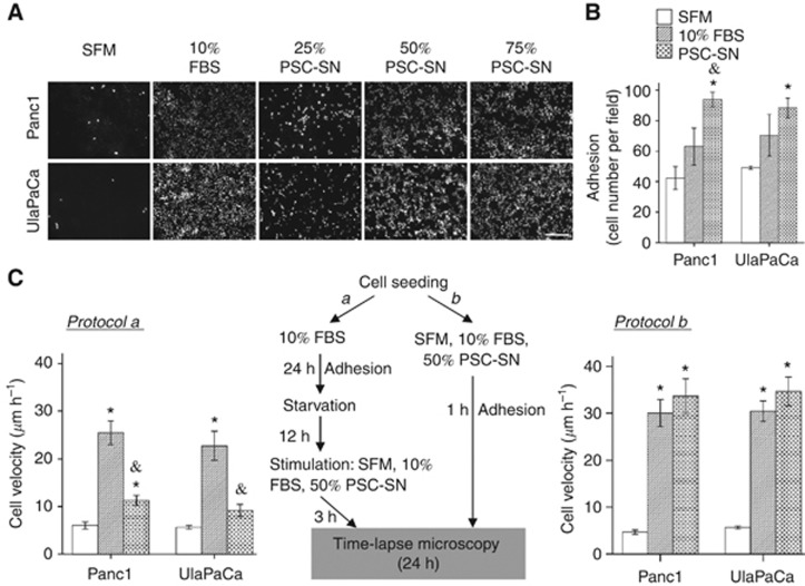
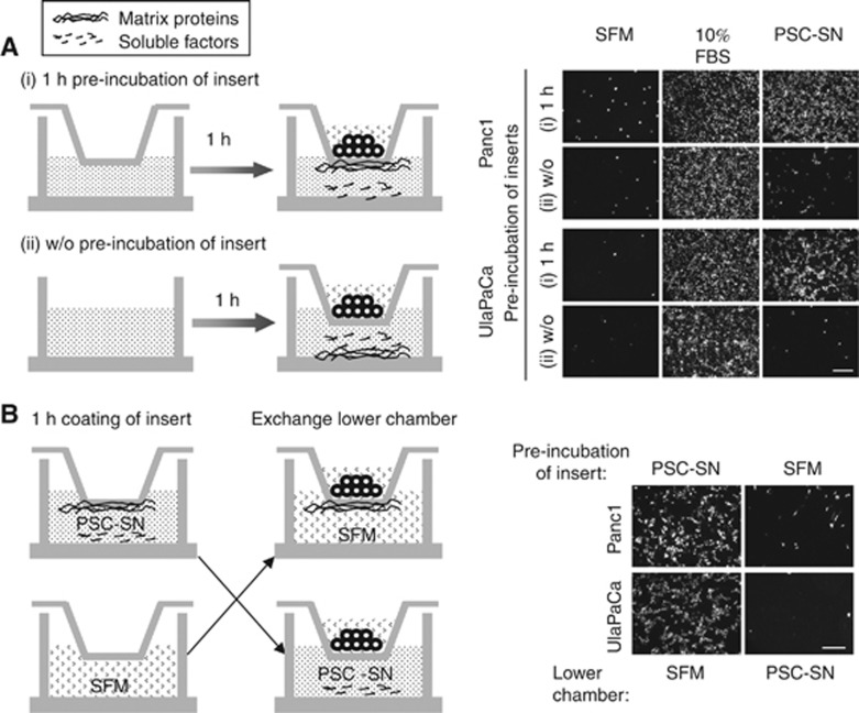
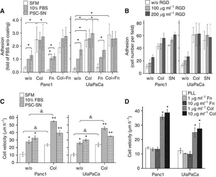
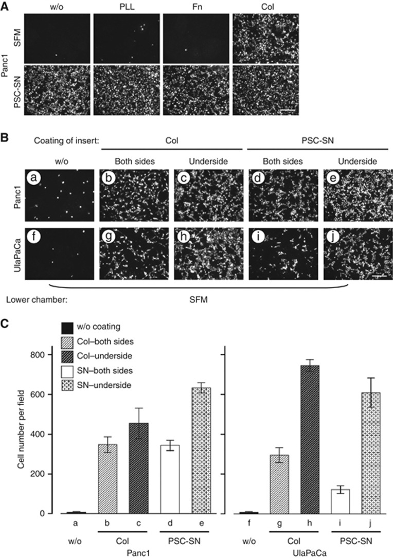
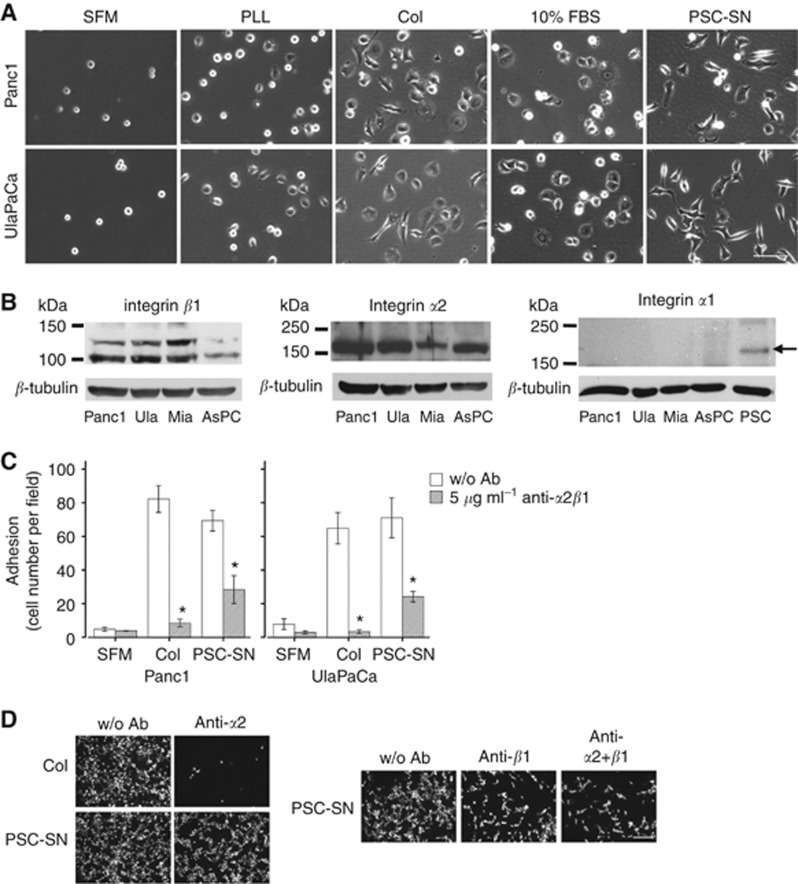
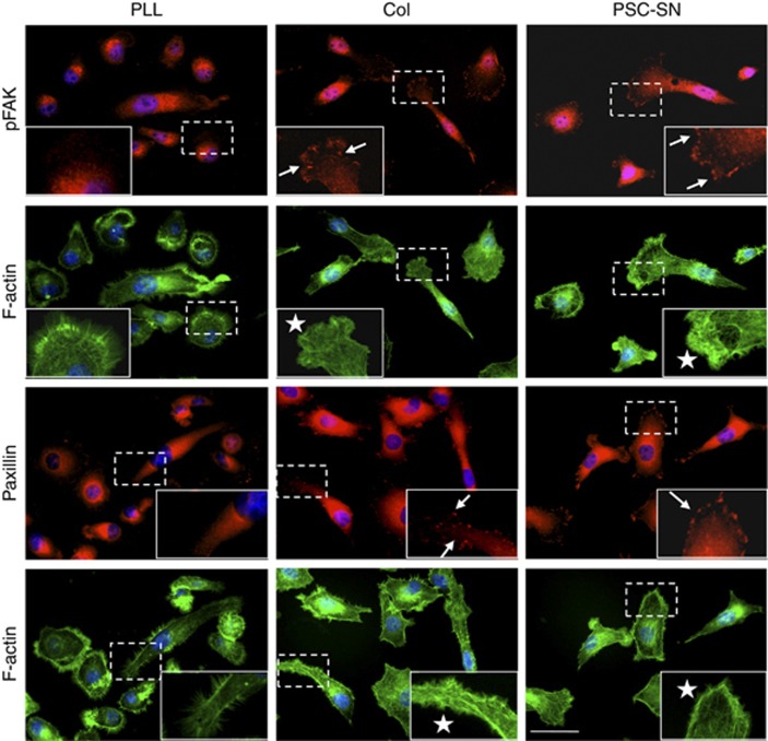
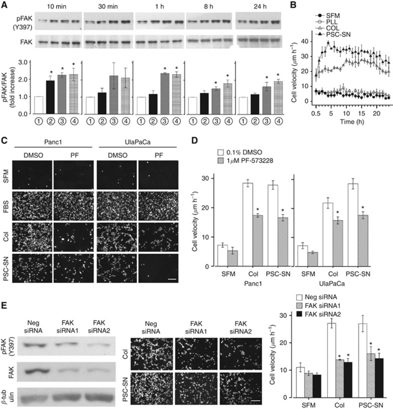
Results: PSC supernatant (PSC-SN) dose-dependently induced the trans-migration of Panc1 and UlaPaCa cells, mainly via haptokinesis and haptotaxis, respectively. In contrast to poly-L-lysine or fibronectin, collagen I resembled PSC-SN with respect to its effect on cancer cell behaviours, including polarised morphology, facilitated adhesion, accelerated motility and stimulated trans-migration. Blocking antibodies against integrin α2/β1 subunits significantly attenuated PSC-SN- or collagen I-promoted cell trans-migration and adhesion. Moreover, both PSC-SN and collagen I induced the formation of F-actin and focal adhesions in cells, which was consistent with the constantly enhanced phosphorylation of focal adhesion kinase (FAK, Tyr397). Inhibition of FAK function by an inhibitor or small interference RNAs significantly diminished the effect of PSC-SN or collagen I on haptotaxis/haptokinesis of pancreatic cancer cells.

Conclusion: Collagen I is the major mediator for PSC-SN-induced haptokinesis of Panc1 and haptotaxis of UlaPaCa by activating FAK signalling via binding to integrin α2β1.


Figures

Figure 1
Figure 1
Effect of PSC-SN on the process of pancreatic cancer cell trans-migration. (A) Modified Boyden chamber assay. The lower compartment was filled with SFM or medium containing 10% FBS or PSC-SN at varying concentrations. Panc1 and UlaPaCa cells were seeded into the inserts and allowed to trans-migrate for 18 h. Representative images for each condition are shown. Scale bars: 200 μm. (B) Cell adhesion assay of Panc1 and UlaPaCa cells. Results are expressed as the mean±s.e.m. of three independent experiments. *P<0.05 compared with corresponding SFM controls. &P<0.05 compared with 10% FBS. (C) Motility of Panc1 and UlaPaCa cells were evaluated by single-cell tracking assays. Schematic depiction of two approaches is shown in the middle. In protocol a, cells were seeded in medium containing 10% FBS and cultured for 24 h, followed by 12-h starvation in SFM (to slow down baseline cell motility), and then stimulated by 50% PSC-SN or 10% FBS or SFM for 3 h. In protocol b, cells were directly seeded in SFM or medium containing 10% FBS or 50% PSC-SN, and allowed to adhere for 1 h. Cell migration in both protocols was recorded over 24 h. Results are expressed as the mean±s.e.m. of three independent experiments. *P<0.05 compared with corresponding SFM controls.
Figure 2
Figure 2
Characterisation of the effect of PSC-SN on haptokinesis/haptotaxis vs chemokinesis/chemotaxis of Panc1 and UlaPaCa cells. Schematic illustration of the experiments is shown on the left. (A) The lower compartment of Boyden chamber was filled with SFM or medium containing 10% FBS or 50% PSC-SN. Group i insert was pre-incubated in the above media for 1 h. This procedure allowed adhesive molecules in the media to coat the underside of the inserts. Group ii was left outside till 1 h later. Cells were then seeded and allowed to trans-migrate for 18 h. (B) Inserts were placed into lower chambers containing SFM or 50% PSC-SN and incubated for 1 h. Thereafter the lower chambers were exchanged, so that PSC-SN-coated inserts placed into SFM whereas SFM-embedded inserts into PSC-SN. Representative images for each condition are shown. Scale bars: 200 μm.
Figure 3
Figure 3
Effect of matrix proteins on the adhesion and motility of pancreatic cancer cells. Data are representative of results from three independent experiments. (A) In all, 24-well plates were left uncoated or coated with collagen I (Col) and/or fibronectin (Fn) as described in the method. Panc1 and UlaPaCa cells were then seeded in SFM or medium containing 10% FBS or 50% PSC-SN, and allowed to adhere for 1 h. Results are expressed as the mean±s.e.m. fold of 10% FBS w/o coating. *P<0.05. (B) Panc1 and UlaPaCa cells were pre-incubated with RGD peptide (100 or 200 μg ml–1) for 1 h and then applied to cell adhesion assay. Results are expressed as the mean±s.e.m. number of adherent cells. (C) Panc1 and UlaPaCa cells were seeded in SFM or medium containing 10% FBS or 50% PSC-SN (SN) with or without pre-coated collagen I. After 1 h adhesion, cell migration was recorded over 24 h. Results are expressed as the mean±s.e.m. *P<0.05 compared with w/o coating SFM control; **P<0.05 compared with collagen I coating SFM control; &P<0.05. (D) In all, 12-well plates were pre-coated with poly-L-lysine (PLL), fibronectin, or collagen I. Panc1 and UlaPaCa were seeded with 10% FBS and allowed to adhere for 1 h. Afterward the culture plates were washed twice with PBS and SFM were filled in. Cell migration was recorded over 30 h. Results are expressed as the mean±s.e.m. *P<0.05 compared with corresponding poly-L-lysine coating SFM controls.
Figure 4
Figure 4
Effect of matrix proteins on the trans-migration of pancreatic cancer cells. (A) Modified Boyden chamber assay of Panc1 cells. The underside of the inserts was left uncoated or coated with poly-L-lysine (PLL) or fibronectin (Fn) or collagen I (Col) as described in the method. The lower compartment of Boyden chamber was filled with SFM or medium containing PSC-SN. Representative images for each condition are shown. (B) Characterisation of the directionality during trans-migration of Panc1 and UlaPaCa cells. Inserts without coating (a, f) were used as a negative control. Other inserts were coated with collagen I or PSC-SN either on both sides (b, g; d, i) or on the underside (c, h; e, j) as described in the Materials and Method. Lower chambers were filled with SFM. Representative images for each condition are shown. (C) Quantification of cell trans-migration under each condition in Figure 4B. Images were taken from seven random fields and cell number was counted with ImageJ 1.44m. Scale bars: 200 μm.
Figure 5
Figure 5
Integrin α2β1 mediates the stimulatory effect of PSC-SN or collagen I on pancreatic cancer cells. (A) Morphology of Panc1 and UlaPaCa cells in cell adhesion assay. In all, 12-well plates were left uncoated or were coated with poly-L-lysine (PLL) or collagen I (Col). Representative phase contrast images for each condition are shown. Scale bars: 100 μm. (B) Thirty microgram of total cell lysates from Panc1, UlaPaCa (Ula), MiaPaCa-2 (Mia) and AsPC-1 cultured with 10% FBS for 24 h were subjected to 8% SDS-PAGE. Antibodies against human α1, α2 and β1 integrin subunits were used to detect the corresponding expression. The band of PSCs (arrow) was shown as a positive control for α1 integrin. β-tubulin was used as a loading control. (C) Panc1 and UlaPaCa cells were pre-incubated with 5 μg ml–1 integrin α2β1 blocking antibody for 1 h, and then were allowed to adhere for 1 h in SFM (with or w/o coated collagen I) or medium containing 50% PSC-SN. Results are expressed as the mean±s.e.m. of three independent experiments. *P<0.05 compared with corresponding controls w/o antibodies. (D) UlaPaCa cells were pre-incubated with 20 μg ml–1 integrin α2 and/or β1 blocking antibodies for 1 h. The cells were then allowed to trans-migrate for 18 h towards pre-coated collagen I on the underside of the inserts or towards 50% PSC-SN. Representative images for each condition are shown. Scale bars: 200 μm.
Figure 6
Figure 6
Fluorescence stainings of pFAK, paxillin and F-actin in Panc1 cells. Glass coverslips were placed in six-well plates and coated with poly-L-lysine (PLL) or collazgen I (Col). Panc1 cells were allowed to adhere in SFM (with PLL/Col coating) or medium containing 50% PSC-SN (w/o coating) for 3 h. The cells were then stained with anti-pFAK (Tyr397) or anti-paxillin (red) and rhodamine-phalloidin (green). Nuclei were stained with Hoechst 33258 (blue). In each image, the boxed region is shown magnified in the insert. Arrows indicate representative focal adhesions, and asterisks indicate representative lamellipodia. Scale bars: 40 μm.
Figure 7
Figure 7
FAK signalling pathway is involved in PSC-SN- or collagen I-induced haptokinesis/haptotaxis of pancreatic cancer cells. (A) Panc1 cells were seeded in SFM (with or w/o coated poly-L-lysine or collagen I) or 50% PSC-SN. The culture was stopped at indicated time points after cell seeding. Both non-adherent and adherent cells were collected and lysed. Twelve microgram of total lysates were subjected to 6% SDS-PAGE, and pFAK (Tyr397) was used to detect the activation of this kinase. The same blots were re-probed with antibody against total FAK as loading controls. Quantification of pFAK was performed by scanning densitometry from three independent experiments, and presented as mean±s.e.m. fold increase above w/o coating SFM. ( SFM, poy-L-lysine, collagen I, 50% PSC-SN). (B) Velocity profile of Panc1 cells migrating under the above conditions was obtained from 24-h single-cell tracking assay. (C) Panc1 and UlaPaCa were pre-incubated with 1% DMSO or 1 μM PF-537228 (PF) for 1 h. The cells were then allowed to trans-migrate for 18 h towards SFM, 10% FBS, pre-coated collagen I (Col) on the underside of the inserts, or 50% PSC-SN. Representative images for each condition are shown. (D) Panc1 and UlaPaCa cells pre-treated with DMSO or PF-537228 were seeded in SFM (with or w/o coated collagen I) or medium containing 50% PSC-SN. Single-cell tracking assay was performed after 1-h adhesion. Results are expressed as the mean±s.e.m. from three independent experiments. *P<0.05 compared with corresponding 0.1% DMSO controls. (E) Panc1 cells were transfected with 40 nM negative siRNA, FAK siRNA1 or FAK siRNA2 for 66 h. Inhibition of FAK was confirmed by western blot using β-tubulin as a loading control. A representative blot from three independent experiments is shown (left panel). Effect of collagen I or PSC-SN on migration of FAK-silenced cells was evaluated in modified Boyden chamber assay (middle panel) and single-cell tracking assay (right panel) as described before. Scale bars: 200 μm.

Similar articles

Cited by

References

    1. Alexander NR, Branch KM, Parekh A, Clark ES, Iwueke IC, Guelcher SA, Weaver AM. Extracellular matrix rigidity promotes invadopodia activity. Curr Biol. 2008;18:1295–1299. - PMC - PubMed
    1. Apte MV, Haber PS, Applegate TL, Norton ID, McCaughan GW, Korsten MA, Pirola RC, Wilson JS. Periacinar stellate shaped cells in rat pancreas: identification, isolation, and culture. Gut. 1998;43:128–133. - PMC - PubMed
    1. Apte MV, Park S, Phillips PA, Santucci N, Goldstein D, Kumar RK, Ramm GA, Buchler M, Friess H, McCarroll JA, Keogh G, Merrett N, Pirola R, Wilson JS. Desmoplastic reaction in pancreatic cancer: role of pancreatic stellate cells. Pancreas. 2004;29:179–187. - PubMed
    1. Apte MV, Pirola RC, Wilson JS. Pancreatic stellate cells: a starring role in normal and diseased pancreas. Front Physiol. 2012;3:344. - PMC - PubMed
    1. Arao S, Masumoto A, Otsuki M. Beta1 integrins play an essential role in adhesion and invasion of pancreatic carcinoma cells. Pancreas. 2000;20:129–137. - PubMed

Publication types

MeSH terms