. 2013 Nov 7;4(11):e908.
doi: 10.1038/cddis.2013.435.

Aven is dynamically regulated during Xenopus oocyte maturation and is required for oocyte survival

Affiliations

Aven is dynamically regulated during Xenopus oocyte maturation and is required for oocyte survival

L O'Shea et al. Cell Death Dis. .

Abstract

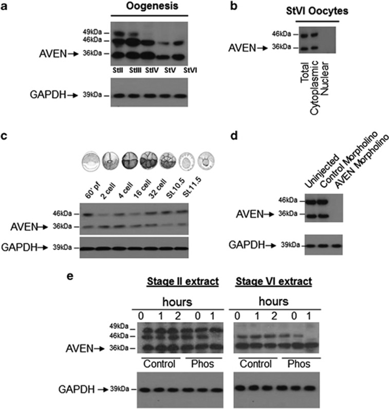
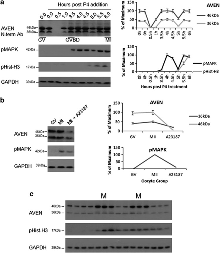
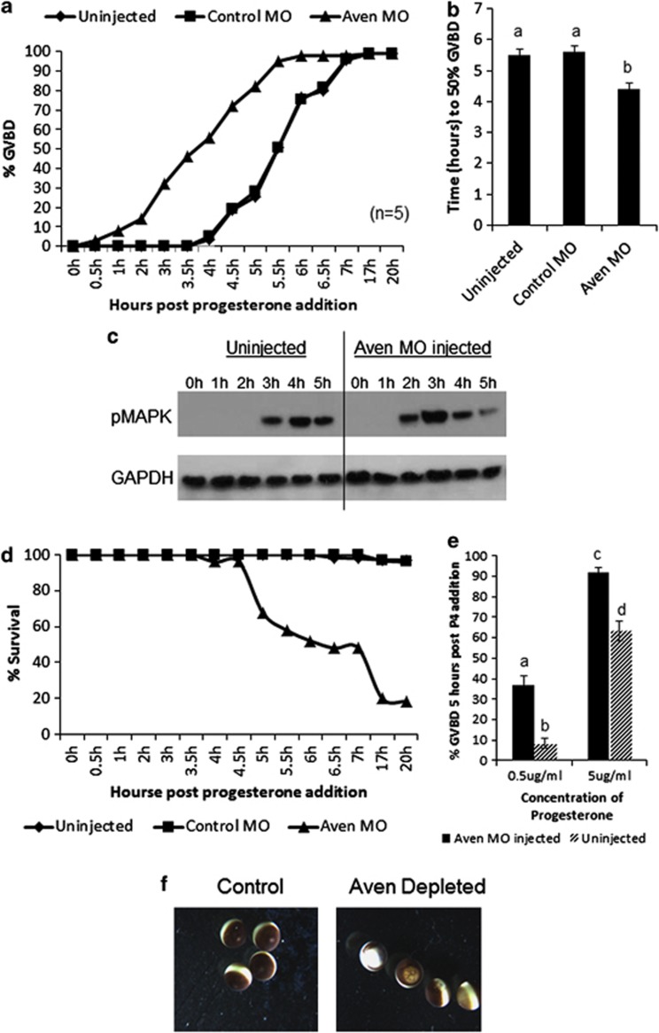
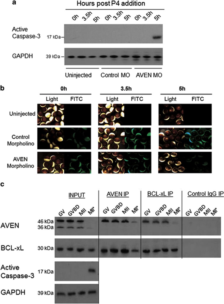
We have analyzed the expression and function of the cell death and cell cycle regulator Aven in Xenopus. Analysis of Xenopus Aven expression in oocytes and embryos revealed a band close to the predicted molecular weight of the protein (36 kDa) in addition to two bands of higher molecular weight (46 and 49 kDa), one of which was determined to be due to phosphorylation of the protein. The protein is primarily detected in the cytoplasm of oocytes and is tightly regulated during meiotic and mitotic cell cycles. Progesterone stimulation of oocytes resulted in a rapid loss of Aven expression with the protein levels recovering before germinal vesicle breakdown (GVBD). This loss of Aven is required for the G2-M1 cell cycle transition. Aven morpholino knockdown experiments revealed that early depletion of the protein increases progesterone sensitivity and facilitates GVBD, but prolonged depletion of Aven results in caspase-3 activation and oocyte death by apoptosis. Phosphorylated Aven (46 kDa) was found to bind Bcl-xL in oocytes, but this interaction was lost in apoptotic oocytes. Thus, Aven alters progesterone sensitivity in oocytes and is critical for oocyte survival.


Figures

Figure 1
Figure 1
AVEN expression and subcellular localization in oocytes. (a) Representative western blot analysis of Aven expression in stage II–stage VI oocyte extracts. GAPDH was used as a loading control (n=5). (b) Western blot analysis of stage VI whole oocyte extract, cytoplasmic extract and nuclear extract (n=3). (c) Aven expression in embryos from single cell to stage 11.5 (n=3). (d) Aven morpholino results in a loss of Aven expression at GVBD (n=6). (e) Phosphatase treatment of Stage II and Stage VI oocytes caused depletion of the 46-kDa Aven band, (n=3)
Figure 2
Figure 2
Dynamic changes in Aven expression during oocyte maturation. (a) Western blot analysis of Aven in oocyte extracts before and at the indicated time points (hours) after progesterone (P4) addition. In all, 64% of oocytes reached GVBD by 3.5  h as determined by the appearance of a white spot (92% at 4 h) and GVBD and MII were confirmed by pMAPK and pHistone-H3 expression. Aven expression is rapidly lost on progesterone addition and recovers by GVBD, error bars correspond to S.D. based on four experiments. (b) Loss of Aven expression following calcium ionophore A23187 induced metaphase release, error bars correspond to S.D., (n=4). (c) Fluctuations in Aven expression in cycling extracts with pHistone-H3 peaks marking the M phase of the cell cycle
Figure 3
Figure 3
AVEN protein depletion in oocytes increases progesterone sensitivity, facilitates rapid GVBD but ultimately results in oocyte death. (a) % GVBD in uninjected, control morpholino (MO)-injected and Aven morpholino (MO)-injected progesterone-treated oocytes (80–120 oocytes per experiment). Shown is a representative experiment from n=5. (b) Columns represent the average time taken for 50% GVBD to occur; error bars correspond to S.D., (n=5). (c) Western blot analysis of pMAPK expression in uninjected and AVEN MO-injected oocytes at the indicated times after progesterone addition. pMAPK is evident 1 h earlier in Aven MO-treated oocytes. (d) Oocyte survival was scored at the indicated times in uninjected, control MO and AVEN MO-injected oocytes. Aven MO-treated embryos gradually die as determined by gross morpholoigcal features. (e) Columns represent % GVBD in uninjected and Aven MO-injected oocytes 5 h after addition of the indicated progesterone concentrations. Loss of Aven increases the sensitivity of oocytes to progesterone. Error bars correspond to S.D., (n=5). (f) The apoptotic morphology of Aven-depleted oocytes as compared with control oocytes represented in d. Columns with different letters indicate significant differences (P<0.05)
Figure 4
Figure 4
Cell death activation in Aven-depleted oocytes. (a) Western blot analysis for active caspase-3 expression in uninjected, control MO-injected and Aven MO-injected, progesterone-treated oocytes (100–120 oocytes per experiment). Active caspase-3 was only detected in Aven-depleted oocytes 5 h after progesterone treatment. Shown is a representative experiment from n=7. (b) Representitive images (light and fluorescence) of Aven MO-injected oocytes undergoing precocious GVBD and subsequent apoptosis at the indicated times after progesterone addition. (c) A comparison of Aven–Bcl-xL interaction in control and apoptotic oocytes. Aven and Bcl-xL immunoprecipates were analyzed by western blot at GV, GVBD, MII and MII*: an MII equivalent time point in irradiated oocytes (n=3). Normal rabbit IgG antibody was used in control immunoprecipitations. Bcl-xL is bound to Aven in immoprecipiated MII-arrested egg extract, whereas in DNA damage-induced apoptotic extracts, little Aven is assoictaed with Bcl-xL

Similar articles

References

    1. Maller JL, Krebs EG. Progesterone-stimulated meiotic cell division in Xenopus oocytes. Induction by regulatory subunit and inhibition by catalytic subunit of adenosine 3':5'-monophosphate-dependent protein kinase. J Biol Chem. 1977;252:1712–1718. - PubMed
    1. Finidori-Lepicard J, Schorderet-Slatkine S, Hanoune J, Baulieu EE. Progesterone inhibits membrane-bound adenylate cyclase in Xenopus laevis oocytes. Nature. 1981;292:255–257. - PubMed
    1. Sadler SE, Maller JL. Progesterone inhibits adenylate cyclase in Xenopus oocytes. Action on the guanine nucleotide regulatory protein. J Biol Chem. 1981;256:6368–6373. - PubMed
    1. Daar I, Yew N, Vande Woude GF. Inhibition of mos-induced oocyte maturation by protein kinase A. J Cell Biol. 1993;120:1197–1202. - PMC - PubMed
    1. Matten W, Daar I, Vande Woude GF. Protein kinase A acts at multiple points to inhibit Xenopus oocyte maturation. Mol Cell Biol. 1994;14:4419–4426. - PMC - PubMed

Publication types

Substances