

Dichloroacetate induces protective autophagy in LoVo cells: involvement of cathepsin D/thioredoxin-like protein 1 and Akt-mTOR-mediated signaling
- PMID: 24201812
- PMCID: PMC3847316
- DOI: 10.1038/cddis.2013.438
Dichloroacetate induces protective autophagy in LoVo cells: involvement of cathepsin D/thioredoxin-like protein 1 and Akt-mTOR-mediated signaling
Abstract
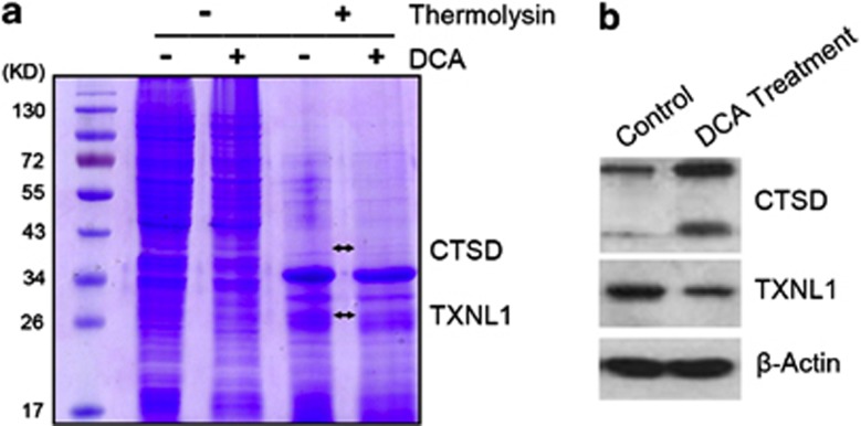
Dichloroacetate (DCA) is an inhibitor of pyruvate dehydrogenase kinase (PDK), and recently it has been shown as a promising nontoxic antineoplastic agent. In this study, we demonstrated that DCA could induce autophagy in LoVo cells, which were confirmed by the formation of autophagosomes, appearance of punctate patterns of LC3 immunoreactivity and activation of autophagy associated proteins. Moreover, autophagy inhibition by 3-methyladenine (3-MA) or Atg7 siRNA treatment can significantly enhance DCA-induced apoptosis. To determine the underlying mechanism of DCA-induced autophagy, target identification using drug affinity responsive target stability (DARTS) coupled with ESI-Q-TOF MS/MS analysis were utilized to profile differentially expressed proteins between control and DCA-treated LoVo cells. As a result, Cathepsin D (CTSD) and thioredoxin-like protein 1 (TXNL1) were identified with significant alterations compared with control. Further study indicated that DCA treatment significantly promoted abnormal reactive oxygen species (ROS) production. On the other hand, DCA-triggered autophagy could be attenuated by N-acetyl cysteine (NAC), a ROS inhibitor. Finally, we demonstrated that the Akt-mTOR signaling pathway, a major negative regulator of autophagy, was suppressed by DCA treatment. To our knowledge, it was the first study to show that DCA induced protective autophagy in LoVo cells, and the potential mechanisms were involved in ROS imbalance and Akt-mTOR signaling pathway suppression.
Figures


Similar articles
-
Proteomic analysis revealed association of aberrant ROS signaling with suberoylanilide hydroxamic acid-induced autophagy in Jurkat T-leukemia cells.Autophagy. 2010 Aug;6(6):711-24. doi: 10.4161/auto.6.6.12397. Epub 2010 Aug 17. Autophagy. 2010. PMID: 20543569
-
ROS-AKT-mTOR axis mediates autophagy of human umbilical vein endothelial cells induced by cooking oil fumes-derived fine particulate matters in vitro.Free Radic Biol Med. 2017 Dec;113:452-460. doi: 10.1016/j.freeradbiomed.2017.10.386. Epub 2017 Oct 28. Free Radic Biol Med. 2017. PMID: 29111231
-
Luteoloside induces G0/G1 arrest and pro-death autophagy through the ROS-mediated AKT/mTOR/p70S6K signalling pathway in human non-small cell lung cancer cell lines.Biochem Biophys Res Commun. 2017 Dec 9;494(1-2):263-269. doi: 10.1016/j.bbrc.2017.10.042. Epub 2017 Oct 9. Biochem Biophys Res Commun. 2017. PMID: 29024631
-
Inhibition of cathepsin S induces autophagy and apoptosis in human glioblastoma cell lines through ROS-mediated PI3K/AKT/mTOR/p70S6K and JNK signaling pathways.Toxicol Lett. 2014 Aug 4;228(3):248-59. doi: 10.1016/j.toxlet.2014.05.015. Epub 2014 May 27. Toxicol Lett. 2014. PMID: 24875536
-
Inhibition of Autophagy Promotes Salinomycin-Induced Apoptosis via Reactive Oxygen Species-Mediated PI3K/AKT/mTOR and ERK/p38 MAPK-Dependent Signaling in Human Prostate Cancer Cells.Int J Mol Sci. 2017 May 18;18(5):1088. doi: 10.3390/ijms18051088. Int J Mol Sci. 2017. PMID: 28524116 Free PMC article.
Cited by
-
Mitochondrial dysfunction generates a growth-restraining signal linked to pyruvate in Drosophila larvae.Fly (Austin). 2019 Mar-Dec;13(1-4):12-28. doi: 10.1080/19336934.2019.1662266. Epub 2019 Sep 17. Fly (Austin). 2019. PMID: 31526131 Free PMC article.
-
Dichloroacetate enhances the antitumor efficacy of chemotherapeutic agents via inhibiting autophagy in non-small-cell lung cancer.Cancer Manag Res. 2018 May 16;10:1231-1241. doi: 10.2147/CMAR.S156530. eCollection 2018. Cancer Manag Res. 2018. PMID: 29844702 Free PMC article.
-
PDK4 promotes vascular calcification by interfering with autophagic activity and metabolic reprogramming.Cell Death Dis. 2020 Nov 17;11(11):991. doi: 10.1038/s41419-020-03162-w. Cell Death Dis. 2020. PMID: 33203874 Free PMC article.
-
Metabolic Response of Pancreatic Carcinoma Cells under Treatment with Dichloroacetate.Metabolites. 2021 May 30;11(6):350. doi: 10.3390/metabo11060350. Metabolites. 2021. PMID: 34070873 Free PMC article.
-
Hydroxytyrosol Acetate Inhibits Vascular Endothelial Cell Pyroptosis via the HDAC11 Signaling Pathway in Atherosclerosis.Front Pharmacol. 2021 Apr 23;12:656272. doi: 10.3389/fphar.2021.656272. eCollection 2021. Front Pharmacol. 2021. PMID: 33967800 Free PMC article.
References
-
- Kundu M, Thompson CB. Autophagy: basic principles and relevance to disease. Ann Rev Pathol. 2008;3:427–455. - PubMed
-
- Choi AM, Ryter SW, Levine B. Autophagy in human health and disease. New Engl J Med. 2013;368:1845–1846. - PubMed
-
- Wang K, Liu R, Li J, Mao J, Lei Y, Wu J, et al. Quercetin induces protective autophagy in gastric cancer cells: involvement of Akt-mTOR- and hypoxia-induced factor 1alpha-mediated signaling. Autophagy. 2011;7:966–978. - PubMed
Publication types
MeSH terms
Substances
LinkOut - more resources
Full Text Sources
Other Literature Sources
Molecular Biology Databases
Research Materials
Miscellaneous
