

Highly efficient tumor transduction and antitumor efficacy in experimental human malignant mesothelioma using replicating gibbon ape leukemia virus
- PMID: 24201868
- PMCID: PMC8185610
- DOI: 10.1038/cgt.2013.67
Highly efficient tumor transduction and antitumor efficacy in experimental human malignant mesothelioma using replicating gibbon ape leukemia virus
Abstract
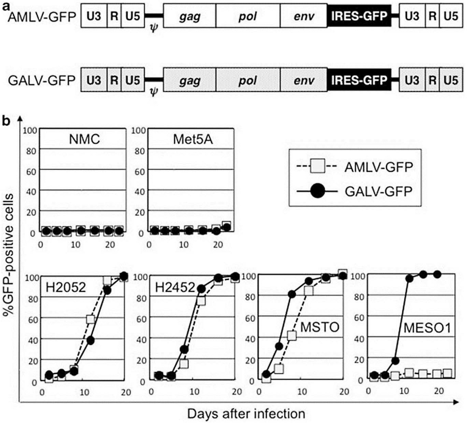
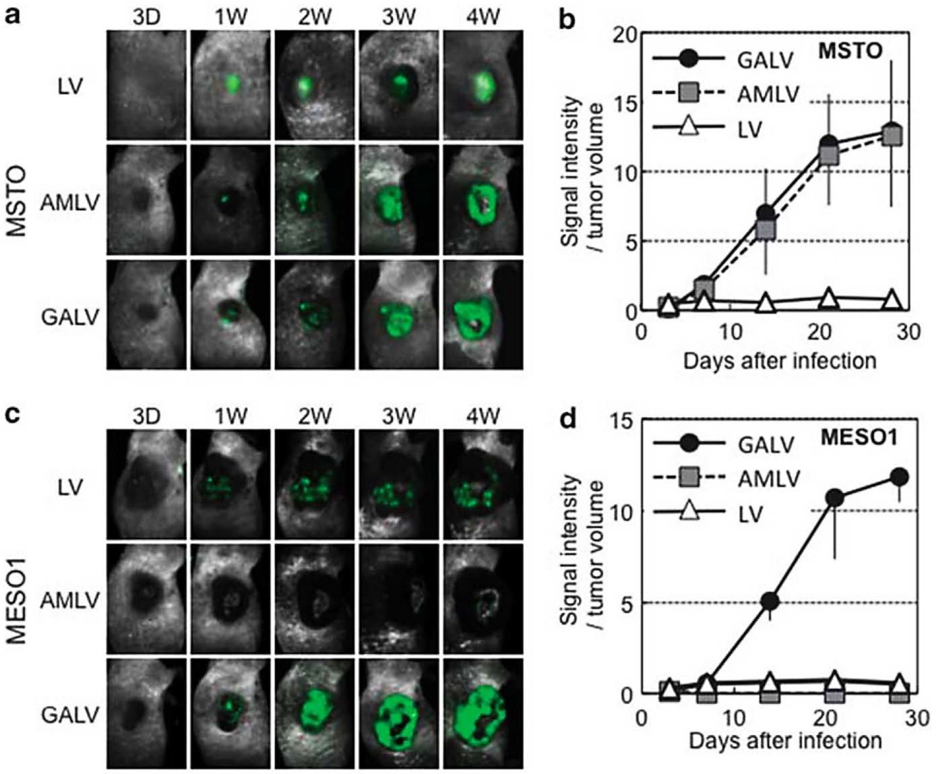
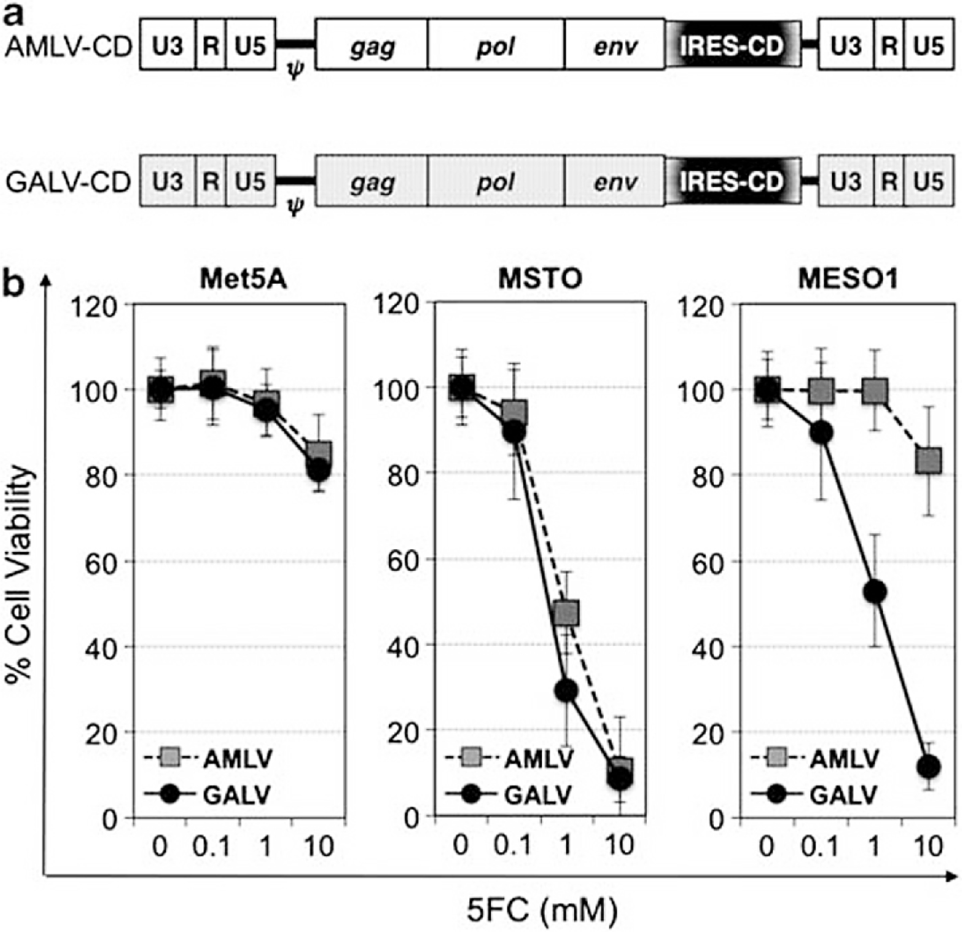
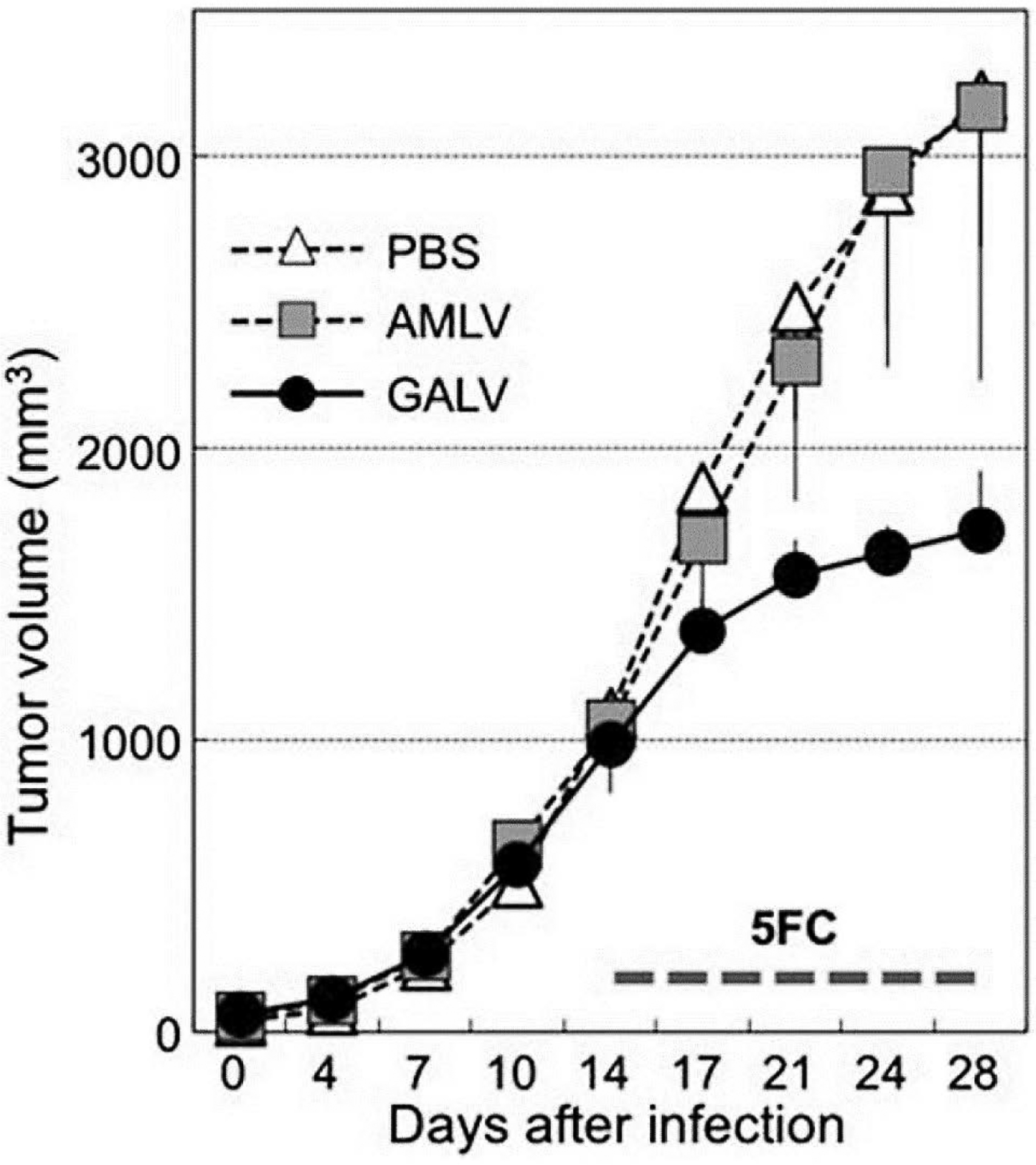
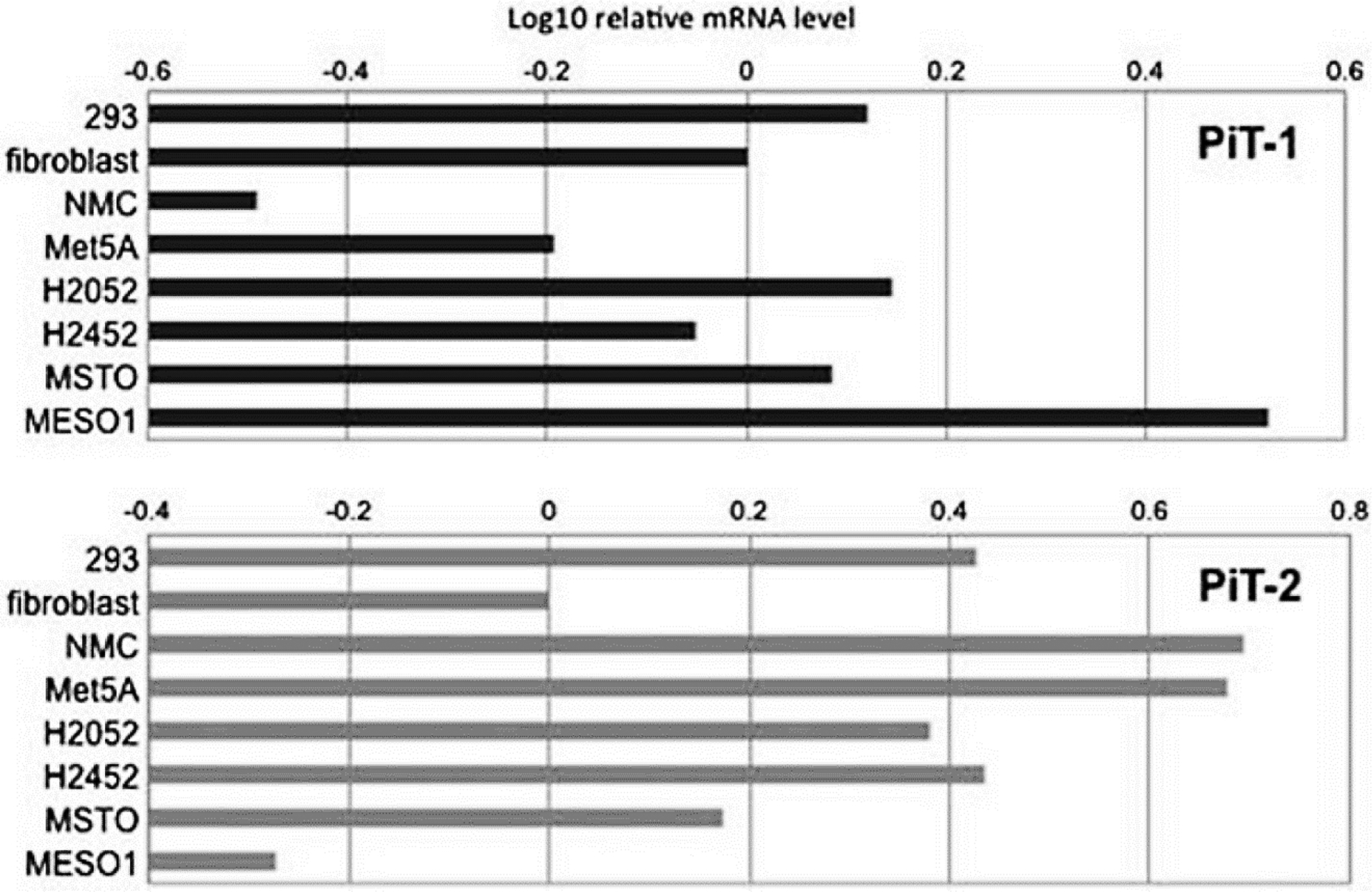
Retroviral replicating vectors (RRVs) have been shown to achieve efficient tumor transduction and enhanced therapeutic benefit in a wide variety of cancer models. Here we evaluated two different RRVs derived from amphotropic murine leukemia virus (AMLV) and gibbon ape leukemia virus (GALV), in human malignant mesothelioma cells. In vitro, both RRVs expressing the green fluorescent protein gene efficiently replicated in most mesothelioma cell lines tested, but not in normal mesothelial cells. Notably, in ACC-MESO-1 mesothelioma cells that were not permissive for AMLV-RRV, the GALV-RRV could spread efficiently in culture and in mice with subcutaneous xenografts by in vivo fluorescence imaging. Next, GALV-RRV expressing the cytosine deaminase prodrug activator gene showed efficient killing of ACC-MESO-1 cells in a prodrug 5-fluorocytosine dose-dependent manner, compared with AMLV-RRV. GALV-RRV-mediated prodrug activator gene therapy achieved significant inhibition of subcutaneous ACC-MESO-1 tumor growth in nude mice. Quantitative reverse transcription PCR demonstrated that ACC-MESO-1 cells express higher PiT-1 (GALV receptor) and lower PiT-2 (AMLV receptor) compared with normal mesothelial cells and other mesothelioma cells, presumably accounting for the distinctive finding that GALV-RRV replicates much more robustly than AMLV-RRV in these cells. These data indicate the potential utility of GALV-RRV-mediated prodrug activator gene therapy in the treatment of mesothelioma.
Conflict of interest statement
CONFLICT OF INTEREST
CRL and NK are paid consultants to Tocagen Inc.
Figures
Similar articles
-
Efficient tumor transduction and antitumor efficacy in experimental human osteosarcoma using retroviral replicating vectors.Cancer Gene Ther. 2019 Feb;26(1-2):41-47. doi: 10.1038/s41417-018-0037-y. Epub 2018 Jul 25. Cancer Gene Ther. 2019. PMID: 30042500 Free PMC article.
-
Retroviral Replicating Vectors Mediated Prodrug Activator Gene Therapy in a Gastric Cancer Model.Int J Mol Sci. 2023 Oct 2;24(19):14823. doi: 10.3390/ijms241914823. Int J Mol Sci. 2023. PMID: 37834271 Free PMC article.
-
Therapeutic Efficacy of Prodrug Activator Gene Therapy Using Retroviral Replicating Vectors for Human Ovarian Cancer.Anticancer Res. 2023 Dec;43(12):5311-5317. doi: 10.21873/anticanres.16734. Anticancer Res. 2023. PMID: 38030176
-
A new look at the origins of gibbon ape leukemia virus.Virus Genes. 2017 Apr;53(2):165-172. doi: 10.1007/s11262-017-1436-0. Epub 2017 Feb 20. Virus Genes. 2017. PMID: 28220345 Review.
-
Clinical development of retroviral replicating vector Toca 511 for gene therapy of cancer.Expert Opin Biol Ther. 2021 Sep;21(9):1199-1214. doi: 10.1080/14712598.2021.1902982. Epub 2021 May 6. Expert Opin Biol Ther. 2021. PMID: 33724117 Free PMC article. Review.
Cited by
-
Efficient tumor transduction and antitumor efficacy in experimental human osteosarcoma using retroviral replicating vectors.Cancer Gene Ther. 2019 Feb;26(1-2):41-47. doi: 10.1038/s41417-018-0037-y. Epub 2018 Jul 25. Cancer Gene Ther. 2019. PMID: 30042500 Free PMC article.
-
Novel Semi-Replicative Retroviral Vector Mediated Double Suicide Gene Transfer Enhances Antitumor Effects in Patient-Derived Glioblastoma Models.Cancers (Basel). 2019 Jul 31;11(8):1090. doi: 10.3390/cancers11081090. Cancers (Basel). 2019. PMID: 31370279 Free PMC article.
-
Retroviral Replicating Vectors Mediated Prodrug Activator Gene Therapy in a Gastric Cancer Model.Int J Mol Sci. 2023 Oct 2;24(19):14823. doi: 10.3390/ijms241914823. Int J Mol Sci. 2023. PMID: 37834271 Free PMC article.
-
Antinociceptive Effect of Intrathecal Injection of Genetically Engineered Human Bone Marrow Stem Cells Expressing the Human Proenkephalin Gene in a Rat Model of Bone Cancer Pain.Pain Res Manag. 2017;2017:7346103. doi: 10.1155/2017/7346103. Epub 2017 Feb 14. Pain Res Manag. 2017. PMID: 28286408 Free PMC article.
-
Oncolytic virotherapy for human malignant mesothelioma: recent advances.Oncolytic Virother. 2015 Sep 10;4:133-40. doi: 10.2147/OV.S66091. eCollection 2015. Oncolytic Virother. 2015. PMID: 27512676 Free PMC article. Review.
References
-
- Ismail-Khan R, Robinson LA, Williams CC Jr, Garrett CR, Bepler G, Simon GR. Malignant pleural mesothelioma: a comprehensive review. Cancer Control 2006; 13: 255–263. - PubMed
-
- van der Most RG, Robinson BW, Nelson DJ. Gene therapy for malignant mesothelioma: beyond the infant years. Cancer Gene Ther 2006; 13: 897–904. - PubMed
-
- Albelda SM, Wiewrodt R, Sterman DH. Gene therapy for lung neoplasms. Clin Chest Med 2002; 23: 265–277. - PubMed
-
- Sterman DH, Recio A, Vachani A, Sun J, Cheung L, DeLong P et al. Long-term follow-up of patients with malignant pleural mesothelioma receiving high-dose adenovirus herpes simplex thymidine kinase/ganciclovir suicide gene therapy. Clin Cancer Res 2005; 11: 7444–7453. - PubMed
Publication types
MeSH terms
Substances
Grants and funding
LinkOut - more resources
Full Text Sources
Other Literature Sources
Medical
Research Materials
Miscellaneous
