. 2014 Apr;17(2):419-27.
doi: 10.1007/s10456-013-9400-7. Epub 2013 Nov 8.

Vascular endothelial growth factor-C enhances radiosensitivity of lymphatic endothelial cells

Affiliations

Vascular endothelial growth factor-C enhances radiosensitivity of lymphatic endothelial cells

Cristina T Kesler et al. Angiogenesis. 2014 Apr.

Abstract

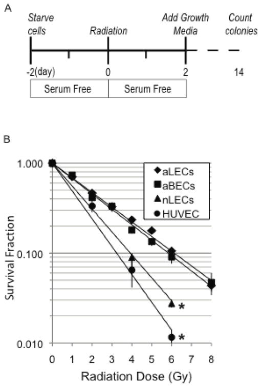
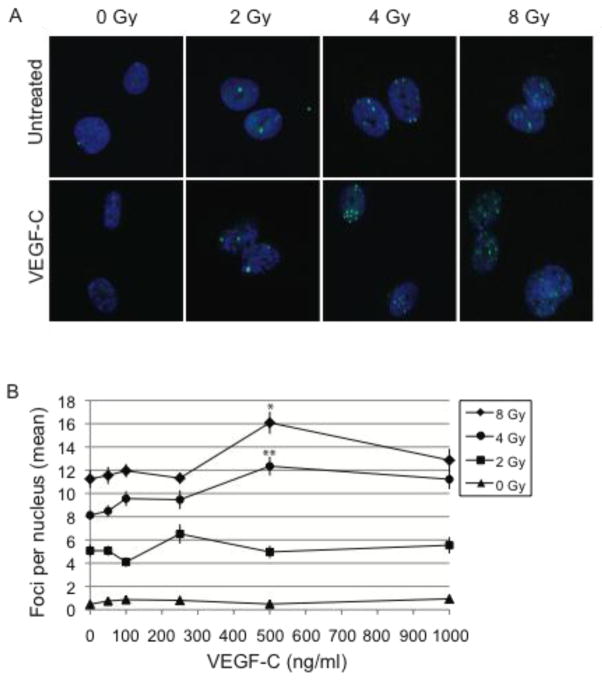
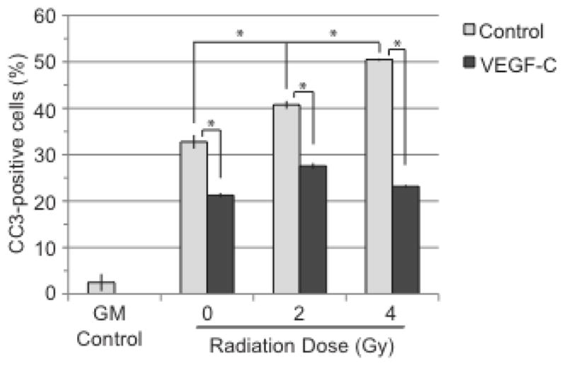
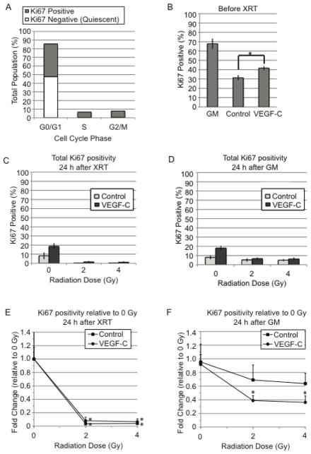
Radiation therapy after lymph node dissection increases the risk of developing painful and incurable lymphedema in breast cancer patients. Lymphedema occurs when lymphatic vessels become unable to maintain proper fluid balance. The sensitivity of lymphatic endothelial cells (LECs) to ionizing radiation has not been reported to date. Here, the radiosensitivity of LECs in vitro has been determined using clonogenic survival assays. The ability of various growth factors to alter LEC radiosensitivity was also examined. Vascular endothelial growth factor (VEGF)-C enhanced radiosensitivity when LECs were treated prior to radiation. VEGF-C-treated LECs exhibited higher levels of entry into the cell cycle at the time of radiation, with a greater number of cells in the S and G2/M phases. These LECs showed higher levels of γH2A.X-an indicator of DNA damage-after radiation. VEGF-C did not increase cell death as a result of radiation. Instead, it increased the relative number of quiescent LECs. These data suggest that abundant VEGF-C or lymphangiogenesis may predispose patients to radiation-induced lymphedema by impairing lymphatic vessel repair through induction of LEC quiescence.


Conflict of interest statement

Conflict of Interest Notification: There are no conflicts of interest related to the work presented in this manuscript.

Figures

Figure 1
Figure 1. Radiosensitivity of adult LECs and adult BECs is similar
Adult and neonatal lymphatic endothelial cells (aLEC and nLEC) were compared to adult blood endothelial cells (aBEC) and human umbilical vein endothelial cells (HUVEC) using clonogenic survival assays. Curves for aLECs and aBECs show no difference in dose-responsive survival fraction between the two. nLECs and are more radiosensitive than their adult counterparts as measured by their reduced survival fractions. * p<0.05 comparing nLECs vs. aLECs and aBECs vs. HUVECs. Statistical comparisons were made using a general linear model as described in the methods.
Figure 2
Figure 2. VEGF-C reduces the survival fraction of LECs
Growth factors were tested using four experimental arms in clonogenic survival assays (a). Dose-responsive radiosensitivity of aLECs in response to VEGF (b), bFGF (c) and VEGF-C (d) were measured. *, p<0.05 comparing SF/SF vs VEGF-C/SF (d). Statistical comparisons were made using a general linear model as described in the methods.
Figure 3
Figure 3. Pre-treatment with VEGF-C increases DNA damage
Radiation-induced DNA damage in LECs was detected by immunofluorescence for H2A.x 20 h after radiation. Sub-nuclear foci formed by H2A.X were imaged using confocal microscopy, scale bar 10 μm. Images shown represent LECs treated with 500 ng/ml VEGF-C (a). Dose-responsive effects were examined by pre-treating LECs with 0, 50, 100, 250, 500 or 1000 ng/ml VEGF-C. The total number of foci per nucleus was counted and averaged. Data were analyzed by ANOVA with Tukey’s Honestly-Significant-Difference Test post-hoc for each radiation dose (b). blue, DAPI; green, H2A.X; *, p<0.05 when compared to 0 ng/ml, 50 ng/ml 100 ng/ml, 250 ng/ml and 1000 ng/ml of VEGF-C; ** p<0.05 when compared to 0 ng/ml, 50 ng/ml, 100 ng/ml and 250 ng/ml of VEGF-C.
Figure 4
Figure 4. VEGF-C does not enhance radiation-induced cell death
LECs were collected 6 h after radiation, stained for cleaved caspase-3 (CC3) and collected by flow cytometry. GM control is represented by LECs that have been grown in regular growth media. All other groups were starved prior to radiation. 0 Gy control groups did not receive radiation. Statistical analysis was performed using the standard t-test. *, p<0.05
Figure 5
Figure 5. Pre-treatment with VEGF-C increases LEC quiescence
Flow cytometry for Ki67-positivity in high passage LECs (p8) showed Ki67 as a distinguishing marker between cycling and quiescent cells in the G0/G1 population (a). The percentage of Ki67 positive cells was determined visually using immunofluorescence microscopy. The control and VEGF-C groups were compared at the time of radiation (b), 24 h after radiation while under starvation (c), and 24 h after GM addition (d). Fold changes relative to 0 Gy control were determined for both groups at 24 h after radiation while under starvation (e) and 24 h after addition of GM (f). Statistical significance for 2 and 4 Gy groups was determined relative to 0 Gy (e,f). Statistical analysis was performed using the standard t-test. GM, growth media; XRT, radiation; *, p<0.05

Similar articles

Cited by

References

    1. Warren AG, et al. Lymphedema: a comprehensive review. Ann Plast Surg. 2007;59(4):464–72. - PubMed
    1. Meek AG. Breast radiotherapy and lymphedema. Cancer. 1998;83(12 Suppl American):2788–97. - PubMed
    1. Hinrichs CS, et al. Lymphedema secondary to postmastectomy radiation: incidence and risk factors. Ann Surg Oncol. 2004;11(6):573–80. - PubMed
    1. Tammela T, et al. Therapeutic differentiation and maturation of lymphatic vessels after lymph node dissection and transplantation. Nat Med. 2007;13(12):1458–66. - PubMed
    1. Gershenwald JE, I, Fidler J. Targeting lymphatic metastasis. Science. 2002;296(5574):1811–2. - PubMed

Publication types

MeSH terms

Substances

LinkOut - more resources