Review
. 2014;8(3):339-48.
doi: 10.1007/s12105-013-0505-5. Epub 2013 Nov 8.

Intraoral basal cell carcinoma, a rare neoplasm: report of three new cases with literature review

Affiliations
Review

Intraoral basal cell carcinoma, a rare neoplasm: report of three new cases with literature review

T R Woods et al. Head Neck Pathol. 2014.

Abstract

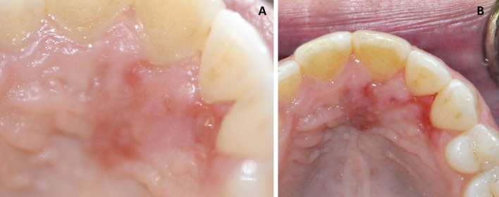
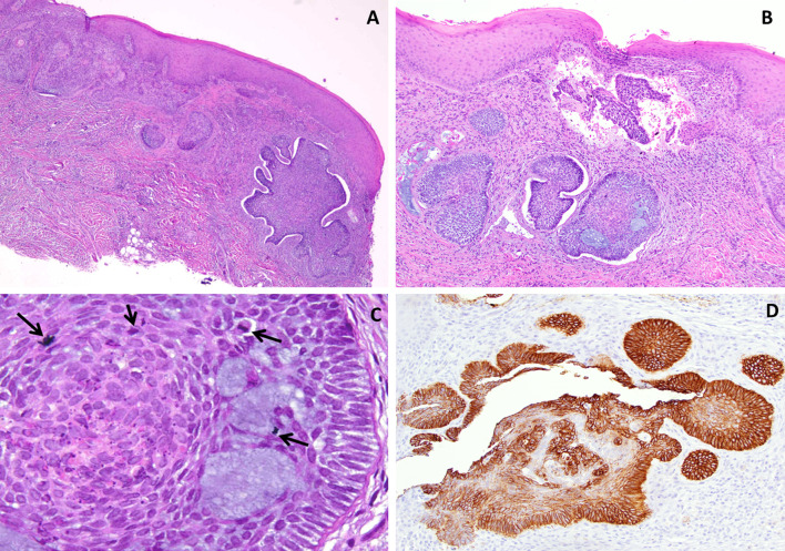
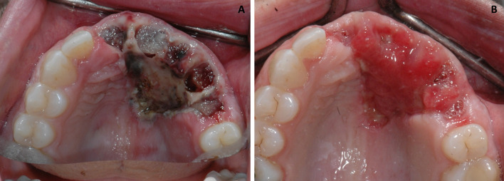
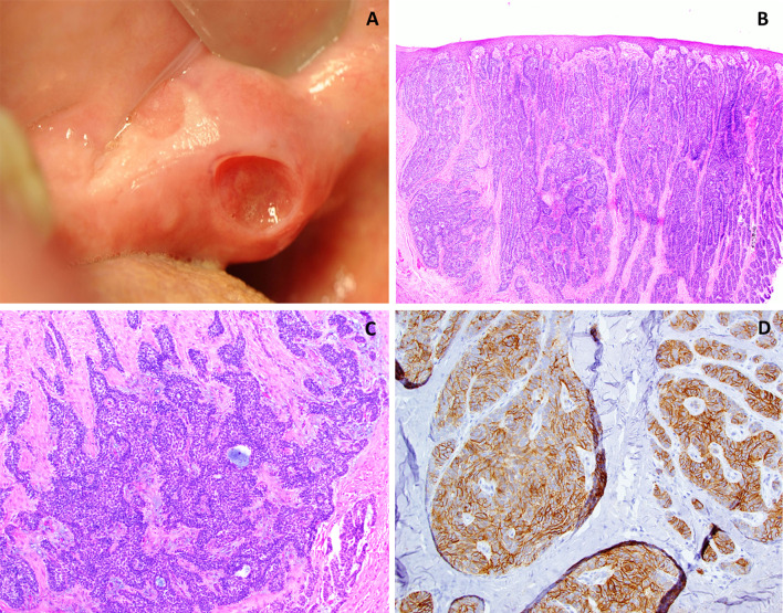
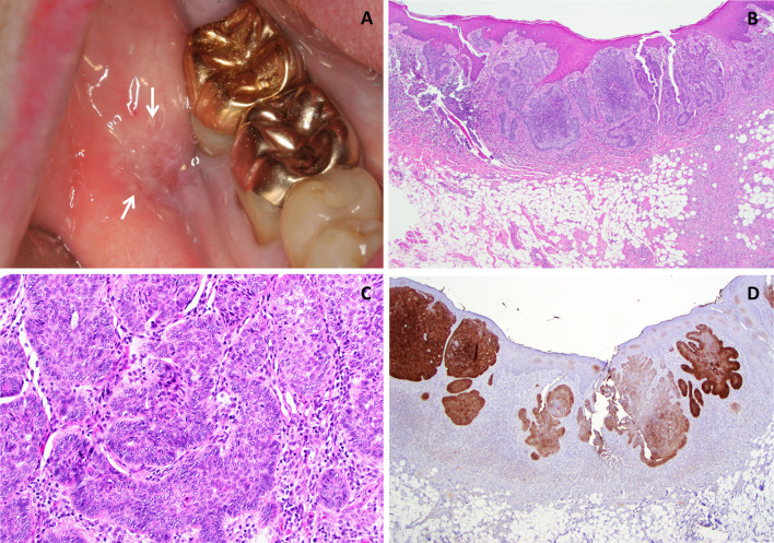
Intraoral basal cell carcinoma (IOBCC) is an extremely rare entity that bears close microscopic resemblance to and is often confused with the peripheral ameloblastoma (PA). Basal cell carcinomas are thought to arise from pluripotential basal cells present within surface epithelium and adnexal structures, so theoretically they can arise within the oral cavity. Many of the early cases reported as IOBCC actually represent PA. Most of the well documented cases arise from the gingiva. The histologic features of basal cell carcinoma that help separate it from a PA include: tumor arising from surface epithelium, scattered mitotic figures and apoptotic cells, presence of mucoid ground substance and tumor infiltrating widely throughout the connective tissue and often exhibiting a prominent retraction artifact. Clinically IOBCC resemble carcinomas, compared to the benign and innocuous appearance of the PA and typically presents as surface ulcerations varying from rodent ulcer to an ulcerated erythroplakia appearance. This contrasts with the classic "bump on the gum" appearance of PAs with usually intact surface and appearing as small discrete, sessile, exophytic lesions. Importantly, the proliferative basaloid epithelium demonstrates positive immunoreactivity for the anti-epithelial antibody, Ber-EP4, a cell surface glycoprotein. The IOBCC has the potential for local recurrence and aggressive behavior and should be treated with wide surgical excision and close clinical follow up. We present 3 rare cases of IOBCC and discuss the salient histologic, immunohistochemical and clinical features.


Figures

Fig. 1
Fig. 1
Case 1. a Clinical presentation at initial visit demonstrating areas of swelling and erythroplakia of anterior palatal mucosa. b Three weeks post-biopsy, recurrent and progressive swelling with erythema, deepening in color was noted
Fig. 2
Fig. 2
Case 1. a Photomicrograph from the excisional biopsy sample demonstrating a basaloid epithelial proliferation arising from the basal surface of the epithelium and invading into the underlying connective tissue (H & E ×4 magnification). b The peripheral cells of the invasive islands displaying a palisaded arrangement with prominent retraction from the surrounding stroma. Small pools of mucoid material are noted as well. (H & E ×10 magnification). c Apoptotic cells and abnormal and increased mitoses were observed scattered amidst pools of basophilic mucoid material (arrows) (H & E x 40 magnification). d Immunohistochemical stain for Ber EP4 exhibiting strong positivity in tumor cells (H & E ×20 magnification)
Fig. 3
Fig. 3
Case 1. a Post surgical view demonstrating wide local excision of lesion with removal of multiple teeth. b Healing surgical site 3 weeks later
Fig. 4
Fig. 4
Case 2. a Clinical photograph demonstrating a raised mass (non-healing biopsy site clearly visible) on the right retromolar pad. b Photomicrograph depicting an invading, infiltrative neoplasm composed of strands and clusters of basaloid appearing epithelial cells arising from the keratinized surface epithelium (H & E ×10 magnification). c Higher magnification demonstrates basophilic tumor islands distributed throughout the connective tissue exhibiting prominent peripheral palisading, and mucoid material (H & E ×20 magnification). d Immunohistochemical stain for Ber EP4 exhibiting positivity within most of the tumor islands (H & E ×20 magnification)
Fig. 5
Fig. 5
Case 3. a A painless non-healing somewhat depressed ulceration present for 6–12 month duration in the buccal mucosa adjacent to teeth #30–#31. b Photomicrograph depicting an invasive basaloid epithelial cell proliferation arising from keratinized surface epithelium. mucosa (H & E ×4 magnification). c Palisading of the peripheral cells as well as occasional apoptotic cells are noted within the invasive islands which are surrounded by chronic inflammatory cells. Retraction from the adjacent stroma is noted (H & E ×20 magnification). d Immunohistochemical stain for Ber EP4 exhibiting strong positivity in tumor cells (H & E ×20 magnification)

References

    1. Wu S, Han J, Li WQ, Li T, Qureshi AA. Basal-cell carcinoma incidence and associated risk factors in US women and men. Am J Epidemiol. 2013;178(6):890. - PMC - PubMed
    1. Luzar B, Bastian BC, Calonje E: Mckee’s Pathology of the Skin. Mckee PH editors. Amsterdam: Elsevier; 2012.
    1. Bath-Hextall F, Leonardi-Bee J, Smith C, Meal A, Hubbard R. Trends in incidence of skin basal cell carcinoma. Additional evidence from a UK primary care database study. Int J Cancer. 2007;121:2105–2108. doi: 10.1002/ijc.22952. - DOI - PubMed
    1. Saint CF. The embryology of the stomodeum and its bearing on the pathology of tumours of the tongue and the salivary glands. S Afr J Clin Sci. 1951;2:1–17. - PubMed
    1. Del Rosario RN, Barr RJ, Jensen JL, Cantos KA. Basal cell carcinoma of the buccal mucosa. Am J Dermatopathol. 2001;23:203–205. doi: 10.1097/00000372-200106000-00007. - DOI - PubMed

Substances

LinkOut - more resources