. 2014 Apr 1;23(7):1687-99.
doi: 10.1093/hmg/ddt558. Epub 2013 Nov 7.

Novel mutations in Lrp6 orthologs in mouse and human neural tube defects affect a highly dosage-sensitive Wnt non-canonical planar cell polarity pathway

Affiliations

Novel mutations in Lrp6 orthologs in mouse and human neural tube defects affect a highly dosage-sensitive Wnt non-canonical planar cell polarity pathway

Redouane Allache et al. Hum Mol Genet. .

Erratum in

  • Hum Mol Genet. 2014 Aug 1;23(15):4185

Abstract

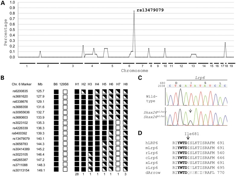
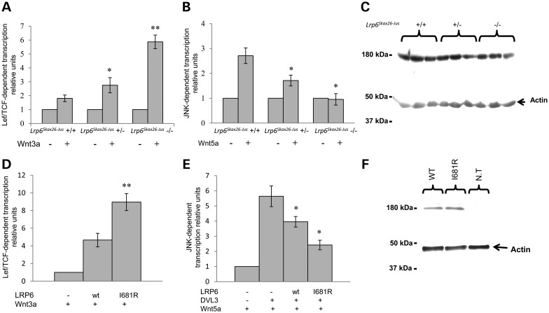
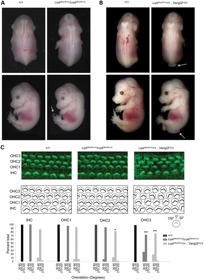
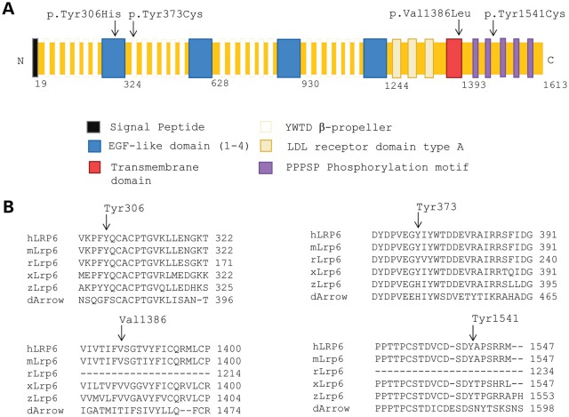
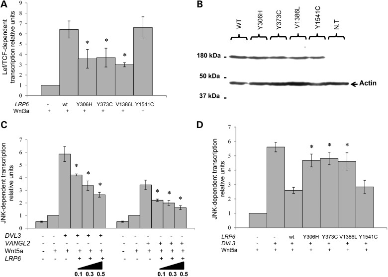
Wnt signaling has been classified as canonical Wnt/β-catenin-dependent or non-canonical planar cell polarity (PCP) pathway. Misregulation of either pathway is linked mainly to cancer or neural tube defects (NTDs), respectively. Both pathways seem to antagonize each other, and recent studies have implicated a number of molecular switches that activate one pathway while simultaneously inhibiting the other thereby partially mediating this antagonism. The lipoprotein receptor-related protein Lrp6 is crucial for the activation of the Wnt/β-catenin pathway, but its function in Wnt/PCP signaling remains largely unknown. In this study, we investigate the role of Lrp6 as a molecular switch between both Wnt pathways in a novel ENU mouse mutant of Lrp6 (Skax26(m1Jus)) and in human NTDs. We demonstrate that Skax26(m1Jus) represents a hypermorphic allele of Lrp6 with increased Wnt canonical and abolished PCP-induced JNK activities. We also show that Lrp6(Skax26-Jus) genetically interacts with a PCP mutant (Vangl2(Lp)) where double heterozygotes showed an increased frequency of NTDs and defects in cochlear hair cells' polarity. Importantly, our study also demonstrates the association of rare and novel missense mutations in LRP6 that is an inhibitor rather than an activator of the PCP pathway with human NTDs. We show that three LRP6 mutations in NTDs led to a reduced Wnt canonical activity and enhanced PCP signaling. Our data confirm an inhibitory role of Lrp6 in PCP signaling in neurulation and indicate the importance of a tightly regulated and highly dosage-sensitive antagonism between both Wnt pathways in this process.


Figures

Figure 1.
Figure 1.
Identification of the gene mutated in Skax26m1Jus. (A) Homozygosity mapping shows linkage of Skax26m1Jus to telomeric position of chromosome 6. The graph shows the percentage of homozygous mice for C57Bl/6J allele for each SNP (n = 149) for each chromosome (n = 19). (B) Haplotype analysis with 16 SNPs places the Skax26m1Jus gene centromeric to rs3023102 at 135.3 Mb on chromosome 6. Black box, C57BL/6J (B6) allele. Open box, 129S6/SvEvTac allele (129S6). Black and open box, heterozygous for B6 and 129S6 alleles. H, haplotype (C) Chromatograms of partial sequences of exon 9 of Lrp6 show a c.2042T>G nucleotide change in Skax26m1Jus/Skax26m1Jus, which leads to p.Ile681Arg in the encoded protein. (D) Partial protein alignments of mouse Lrp6 with five orthologs. The p.Ile681Arg variant maps to a highly a conserved region following an YWTD repeat (indicated in bold). Accession numbers: Homo sapiens LRP6 (hLRP6), NP_002327.2; Mus musculus Lrp6 (mLrp6), NP_032540.2; Rattus norvegicus Lrp6 (rLrp6), NP_001101362.1; Gallus gallus Lrp6 (cLrp6), XP_417286.3; Danio rerio Lrp6 (zLrp6), NP_001128156.1; Xenopus tropicalis Lrp6 (xLrp6), NP_001079233.1; Drosophila melanogaster Lrp6, (dArrow), NP_524737.2.
Figure 2.
Figure 2.
Functional validation of the mutant Lrp6p.Ile681Arg in wild-type, heterozygous and homozygous Lrp6Skax26-Jus littermates and in HEK293T cells. (A) TCF/LEF-1 activity was measured in E13.5mouse embryonic fibroblasts (MEFs) in the presence of Wnt3a. Heterozygous (n = 7) and homozygous (n = 5) MEFs showed a significantly increased reporter activity as compared with wild-type fibroblasts (n = 5) (t-test, P < 0.05 and P < 0.00001, respectively). (B) JNK-AP-1 activity was measured in E13.5 MEFs in the presence of Wnt5a. JNK activity was reduced by half in heterozygous MEFs (n = 7) (P < 0.05) and completely absent in homozygous cells (n = 4) (P < 0.05) as compared with wild-type littermates (n = 4). (C) Representative western blots of three E13.5 embryos from each genotype (+/+, Lrp6Skax26-Jus/+, Lrp6Skax26-Jus/Lrp6Skax26-Jus) show comparable levels of Lrp6 protein expression. (D) TCF/LEF-1 activity was significantly increased in cells transfected with Lrp6p.Ile681Arg cDNA as compared with wild-type Lrp6 (P < 0.005). (E) Co-transfection of the Lrp6p.Ile681Arg cDNA with DVL3 resulted in a significant increase in its ability to inhibit Wnt5a-induced JNK activation (P < 0.05). (F) Representative western blot of HEK293T cells transfected with Lrp6p.Ile681Arg cDNA shows a protein expression level that was comparable with the wild-type.
Figure 3.
Figure 3.
Genetic interaction studies between Lrp6Skax26-Jus and Vangl2Lp. (A) Lrp6Skax26-Jus/Lrp6Skax26-JusE18.5 embryos displayed a kinky tail (arrow) but no neural tube defects (NTDs) as compared with their wild-type littermates. (B) Lrp6Skax26-Jus/+; Vangl2Lp/+ double heterozygotes showed spina bifida (arrow) as compared with their wild-type littermates. (C) Comparison of hair bundle orientation at the apical region of the organ of Corti in wild-type, Lrp6Skax26-Jus/Lrp6Skax26-Jus and Lrp6Skax26-Jus/+; Vangl2Lp/+ at E18.5 Top, Stereocilia labeled with phalloidin (green). Middle, diagrams showing the scoring of hair bundle orientation for the images above. IHC, inner hair cells; OHC1, inner row of outer hair cells; OHC2, central row of outer hair cells; OHC3, outer row of outer hair cells. The genotype is indicated above each column of panels. Bottom, quantification of the IHC, OHC1, OHC2 and OHC3 bundle orientations for each of the three genotypes indicated above based on phalloidin staining. The convention for angular measurements is shown in the top right corner of the panel. Lrp6Skax26-Jus /Lrp6Skax26-Jus showed significant defects in OHC3 orientation and Lrp6Skax26-Jus/+; Vangl2Lp/+ showed significant defects in OHC2 and OHC3 orientation as compared with their wild-type littermates. For statistical analysis, the χ2 test was used to compare the distributions of the various subgroups of hair cells (*P < 0.01; **P < 0.001).
Figure 4.
Figure 4.
Rare novel mutations in LRP6 in human neural tube defects (NTDs). (A) A schematic diagram of LRP6 showing the approximate locations of the 4 NTD-associated mutations p.Tyr306His, p.Tyr373Cys, p.Val1386Leu and p.Tyr1541Cys. (B) A partial alignment of human LRP6 with five other orthologous sequences. The LRP6 variants found in NTD patients affect conserved residues (indicated by arrows). Accession numbers: Homo sapiens LRP6 (hLRP6), NP_002327.2; Mus musculus Lrp6 (mLrp6), NP_032540.2; Rattus norvegius Lrp6 (rLrp6), NP_001101362.1; Gallus gallus Lrp6 (cLrp6), XP_417286.3; Danio rerio Lrp6 (zLrp6), NP_001128156.1; Xenopus tropicalis Lrp6 (xLrp6), NP_001079233.1; Drosophila melanogaster Lrp6, (dArrow), NP_524737.2.
Figure 5.
Figure 5.
Functional validation of NTD-associated mutations in LRP6 using TCF/LEF-1-responsive Wnt/β-catenin and JNK-AP-1 reporter assays. (A) TCF/LEF-1 activity was significantly decreased in cells transfected with each of the LRP6p.Tyr306His, LRP6p.Tyr373Cys and LRP6p.Val1386Leu cDNAs, as compared with wild-type LRP6 (t-test, *P < 0.05). This activity was not affected in HEK293T cells transfected with the LRP6p.Tyr1541Cys cDNA. (B) A representative western blot analysis of HEK293T cells transfected with each of the four variants shows protein expression levels for all LRP6 variants that were comparable with the wild-type. (C) JNK-AP1-1 activity was significantly inhibited by co-transfection of LRP6 with either DVL3 or VANGL2 in the presence of Wnt5a in a dose-dependent manner (t-test, *P < 0.05). (D) Co-transfection of each of LRP6p.Tyr306His, LRP6p.Tyr373Cys and LRP6p.Val1386Leu cDNAs with DVL3in the presence of Wnt5a resulted in less inhibition of JNK-AP1-1 activity as compared with wild-type LRP6 (t-test, *P < 0.05). LRP6p.Tyr1541Cys behaved like the wild-type LRP6.

References

    1. Niehrs C. The complex world of WNT receptor signalling. Nat. Rev. Mol. Cell Biol. 2012;13:767–779. - PubMed
    1. Chien A.J., Conrad W.H., Moon R.T. A Wnt survival guide: from flies to human disease. J. Invest. Dermatol. 2009;129:1614–1627. - PMC - PubMed
    1. Maung S.M.T.W., Jenny A. Planar cell polarity in Drosophila. Organogenesis. 2011;7:165–179. - PMC - PubMed
    1. Montcouquiol M., Crenshaw E.B., 3rd, Kelley M.W. Noncanonical Wnt signaling and neural polarity. Annu. Rev. Neurosci. 2006;29:363–386. - PubMed
    1. Tada M., Heisenberg C.-P. Convergent extension: using collective cell migration and cell intercalation to shape embryos. Dev. Camb. Engl. 2012;139:3897–3904. - PubMed

Publication types

MeSH terms

Substances