. 2013 Dec 18;105(24):1897-906.
doi: 10.1093/jnci/djt306. Epub 2013 Nov 7.

An international Ki67 reproducibility study

Affiliations

An international Ki67 reproducibility study

Mei-Yin C Polley et al. J Natl Cancer Inst. .

Abstract

Background: In breast cancer, immunohistochemical assessment of proliferation using the marker Ki67 has potential use in both research and clinical management. However, lack of consistency across laboratories has limited Ki67's value. A working group was assembled to devise a strategy to harmonize Ki67 analysis and increase scoring concordance. Toward that goal, we conducted a Ki67 reproducibility study.

Methods: Eight laboratories received 100 breast cancer cases arranged into 1-mm core tissue microarrays-one set stained by the participating laboratory and one set stained by the central laboratory, both using antibody MIB-1. Each laboratory scored Ki67 as percentage of positively stained invasive tumor cells using its own method. Six laboratories repeated scoring of 50 locally stained cases on 3 different days. Sources of variation were analyzed using random effects models with log2-transformed measurements. Reproducibility was quantified by intraclass correlation coefficient (ICC), and the approximate two-sided 95% confidence intervals (CIs) for the true intraclass correlation coefficients in these experiments were provided.

Results: Intralaboratory reproducibility was high (ICC = 0.94; 95% CI = 0.93 to 0.97). Interlaboratory reproducibility was only moderate (central staining: ICC = 0.71, 95% CI = 0.47 to 0.78; local staining: ICC = 0.59, 95% CI = 0.37 to 0.68). Geometric mean of Ki67 values for each laboratory across the 100 cases ranged 7.1% to 23.9% with central staining and 6.1% to 30.1% with local staining. Factors contributing to interlaboratory discordance included tumor region selection, counting method, and subjective assessment of staining positivity. Formal counting methods gave more consistent results than visual estimation.

Conclusions: Substantial variability in Ki67 scoring was observed among some of the world's most experienced laboratories. Ki67 values and cutoffs for clinical decision-making cannot be transferred between laboratories without standardizing scoring methodology because analytical validity is limited.


Figures

Figure 1.
Figure 1.
Bland–Altman plots for all pairs of days by laboratory (Experiment 1: same laboratory, different days). The y-axis (difference in Ki67) represents the Ki67 value (percentage of positively stained tumor cells) of the former day minus that of the latter day. The middle dashed line represents the average of the differences across all observations. Hence, a middle dashed line greater than 0 would indicate that the average Ki67 value of the former day is greater than that of the latter day, and vice versa. When 2 days have the same Ki67 value, random jittering is used to displace the points vertically to aid visualization. All plots were based on 50 data points, except for those for Laboratory B (n = 46), Laboratory G (n = 49), and Laboratory H (n = 49).
Figure 2.
Figure 2.
Side-by-side boxplots of Ki67 distribution from Experiments 2A and 2B. A) Centrally stained, local scoring method. B) Locally stained, local scoring method. Laboratories are reordered according to increasing median Ki67 value. In Experiment 2A, the median of Ki67 ranged from 10% (Laboratories B and G) to 28% (Laboratory D). In Experiment 2B, the median of Ki67 ranged from 5% (Laboratory G) to 33% (Laboratory F). Note that the bottom and top of the box in each boxplot represent the first (Q1) and third (Q3) quartiles, and the bold line inside the box represents the median of the distribution. The two bars outside the box represent the lowest datum still within 1.5 × (Q3 − Q1) of Q1, and the highest datum still within 1.5 × (Q3 − Q1) of Q3 (ie, 1.5 × the interquartile range). Any data not within the two bars are outliers and are represented with empty circles.
Figure 3.
Figure 3.
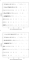
Dot plot of Ki67 measurements by laboratory from Experiments 2A and 2B. A) Centrally stained, local scoring method. B) Locally stained, local scoring method. Laboratories are reordered according to increasing median Ki67 value (percentange of positively stained tumor cells). When multiple observations have the same Ki67 value, random jittering is used to displace the points vertically to aid visualization.
Figure 4.
Figure 4.
Pairwise Bland–Altman plots showing high interlaboratory reproducibility among three laboratories (Laboratories D, E, and H). A) Experiment 2A: central staining, local scoring method. B) Experiment 2B: local staining, local scoring method. The y-axis (difference in Ki67) represents the Ki67 value (percentage of positively stained tumor cells) of the former laboratory minus that of the latter laboratory. The middle dashed line represents the average of the differences across all observations. Hence, a middle dashed line greater than 0 would indicate that the average Ki67 value of the former laboratory is greater than that of the latter laboratory, and vice versa.
Figure 5.
Figure 5.
Degree of concordance (bolded box) between Laboratory B and Laboratory D at the St. Gallen–recommended Ki67 percentage of positively stained tumor cells cutoff of 13.5%. At a hypothetical 13.5% cutoff, there are 32.3% cases that Laboratory D would call high Ki67 but Laboratory B would call low Ki67. The plot is based on 96 cores (three cores were not scored by one of the labs, and one core was not scored by both labs). When more than one core obtained the same paired Ki67 measurements from the two labs, random jittering is used to displace the points vertically to aid visualization (the small amount of noise added to break ties was generated from a uniform distribution between -d/5 and d/5 where d is the smallest difference among the original values within each lab).

References

    1. Dowsett M, Nielsen TO, A’Hern R, et al. Assessment of Ki67 in breast cancer: recommendations from the International Ki67 in Breast Cancer working group. J Natl Cancer Inst. 2011;103(22):1656–1664 - PMC - PubMed
    1. Goldhirsch A, Wood WC, Coates AS, Gelber RD, Thurlimann B, Senn HJ. Strategies for subtypes--dealing with the diversity of breast cancer: highlights of the St. Gallen International Expert Consensus on the Primary Therapy of Early Breast Cancer 2011. Ann Oncol. 2011;22(8):1736–1747 - PMC - PubMed
    1. Yerushalmi R, Woods R, Ravdin PM, Hayes MM, Gelmon KA. Ki67 in breast cancer: prognostic and predictive potential. Lancet Oncol. 2010;11(2):174–183 - PubMed
    1. Viale G, Giobbie-Hurder A, Regan MM, et al. Prognostic and predictive value of centrally reviewed Ki-67 labeling index in postmenopausal women with endocrine-responsive breast cancer: results from Breast International Group Trial 1–98 comparing adjuvant tamoxifen with letrozole. J Clin Oncol. 2008;26(34):5569–5575 - PMC - PubMed
    1. Dowsett M, Ebbs SR, Dixon JM, et al. Biomarker changes during neoadjuvant anastrozole, tamoxifen, or the combination: influence of hormonal status and HER-2 in breast cancer—a study from the IMPACT trialists. J Clin Oncol. 2005;23(11):2477–2492 - PubMed

Publication types