. 2014 Mar;25(3):574-86.
doi: 10.1681/ASN.2013040337. Epub 2013 Nov 7.

Mutations in PCBD1 cause hypomagnesemia and renal magnesium wasting

Affiliations

Mutations in PCBD1 cause hypomagnesemia and renal magnesium wasting

Silvia Ferrè et al. J Am Soc Nephrol. 2014 Mar.

Abstract

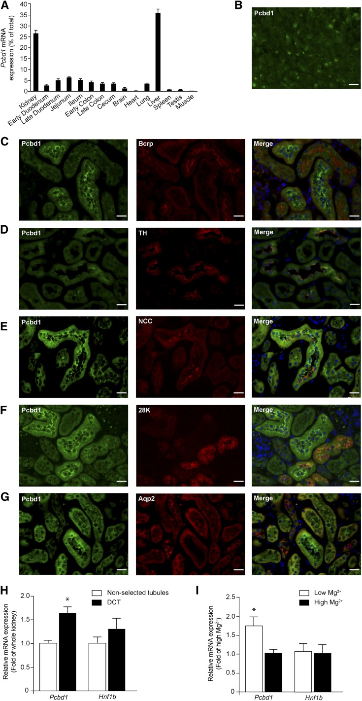
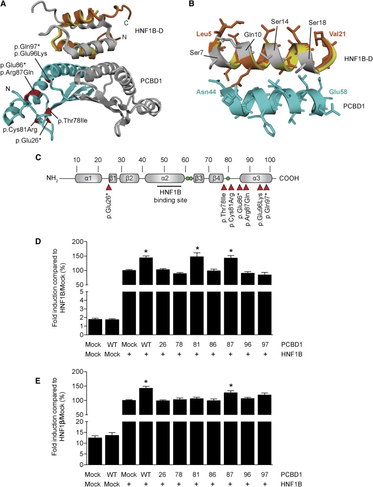
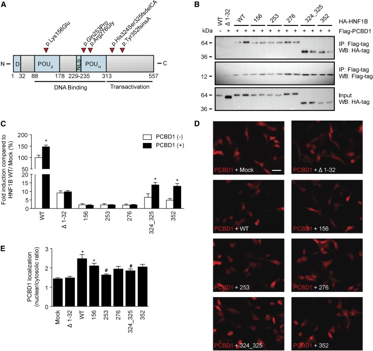
Mutations in PCBD1 are causative for transient neonatal hyperphenylalaninemia and primapterinuria (HPABH4D). Until now, HPABH4D has been regarded as a transient and benign neonatal syndrome without complications in adulthood. In our study of three adult patients with homozygous mutations in the PCBD1 gene, two patients were diagnosed with hypomagnesemia and renal Mg(2+) loss, and two patients developed diabetes with characteristics of maturity onset diabetes of the young (MODY), regardless of serum Mg(2+) levels. Our results suggest that these clinical findings are related to the function of PCBD1 as a dimerization cofactor for the transcription factor HNF1B. Mutations in the HNF1B gene have been shown to cause renal malformations, hypomagnesemia, and MODY. Gene expression studies combined with immunohistochemical analysis in the kidney showed that Pcbd1 is expressed in the distal convoluted tubule (DCT), where Pcbd1 transcript levels are upregulated by a low Mg(2+)-containing diet. Overexpression in a human kidney cell line showed that wild-type PCBD1 binds HNF1B to costimulate the FXYD2 promoter, the activity of which is instrumental in Mg(2+) reabsorption in the DCT. Of seven PCBD1 mutations previously reported in HPABH4D patients, five mutations caused proteolytic instability, leading to reduced FXYD2 promoter activity. Furthermore, cytosolic localization of PCBD1 increased when coexpressed with HNF1B mutants. Overall, our findings establish PCBD1 as a coactivator of the HNF1B-mediated transcription necessary for fine tuning FXYD2 transcription in the DCT and suggest that patients with HPABH4D should be monitored for previously unrecognized late complications, such as hypomagnesemia and MODY diabetes.


Figures

Figure 1.
Figure 1.
Pcbd1 is expressed in the DCT of the kidney. (A) Tissue expression pattern of the Pcbd1 transcript. Pcbd1 mRNA expression level was measured in a panel of mouse tissues by quantitative RT-PCR and normalized for Gapdh1 expression. Data represent the mean of three individual experiments±SEM and are expressed as the percentage of the total tissue expression. (B) Immunohistochemical analysis of Pcbd1 in mouse pancreas tissue. Scale bar, 20 μm. (C–G) Mouse kidney sections were costained for Pcdb1 (green) and (C) Bcrp (red), (D) TH (red), (E) NCC (red), (F) 28K (red), or (G) Aqp2 (red). Nuclei are shown by 4′,6-diamidino-2-phenylindole staining (blue). TH, Tamm Horsfall; 28K, calbindin-D28K. Scale bar, 20 μm. (H) Kidney expression pattern of Pcbd1 shows the highest expression in DCT. The mRNA expression levels of Pcbd1 and Hnf1b in (black bars) COPAS-selected mouse DCT and (white bars) control (nonselected) kidneys were measured by quantitative RT-PCR and normalized for Gapdh expression. Data represent the mean of three individual experiments±SEM and are expressed as fold difference compared with the expression in nonselected tubules. *P<0.05 versus total kidney. (I) DCT expression of Pcbd1 is regulated by dietary Mg2+ intake. The mRNA expression levels of Pcbd1 and Hnf1b in COPAS-selected mouse DCT kidney tubules from mice fed with (white bars) low and (black bars) high Mg2+-containing diets were measured by real-time RT-PCR and normalized for Gapdh expression. Data represent the mean of four individual experiments±SEM and are expressed as fold difference compared with the expression in high Mg2+-containing diets. *P<0.05 versus high Mg2+. Aqp2, aquaporin 2; Bcrp, breast cancer resistance protein.
Figure 2.
Figure 2.
PCBD1 coactivates HNF1B-induced FXYD2 and PKHD1 promoter activity. (A) Homology model of the PCBD1–HNF1B dimerization domain (HNF1B–D) tetramer modeled using the structure of the PCBD1–HNF1A dimerization domain (HNF1A–D) complex (Protein Data Bank ID code 1F93). (Light blue and grey) The PCBD1 dimer binds the (orange and grey) HNF1B dimer through helix sequences. The HNF1A–D monomer is shown in yellow. Residues in the PCBD1 protein that were found mutates in patients affected by hyperphenylalaninemia are depicted in red. (B) Homology model of the interaction site within the PCBD1–HNF1B dimerization domain (HNF1A–D) complex. (Orange) The bound HNF1B monomer forms a helix bundle with the (light blue) PCBD1 monomer. The HNF1A–D monomer is shown in yellow. The residues that differ between HNF1B–D and HNF1A–D are visualized in grey. (C) Linear representation of the secondary structure elements of the human PCBD1 protein. Red arrowheads indicate the positions of the patient mutations described in the literature. Green balls indicate the histidine residues involved in the dehydratase active site (His61, His62, and His79). A luciferase assay was performed in HEK293 cells transiently cotransfected with a (D) Firefly luciferase FXYD2 and (E) PKHD1 promoter construct and HNF1B or mock DNA in the presence of wild-type or mutant PCBD1. A Renilla luciferase construct was cotransfected to correct for transfection efficiency. Firefly/Renilla luciferase ratios were determined as a measure of promoter activity. Results are depicted as percentage compared with HNF1B-Mock transfected cells. *P<0.05 versus HNF1B/Mock (n=0). Mock, mock DNA; WT, wild type; 26, p.Glu26+; 78, p.Thr78lle; 81, p.Cys81Arg; 86, p.Glu86*; 87, Arg87Gln; 96, p.Glu96Lys; 97, p.Gln97*.
Figure 3.
Figure 3.
Several PCBD1 mutations lead to protein degradation HEK293 cells. (A) Immunocytochemistry analysis of the subcellular localization of HA-tagged PCDB1 wild type or HA-tagged PCDB1 mutants when coexpressed in a 1:1 ratio with FLAG-tagged HNF1B or mock DNA in HEK293 cells. Red signal represents immunodetected HA epitopes. Nuclei stained with 4′,6-diamidino-2-phenylindole are shown in blue. Scale bar, 10 μm. Representation immunocytochemical images are shown. (B) HA-tagged PCDB1 wild-type, PCDB1 mutant, or mock DNA were transiently expressed in HEK293 cells with or without FLAG-tagged HNF1B. Immunoprecipitations on nuclear extracts using an anti-FLAG antibody were separated by SDS-PAGE, and Western blots were probed with (top) anti-HA and (middle) anti-FLAG antibodies. (Bottom) HA-PCDB1 input (25%) expression was also included in the analysis. The immunoblots shown are representative of three independent experiments. (C) Western blot analysis of HA-tagged PCDB1 mutants expressed in HEK293 cells treated with (+) or without (−) 10 nM MG-132 for 24 hours. A representative immunoblot is shown. IP, immunoprecipitation; Mock, mock DNA; WB, Western blot; WT, wild-type; 26, p.Glu26+; 78, p.Thr78lle; 81, p.Cys81Arg; 86, p.Glu86*; 87, Arg87Gln; 96, p.Glu96Lys; 97, p.Gln97*.
Figure 4.
Figure 4.
HNF1B mutations result in cytosolic localization of PCBD1. Effect of HNF1B mutations on PCDB1 binding, transcription coactivation, and subcellular localization. (A) Linear representation of the human HNF1B protein. Red arrowheads indicate patient mutations that were tested in this study. D, dimerization domain; NLS, nuclear localization signal; POUH, atypical POU homeodomain; POUS, POU-specific domain. (B) HA-tagged HNF1B wild-type (WT), HNF1B mutants, or HNF1B lacking the extracts using an anti-FLAG antibody were separated by SDS-PAGE, and Western blots (WBs) were probed with (top) anti-HA or (middle) anti-FLAG antibodies. (Lower) HA-HNF1B input (25%) expression was also included in the analysis. The immunoblots shown are representative for three independent experiments. IP, immunoprecipitation. (C) A luciferase assay was performed in HEK293 cells transiently cotransfected with a Firefly luciferase FXYD2 promoter construct and each of the HNF1B variants (black bars) with and (white bars) without PCDB1. A Renilla luciferase construct was cotransfected to correct for transfection efficiency. Firefly/Renilla luciferase ratios were determined as a measure of promoter activity. Results are depicted as percentage compared with HNF1B/Mock transfected cells. *P<0.05 compared with the HNF1B/Mock condition (n=9). (D) Immunocytochemistry analysis of the subcellular localization of FLAG-tagged PCDB1 when coexpressed in a 1:1 ratio with HA-tagged HNF1B WTs, HA-tagged HNF1B mutants, or mock DNA HEK293 cells. Red signal represents immunodetected FLAG epitopes. Scale bar, 20 μm. The immunocytochemical images shown are representative for three independent experiments. (E) Quantification of the nuclear versus cytosolic localization of PCDB1 when coexpressed in a 1:1 ratio with mock DNA (n=24), HNF1B Δ1–32 (n=33), WT (n=41), 156 (n=38), 253 (n=35), 276 (n=33), 324_325 (n=37), or 352 (n=36). *P<0.05 compared with Mock. #P<0.05 compared with WT. Mock, mock DNA; Δ1–32, HNF1B lacking the dimerization domain; 156, p.Lys156Glu; 253, p.Gln253Pro; 276, p.Arg276Gly; 324_325, His324Ser325fsdelCA; 352, p.Tyr352fsinsA.

References

    1. Adalat S, Woolf AS, Johnstone KA, Wirsing A, Harries LW, Long DA, Hennekam RC, Ledermann SE, Rees L, van’t Hoff W, Marks SD, Trompeter RS, Tullus K, Winyard PJ, Cansick J, Mushtaq I, Dhillon HK, Bingham C, Edghill EL, Shroff R, Stanescu H, Ryffel GU, Ellard S, Bockenhauer D: HNF1B mutations associate with hypomagnesemia and renal magnesium wasting. J Am Soc Nephrol 20: 1123–1131, 2009 - PMC - PubMed
    1. Edghill EL, Bingham C, Ellard S, Hattersley AT: Mutations in hepatocyte nuclear factor-1beta and their related phenotypes. J Med Genet 43: 84–90, 2006 - PMC - PubMed
    1. Faguer S, Decramer S, Chassaing N, Bellanné-Chantelot C, Calvas P, Beaufils S, Bessenay L, Lengelé JP, Dahan K, Ronco P, Devuyst O, Chauveau D: Diagnosis, management, and prognosis of HNF1B nephropathy in adulthood. Kidney Int 80: 768–776, 2011 - PubMed
    1. Glaudemans B, Knoers NV, Hoenderop JG, Bindels RJ: New molecular players facilitating Mg(2+) reabsorption in the distal convoluted tubule. Kidney Int 77: 17–22, 2010 - PubMed
    1. Meij IC, Koenderink JB, van Bokhoven H, Assink KF, Groenestege WT, de Pont JJ, Bindels RJ, Monnens LA, van den Heuvel LP, Knoers NV: Dominant isolated renal magnesium loss is caused by misrouting of the Na(+),K(+)-ATPase gamma-subunit. Nat Genet 26: 265–266, 2000 - PubMed

Publication types

MeSH terms

Supplementary concepts

LinkOut - more resources