. 2013 Oct;9(10):e1003728.
doi: 10.1371/journal.ppat.1003728. Epub 2013 Oct 24.

Plasmacytoid dendritic cells contribute to systemic but not local antiviral responses to HSV infections

Affiliations

Plasmacytoid dendritic cells contribute to systemic but not local antiviral responses to HSV infections

Melissa Swiecki et al. PLoS Pathog. 2013 Oct.

Abstract

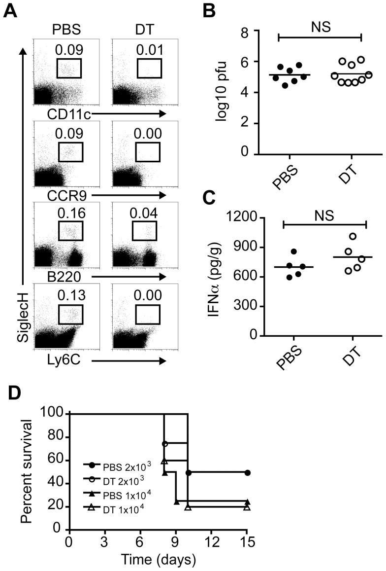
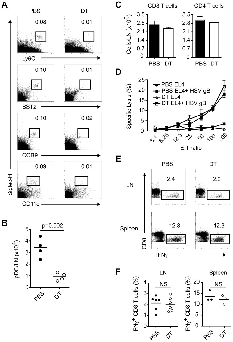
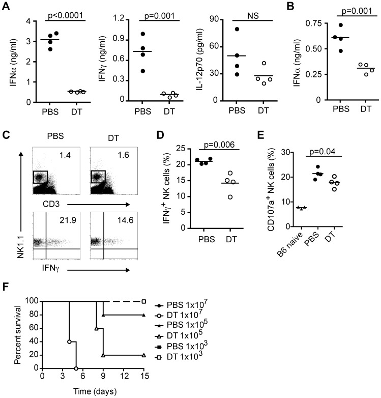
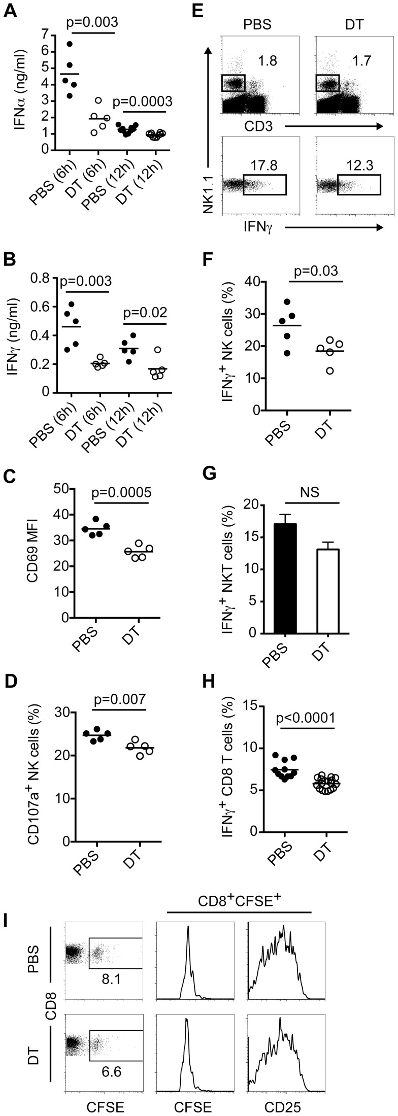
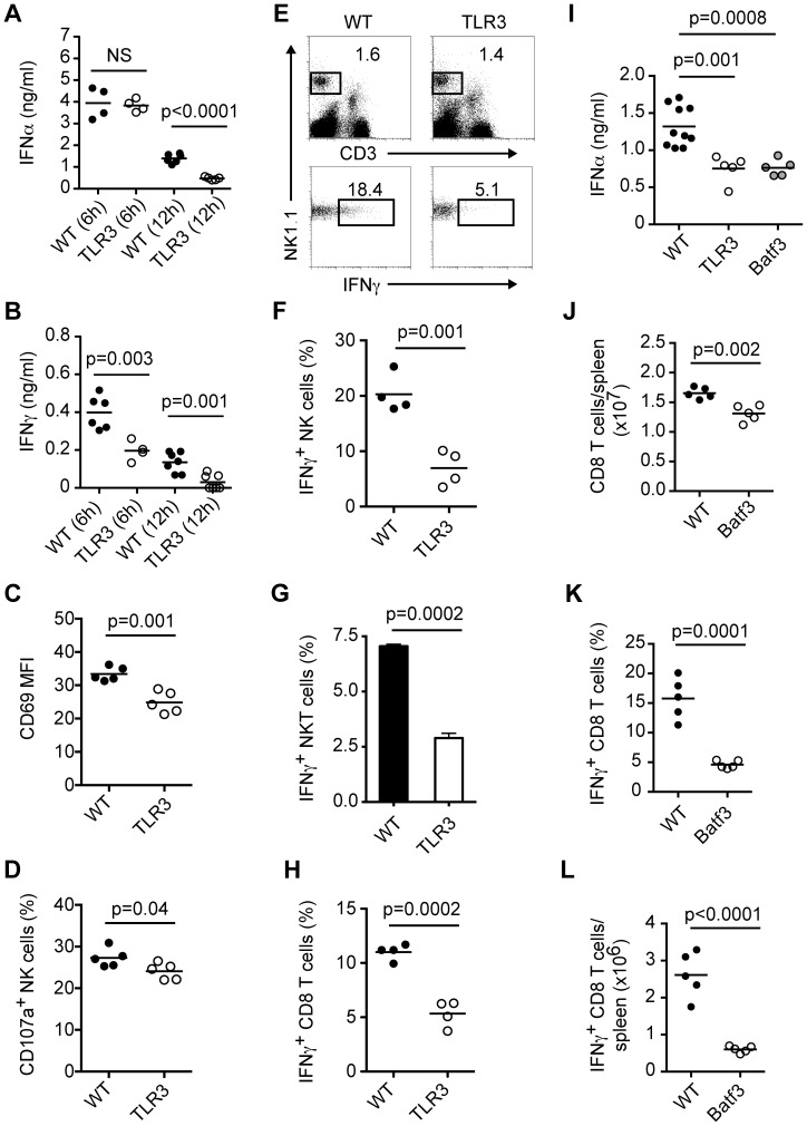
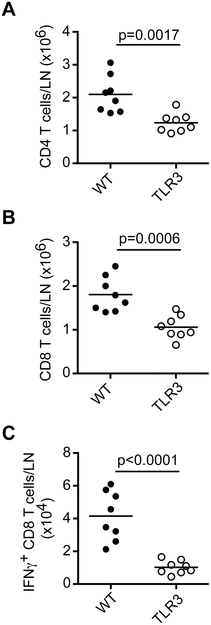
Plasmacytoid dendritic cells (pDC) produce type I interferons (IFN-I) and proinflammatory cytokines in response to viruses; however, their contribution to antiviral immunity in vivo is unclear. In this study, we investigated the impact of pDC depletion on local and systemic antiviral responses to herpes simplex virus (HSV) infections using CLEC4C-DTR transgenic mice. We found that pDC do not appear to influence viral burden or survival after vaginal HSV-2 infection, nor do they seem to contribute to virus-specific CD8 T cell responses following subcutaneous HSV-1 infection. In contrast, pDC were important for early IFN-I production, proinflammatory cytokine production, NK cell activation and CD8 T cell responses during systemic HSV-2 and HSV-1 infections. Our data also indicate that unlike pDC, TLR3-expressing cells are important for promoting antiviral responses to HSV-1 regardless of the route of virus administration.


Conflict of interest statement

The authors have declared that no competing interests exist.

Figures

Figure 1
Figure 1. Impact of pDC on vaginal HSV-2 infection.
CLEC4C-DTR Tg mice were injected subcutaneously with Depo-Provera or medroxyprogesterone acetate 7 days before vaginal HSV-2 infection. Mice were injected i.p. with PBS or DT 24 h before infection and every other day after. (A) Dotplots show pDC frequencies in lumbar lymph nodes of PBS or DT-treated mice at the time of infection. HSV-2 titers (B) and IFN-α levels (C) in vaginal/cervical tissues on day 2 p.i. after inoculation with 1×10³ pfu. (D) Control or pDC-depleted mice were infected with 2×10³ or 1×10⁴ pfu of HSV-2 and survival was monitored for 15 days. Data are combined from or are representative of two independent experiments. Not significant (NS).
Figure 2
Figure 2. Contribution of pDC to virus-specific CD8 T cell responses after local HSV-1 infection.
Control (PBS) and pDC-depleted (DT) mice were infected in the footpad with HSV-1. Draining popliteal lymph nodes (DLN) and spleens were analyzed on day 7 p.i. (A) Dotplots show pDC frequencies in popliteal lymph nodes of PBS or DT-treated mice at the time of infection. (B) pDC numbers in DLN of PBS or DT-treated mice on day 7 p.i. (C) Total numbers of CD8 and CD4 T cells in DLN. (D) Antigen-specific lysis using DLN cells as effectors and HSV gB peptide-pulsed or unpulsed EL4 cells as targets was assessed in standard 4 h 51Cr release assays. (E, F) Frequencies of CD8 T cells producing IFN-γ in DLN and spleens after restimulation with HSV gB peptide. Data are combined from or are representative of two independent experiments.
Figure 3
Figure 3. pDC produce IFN-I and activate NK cells during systemic HSV-2 infection.
CLEC4C-DTR Tg mice were depleted (DT) or not (PBS) of pDC then infected i.v. with HSV-2 (5×10⁶ pfu). (A) Serum IFN-α, IFN-γ and IL-12p70 levels were measured 8 h p.i. (B) Serum IFN-α levels at 12 h p.i. in control and pDC-depleted mice. Frequencies of IFN-γ-producing NK cells (NK1.1+CD3) (C, D) and CD107a+ NK cells (E) in spleens were determined 12 h p.i. (F) PBS and DT-treated mice were infected i.v. with different doses of HSV-2. Survival was monitored for 15 days. Data are representative of two independent experiments. Statistical significance is indicated by p values. Not significant (NS).
Figure 4
Figure 4. pDC promote antiviral responses during systemic HSV-1 infection.
Control (PBS) and pDC-depleted (DT) mice were infected i.v. with HSV-1 (1×10⁷ pfu). Serum IFN-α (A) and IFN-γ (B) were measured at 6 and 12 h p.i. (C) CD69 expression on NK cells (NK1.1+CD3) from spleens of PBS or DT-treated mice 12 h p.i. Frequencies of CD107a+ (D) and IFN-γ-producing NK cells (E, F) in spleens of HSV-1 infected mice 12 h p.i. (G) Frequencies of IFN-γ-producing NKT cells (NK1.1+CD3+) in spleens of HSV-1 infected mice 12 h p.i. (H) Frequencies of IFN-γ-producing CD8 T cells in spleens on day 7 p.i. after restimulation with HSV gB peptide. (I) CFSE-labeled CD8 T cells from gBT-I mice were injected i.v. into CLEC4C-DTR Tg mice. Mice were depleted or not of pDC and infected i.v. with HSV-1 (1×10⁷ pfu). Spleens were analyzed on day 3 p.i. for numbers of transferred CD8 T cells, CFSE dilution and CD25 levels. Dotplots and histograms are representative of four mice per group. Data are representative of one (I) or two-three independent experiments (A–H). Statistical significance is indicated by p values. Not significant (NS).
Figure 5
Figure 5. TLR3-expressing cells are necessary for robust antiviral responses during systemic HSV-1 infection.
(A–H) WT and TLR3−/− mice were infected i.v. with HSV-1 (1×10⁷ pfu). Serum IFN-α (A) and IFN-γ (B) were measured at 6 and 12 h p.i. (C) CD69 expression on NK cells (NK1.1+CD3) from spleens of WT and TLR3−/− mice 12 h p.i. Frequencies of CD107a+ (D) and IFN-γ-producing NK cells (E, F) in spleens of HSV-1 infected mice 12 h p.i. (G) Frequencies of IFN-γ-producing NKT cells (NK1.1+CD3+) in spleens of HSV-1 infected mice 12 h p.i. (H) Frequencies of IFN-γ-producing CD8 T cells in spleens on day 7 p.i. after restimulation with HSV gB peptide. (I) WT, TLR3−/− and Batf3−/− mice were infected i.v. with HSV-1 (1×10⁷ pfu). Serum IFN-α levels were measured 12 h p.i. (J–L) WT and Batf3−/− mice were infected i.v. with HSV-1 (1×10⁷ pfu). (J) Total CD8 T cells in spleens on day 7 p.i. Frequencies (K) and numbers (L) of IFN-γ-producing CD8 T cells in spleens on day 7 p.i. after restimulation with HSV gB peptide. Data are representative of two or three independent experiments. Statistical significance is indicated by p values. Not significant (NS).
Figure 6
Figure 6. TLR3-expressing cells promote CD8 T cell responses to local HSV-1 infection.
WT and TLR3−/− mice were infected in the footpad with HSV-1 (1×10⁵ pfu). Total CD4 (A) and CD8 (B) T cells were analyzed in DLN on day 7 p.i. (C) Numbers of IFN-γ-producing CD8 T cells in DLN on day 7 p.i. after restimulation with HSV gB peptide. Data are combined from two independent experiments. Statistical significance is indicated by p values.

References

    1. Fitzgerald-Bocarsly P, Dai J, Singh S (2008) Plasmacytoid dendritic cells and type I IFN: 50 years of convergent history. Cytokine Growth Factor Rev 19: 3–19. - PMC - PubMed
    1. Blasius AL, Beutler B (2010) Intracellular toll-like receptors. Immunity 32: 305–315. - PubMed
    1. Trinchieri G (2010) Type I interferon: friend or foe? J Exp Med 207: 2053–2063. - PMC - PubMed
    1. Gilliet M, Cao W, Liu YJ (2008) Plasmacytoid dendritic cells: sensing nucleic acids in viral infection and autoimmune diseases. Nat Rev Immunol 8: 594–606. - PubMed
    1. Reizis B, Bunin A, Ghosh HS, Lewis KL, Sisirak V (2011) Plasmacytoid Dendritic Cells: Recent Progress and Open Questions. Annu Rev Immunol 29: 163–183. - PMC - PubMed

Publication types

Substances