. 2013 Oct;9(10):e1003885.
doi: 10.1371/journal.pgen.1003885. Epub 2013 Oct 31.

PNUTS/PP1 regulates RNAPII-mediated gene expression and is necessary for developmental growth

Affiliations

PNUTS/PP1 regulates RNAPII-mediated gene expression and is necessary for developmental growth

Anita Ciurciu et al. PLoS Genet. 2013 Oct.

Abstract

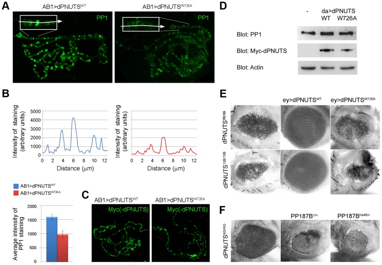
In multicellular organisms, tight regulation of gene expression ensures appropriate tissue and organismal growth throughout development. Reversible phosphorylation of the RNA Polymerase II (RNAPII) C-terminal domain (CTD) is critical for the regulation of gene expression states, but how phosphorylation is actively modified in a developmental context remains poorly understood. Protein phosphatase 1 (PP1) is one of several enzymes that has been reported to dephosphorylate the RNAPII CTD. However, PP1's contribution to transcriptional regulation during animal development and the mechanisms by which its activity is targeted to RNAPII have not been fully elucidated. Here we show that the Drosophila orthologue of the PP1 Nuclear Targeting Subunit (dPNUTS) is essential for organismal development and is cell autonomously required for growth of developing tissues. The function of dPNUTS in tissue development depends on its binding to PP1, which we show is targeted by dPNUTS to RNAPII at many active sites of transcription on chromosomes. Loss of dPNUTS function or specific disruption of its ability to bind PP1 results in hyperphosphorylation of the RNAPII CTD in whole animal extracts and on chromosomes. Consistent with dPNUTS being a global transcriptional regulator, we find that loss of dPNUTS function affects the expression of the majority of genes in developing 1(st) instar larvae, including those that promote proliferative growth. Together, these findings shed light on the in vivo role of the PNUTS-PP1 holoenzyme and its contribution to the control of gene expression during early Drosophila development.


Conflict of interest statement

The authors have declared that no competing interests exist.

Figures

Figure 1
Figure 1. dPNUTS is a nuclear protein that colocalises with transcriptionally active RNAPII on salivary gland polytene chromosomes.
A) Distribution of dPNUTS transcripts detected by RNA in situ hybridization; dPNUTS transcripts are maternally provided (top left) and are ubiquitously distributed in embryos at cellularisation (top right). At gastrulation, dPNUTS mRNA levels are enriched in the germband and in the fore- and hind-gut (fg and hg, respectively). Later, dPNUTS is highly expressed in the brain (br) and ventral nerve cord (vnc). Embryonic stage and approximate age, hours post fertilization (hpf), are indicated. B) 3rd instar wing discs stained to reveal the distribution of ectopically expressed Myc-tagged dPNUTS (green in merge), Histone H3S10ph (red in merge, marking mitotic nuclei) and DNA. C) Images of whole mount salivary gland and magnified images of an individual nucleus (below), stained to show the localization of Myc-tagged dPNUTS (green in merge) and DNA (magenta in merge). D) Line scans of images in C) reveal that Myc-tagged dPNUTS is localised to interbands that stain weakly for DNA. Fluorescence intensity of anti-Myc antibody and TOPRO-3 staining was measured along a line through the indicated chromosomal region in the images shown. The profile plot below shows that the peaks of Myc-PNUTS and DNA of staining do not overlap. E) Polytene chromosomes from salivary gland squashes showing that dPNUTS localises to a number of discrete bands that are broadly distributed. F) Merging of the green signal representing dPNUTS with the red signal representing RNAPII Ser2-P (H5) identifies sites where these two proteins co-localize (example indicated with arrow). The relative signals of dPNUTS and RNAPII Ser2-P vary between sites, but the majority dPNUTS loci colocalize with RNAPII Ser2-P staining (star indicates example where only dPNUTS staining is visible).
Figure 2
Figure 2. dPNUTS loss of function results in larval growth arrest and defective tissue development.
A) Genomic region showing dPNUTS locus flanked by dribble and ninaA. Coding regions of the genes is represented by shading. dPNUTS produces two transcripts dPNUTS and dPNUTS-S. ninaA is a non-essential gene that is expressed solely in the eye to regulate rhodopsin synthesis , . dPNUTSKG572 contains a P element insertion in an untranslated region of dPNUTS. The extent of deletions in dPNUTS9B and dPNUTS13B resulting from imprecise excision of this element is indicated, together with the genomic sequence of the breakpoints. The dPNUTS genomic rescue construct, which contains the coding region of ninaA, and the 5′ end of dribble, is indicated. B) Levels of dPNUTS and dPNUTS-S transcripts produced in homozygous dPNUTSexKG, dPNUTSKG572, dPNUTS9B and dPNUTS13B larvae, as determined by qRT-PCR. dPNUTSeexKG is a revertant strain in which the P element had precisely excised. C) Images of homozygous mutant and control (heterozygous sibling) larvae at different time points after egg laying as indicated. D) Graph showing percentage of surviving larvae over time for each genotype, as indicated. E) Images of adult female eyes. Homozygous dPNUTS mutant eyes are smaller than controls (isogenic w1118 strain), but are able to form some facets, unlike eyes expressing the proapototic gene hid under GMR control.
Figure 3
Figure 3. dPNUTS mutant clones reveal a cell autonomous growth defect in developing tissues.
Clones (marked by absence of GFP) of either wild-type or dPNUTS13B mutant cells are shown in wing imaginal discs obtained from 3rd instar larvae. Clones were induced in a wild type (A–J) or Minute (M) mutant background (K–R) 48 hr, 72 hr or 96 hr prior to dissection, as indicated. The parental (p) genotypes are indicated, along with the genotype of clones (c) generated by FLP-mediated mitotic recombination and are coded with grayscale to indicate the relative level of GFP expression. A–J, wing discs were stained for activated caspase shown in blue, and in cross sections (E–J), apico-lateral junctions are marked by discs-large staining in red; GFP is shown in green. In cross sections Q–R, DNA is shown in red. Arrowheads in panel L indicate the presence of dPNUTS13B GFP-negative clones in a Minute (M) mutant background. +, M, GFP/+, M, GFP twinspot clones, indicated by arrowhead in panel R, were almost never observed because of a severe growth defect.
Figure 4
Figure 4. dPNUTS mutants deregulate the expression of the majority of genes in 1st instar larvae, including highly expressed genes involved in cellular metabolism and proliferative growth.
A) MA plot of RNA-Seq data in which the log2 of the ratio of abundance of each transcript between dPNUTS13B (13B) mutant and w1118 (WT) control (M) is plotted against the log2 geometric average of abundance (FPKM) in both conditions (A). Transcripts with an FPKM of less than 0.25, which are a source of noise in these plots, are not shown for clarity. Transcripts that are differentially expressed (DE) by <0.67 or >1.5 fold are shown in grey; unaffected transcripts are shown in white. Loci corresponding to enriched Gene Ontology (GO) terms amongst the differentially expressed genes relative to the entire genome are highlighted in yellow (enriched amongst overexpressed genes) or red (enriched amongst underexpressed genes). Log2 median expression for genes expressed in WT and 13B is indicated with a dashed line. Log2 median expression for genes belonging to GO categories is given in the legend. A complete list of GO categories is provided in the Supplementary information. B) Expression levels of the indicated genes in dPNUTS9B/dPNUTS9B and dPNUTS13B/dPNUTS13B mutant larvae, relative to control (w1118) larvae, determined by qRT-PCR. Error bars represent the SE (n≥3 biological replicates). The GO categories to which the genes belong are shown at the top.
Figure 5
Figure 5. dPNUTS binds to and co-localises with PP1 on chromosomes.
A) Predicted domain structure of dPNUTS and dPNUTS-S proteins, indicating the position of the putative PP1-binding motif (residues 722–726), which is located in the shortest yeast two-hybrid interacting clone (SIC) of dPNUTS. B) Beta-galactosidase assays showing binding of dPNUTS but not dPNUTS-S to all four D. melanogaster PP1 isoforms in the yeast two-hybrid system. C) dPNUTSWT, but not dPNUTSW726A, co-precipitates PP1 from nuclear extracts from adult flies. da-GAL4 UAS-HM-dPNUTSWT and da-GAL4 UAS-HM-dPNUTSW726A fly extracts were subjected to immunoprecipitation (IP) with Myc antibodies followed by immunoblotting with PP1 antibodies. Blots of total lysates confirmed levels of HM-tagged dPNUTS and PP1. D) dPNUTS and PP1 colocalise at many sites on polytene chromosomes. Inset is an enlarged view of the end of the X chromosome where this is clearly visible. Plot of fluorescence intensity of anti-PP1 and dPNUTS antibody staining, measured along a line through the indicated chromosomal region, reveal the degree of colocalisation between PP1 and dPNUTS.
Figure 6
Figure 6. PP1 localisation is regulated by dPNUTS and binding to PP1 is important for dPNUTS function.
A) Images of polytene chromosome squashes from salivary glands expressing either dPNUTSWT or dPNUTSW726A stained with PP1 (in green). Inset are enlarged views of the distal end of the X chromosome. Arrows indicate approximate lines along which quantitation of fluorescence (see B) was performed. B) Plots of line scans through the chromosomal region indicated in A, showing levels of PP1 staining in salivary glands expressing either dPNUTSWT or dPNUTSW726A. Bar graphs represent the average fluorescence in this region from 6 independent images/genotype. Genotypes are indicated by the colour key. C) Levels and distribution of Myc-dPNUTS on polytene chromosome squashes from salivary glands expressing either dPNUTSWT or dPNUTSW726A, as revealed by anti-Myc staining (green). D) Western blots showing levels of PP1 and Myc-dPNUTS, relative to Actin, in extracts from animals ectopically expressing dPNUTSWT or dPNUTSW726A under the control of da-GAL4 (da>dPNUTSWT and da>dPNUTSW726A, respectively) compared to w1118 control (−). E) Images of adult female eyes showing that the severely reduced eye phenotype of homozygous dPNUTS mutant eyes is fully rescued by ectopic expression of dPNUTSWT but not dPNUTSW726A. F) Homozygous dPNUTSKG572 mutant eyes show a weaker phenotype than either dPNUTS9B or dPNUTS13B, and this can be enhanced by loss of one copy of PP187B.
Figure 7
Figure 7. dPNUTS complexes with and regulates RNAPII phosphorylation.
A) dPNUTS complexes contain RNAPII and PP1; inhibition of PP1 activity in dPNUTS complexes leads to hyperphosphorylation of RNAPII. dPNUTS-S and dPNUTS were immunoprecipitated (IP) from embryonic nuclear extracts and precipitates were probed with ARNA-3 anti-RNAPII antibody. Lane 1, neither hypo- or hyper-phosphorylated RNAPII (RNAPIIa and RNAPIIo respectively) precipitate with pre-immune serum; Lane 2, both RNAPIIa and RNAPIIo precipitate with dPNUTS-S; Lane 3, RNAPIIa, but almost no RNAPIIo, is detected in dPNUTS precipitates. Lane 4, pre-immune serum does not precipitate RNAPII; Lane 5, Inhibitor 2 does not affect the ability of RNAPIIa and RNAPIIo to associate with dPNUTS-S (compare Lane 2); Lane 6, inhibition of PP1 results in conversion of RNAPIIa to RNAPIIo in dPNUTS precipitates (compare Lane 3). Ratios of RNAPIIa and RNAPIIo levels, as derived from densitometry measurements of the respective bands, are shown above the blots. B) Western Blot showing levels of RNAPII CTD Ser5-P (4H8) in extracts from either 1st (L1) or 2nd (L2) instar larvae of the indicated genotypes: homozygous revertant dPNUTSexKG/dPNUTSexKG (exKG/exKG); homozygous null mutant dPNUTS9B/dPNUTS9B (9B/9B) or dPNUTS13B/dPNUTS13B (13B/13B); isogenic control strain w1118/w1118; homozygous hypomorphic mutant dPNUTSKG572/dPNUTSKG572 (KG/KG). 1st instar larval samples from dPNUTS9B/9B and dPNUTS13B/13B were independent extracts run in parallel on the same gel. Blot with anti-Actin antibody shows relative loading. C) Precipitation of RNAPII Ser5-P with dPNUTSW726A but not dPNUTSWT. dPNUTS complexes from Drosophila embryonic nuclear extracts expressing Myc-tagged dPNUTSWT or dPNUTSW726A under the control of da-GAL4 were isolated by immunoprecipitation with anti-Myc antibody. Control precipitations were performed on w1118 extracts (−). This was followed by immunoblotting with anti-RNAPII CTD Ser5-P (4H8) antibody to test for co-immunoprecipitation. Lower panels show immunoblot analyses of total lysates, confirming the levels of total RNAPII and Myc-dPNUTS. D) Levels of RNAPII CTD Ser5-P (H14) on polytene chromosome squashes from salivary glands expressing either histone-H2B YFP or Myc-dPNUTSW726A prepared on the same slide to ensure identical staining conditions (H14 staining in green; DNA staining in magenta). Insets are enlarged views of the distal end of the X chromosome. Arrows indicate approximate lines along which quantitation of fluorescence (in E) was performed. E) Representative line scans through the regions illustrated in D, showing levels of RNAPII CTD Ser5-P staining in the two genotypes. Bar graphs represent the average fluorescence in this region from 6 independent images/genotype. Genotypes are indicated by the colour key.

References

    1. Tennessen JM, Thummel CS (2011) Coordinating growth and maturation - insights from Drosophila. Curr Biol 21: R750–757. - PMC - PubMed
    1. Brookes E, Pombo A (2009) Modifications of RNA polymerase II are pivotal in regulating gene expression states. EMBO Rep 10: 1213–1219. - PMC - PubMed
    1. Phatnani HP, Greenleaf AL (2006) Phosphorylation and functions of the RNA polymerase II CTD. Genes Dev 20: 2922–2936. - PubMed
    1. Hsin JP, Manley JL (2012) The RNA polymerase II CTD coordinates transcription and RNA processing. Genes Dev 26: 2119–2137. - PMC - PubMed
    1. Bataille AR, Jeronimo C, Jacques PE, Laramee L, Fortin ME, et al. (2012) A universal RNA polymerase II CTD cycle is orchestrated by complex interplays between kinase, phosphatase, and isomerase enzymes along genes. Mol Cell 45: 158–170. - PubMed

Publication types

MeSH terms

Associated data

LinkOut - more resources