. 2013 Oct;9(10):e1003894.
doi: 10.1371/journal.pgen.1003894. Epub 2013 Oct 31.

Genome-wide high-resolution mapping of UV-induced mitotic recombination events in Saccharomyces cerevisiae

Affiliations

Genome-wide high-resolution mapping of UV-induced mitotic recombination events in Saccharomyces cerevisiae

Yi Yin et al. PLoS Genet. 2013 Oct.

Abstract

In the yeast Saccharomyces cerevisiae and most other eukaryotes, mitotic recombination is important for the repair of double-stranded DNA breaks (DSBs). Mitotic recombination between homologous chromosomes can result in loss of heterozygosity (LOH). In this study, LOH events induced by ultraviolet (UV) light are mapped throughout the genome to a resolution of about 1 kb using single-nucleotide polymorphism (SNP) microarrays. UV doses that have little effect on the viability of diploid cells stimulate crossovers more than 1000-fold in wild-type cells. In addition, UV stimulates recombination in G1-synchronized cells about 10-fold more efficiently than in G2-synchronized cells. Importantly, at high doses of UV, most conversion events reflect the repair of two sister chromatids that are broken at approximately the same position whereas at low doses, most conversion events reflect the repair of a single broken chromatid. Genome-wide mapping of about 380 unselected crossovers, break-induced replication (BIR) events, and gene conversions shows that UV-induced recombination events occur throughout the genome without pronounced hotspots, although the ribosomal RNA gene cluster has a significantly lower frequency of crossovers.


Conflict of interest statement

The authors have declared that no competing interests exist.

Figures

Figure 1
Figure 1. Mechanisms of homologous recombination.
The two interacting DNA molecules are shown as double-stranded. The recombination event is initiated by a double-stranded DNA break (DSB) on the black molecule, and the broken ends are resected 5′ to 3′. In this depiction, the left processed end invades the red molecule, forming a heteroduplex. Dotted lines show DNA synthesis associated with the recombination event. A. Synthesis-dependent strand annealing (SDSA). The 3′ end of the invading strand is used as a primer for DNA synthesis, extending the D-loop. Dissociation of the D-loop, followed by re-annealing of the two broken ends results in a region of heteroduplex (outlined in blue) with flanking markers in the parental non-crossover (NCO) configuration. B. Double-strand break repair (DSBR). Following expansion of the D-loop, pairing occurs between the displaced strand and the right end of the broken chromosome, resulting in two regions of heteroduplex. The resulting double Holliday junction can be resolved by topoisomerase-mediated dissolution or by cleavage of the Holliday junctions to yield an NCO or a crossover (CO). C. Break-induced replication (BIR). In this pathway, the right broken chromosome is lost and the left molecule invades the homologous chromosome, resulting in duplication of sequences distal to the point of invasion.
Figure 2
Figure 2. Patterns of mitotic gene conversions and crossovers.
Chromosomes are shown as thick lines with the two homologs in red or black. Circles indicate the centromeres and blue boxes show gene conversions. A. Conversion and crossover associated with a DSB formed in S or G2 on one of the black chromatids. Since the broken chromosome acts as the recipient in a gene conversion event , sequence information derived from the red chromatid is non-reciprocally transferred in conjunction with the crossover. This type of conversion is termed “3∶1” since, at the site of the conversion, three of the four chromosomes of the two daughter cells (D1 and D2) have information derived from one of the homologs. If chromatids 1 and 3, and 2 and 4 co-segregate, any markers distal to the crossover will be homozygous. B. S or G2 conversion event unassociated with a crossover. If the DSB is repaired to generate a conversion unassociated with a crossover, a region of 3∶1 segregation will be observed, but the markers distal to the conversion event will remain heterozygous. C. Break-induced replication. The chromosome fragment centromere-distal to the DSB is lost, and the centromere-containing fragment invades and replicates the red chromosome to the telomere. D. Repair of a G1-induced DSB. A black chromosome with a G1-induced DSB is replicated to yield two sister-chromatids with breaks at the same positions. Gene conversion events are generated at the sites of repair, and the repair of one of the DSBs is associated with a crossover. Within the chromosomes of the two daughter cells, there is a region that is derived from only the red homolog (a “4∶0” conversion tract shown in the blue rectangle).
Figure 3
Figure 3. A system for detecting mitotic crossovers by a colony sectoring assay.
G1-synchronized diploid cells were treated with UV and immediately plated on solid medium. The diploid is homozygous for the ade2-1 mutation, an ochre mutation that when unsuppressed results in a red colony. The diploid has one copy of the ochre suppressor gene SUP4-o inserted near the telomere of chromosome IV on the black homolog. Strains with zero, one, and two copies of SUP4-o form red, pink, and white colonies, respectively. A. Crossover in G2 of the first division following irradiation. A DSB in one chromatid repaired during G2 will generate a red/white sectored colony, the white sector derived from daughter cell 1 (D1) and the red sector derived from daughter cell 2 (D2). B. Crossover delayed to G2 of the second division. If DNA damage induced in G1 is not repaired during the first division, a pink/white/red sectored colony would be generated. The abbreviation “GD” indicates the granddaughter of the irradiated cell.
Figure 4
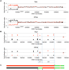
Figure 4. Mapping crossovers on chromosome V by SNP arrays.
Genomic DNA was isolated from the red and white sectors of a sectored colony derived from UV-treated cells. The ratio of hybridization of SNP-specific oligonucleotides (relative to the hybridization levels of DNA from a fully heterozygous strain) for each sample was measured (details in text) and is shown on the Y axis. A hybridization level of about 1 indicates that the strain was heterozygous. The X axis shows the SGD coordinates of chromosome V beginning at the left telomere. Black and red lines indicate the normalized hybridization ratio to the YJM789- and W303a-specific oligonucleotides, respectively. The crossover and conversions associated with the sectors are diagrammed with the upper and lower panels showing patterns of LOH in the red and white sectors, respectively. A. Low-resolution depiction of the LOH events in the red and white sectors. The transition is at about SGD coordinate 105 kb. B. High-resolution depiction of LOH events (same event as shown in Figure 4A). Black squares show hybridization to YJM789-specific oligonucleotides and red diamonds show hybridization to W303a-specific oligonucleotides. The red sector has a single transition between heterozygous and homozygous SNPs, whereas the white sector has three transitions. The pattern of transitions is consistent with a 3∶1/4∶0 hybrid tract associated with a crossover. C. Summary of patterns of heterozygous and homozygous markers with the top line showing the red sector and the bottom line showing the white sector. The black, red, and green lines indicate regions homozygous for YJM789-derived SNPs, homozygous for W303a-derived SNPs, and heterozygous regions, respectively. The orange circles show the position of the centromeres. The lengths of the line segments showing the LOH region associated with the crossover and the heterozygous region centromere-proximal to the crossover are not shown to scale.
Figure 5
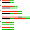
Figure 5. Common patterns of conversions and crossovers derived from UV-treated cells.
The key is described in Figure 4C. A. Simple crossover unassociated with conversion. B. Crossover associated with a 3∶1 conversion event. C. Crossover associated with a 3∶1/4∶0 hybrid conversion tract. D. Complex crossover/conversion event. E. 3∶1 conversion unassociated with a crossover. F. BIR event.
Figure 6
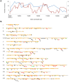
Figure 6. Patterns of selected events on chromosome V and unselected events throughout the genome.
A. Selected events on the left arm of chromosome V. All events were based on red/white sectored colonies. The blue and red lines represent spontaneous and UV-induced (15 J/m2), respectively. The X-axis shows the number of events that include specific SNPs with the left scale indicating the number of spontaneous events and the right scale showing the number of UV-induced events. The Y axis shows the SGD coordinates on V between the can1-100/SUP4-o markers (near coordinate 33 kb) and CEN5 (near coordinate 152 kb). B. Location of unselected conversion and crossovers. Events on the left arm of chromosome V are not shown, since these events were selected. Centromeres are shown as black circles, and SNPs are indicated as short orange bars. Gene conversion tracts unassociated with crossovers are depicted by black line segments, with the length of the line approximately equivalent to the tract length. Red arrows show the positions of conversions associated with crossovers, and BIR events are indicated by green arrows. The lengths of the chromosomes are shown to scale; chromosome I is about 230 kb.
Figure 7
Figure 7. System to monitor crossovers on chromosome XII.
A diploid was constructed with heterozygous markers immediately centromere-proximal to the ribosomal RNA (rRNA) gene clusters (HYG), within the rRNA gene cluster (TRP1), and immediately centromere-distal to the cluster (URA3) . G1-synchronized cells were treated with UV, plated on solid medium and grown non-selectively. The resulting colonies were replica-plated to medium lacking uracil, tryptophan, or containing hygromycin as described in the text. A. Sectoring pattern expected for a crossover centromere-proximal to the HYG marker. B. Sectoring pattern expected for a crossover within the rRNA gene cluster centromere-proximal to TRP1. C. Sectoring pattern expected for a crossover within the rRNA gene cluster centromere-distal to TRP1.
Figure 8
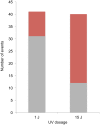
Figure 8. Selected SCB and DSCB conversions in strains treated with 1/m2 and 15 J/m2.
SCB and DSCB events are indicated in gray and red, respectively.
Figure 9
Figure 9. Mechanisms for generating UV-induced recombinogenic DSBs.
At the top part of the figure, chromosomal DNA molecules are depicted as unreplicated double-stranded DNA molecules. Newly-synthesized DNA is depicted as gray dashed lines. UV-induced pyrimidine dimers are shown as triangles, and centromeres of replicated chromosomes are shown as ovals. A. Excision of a dimer results in a small gap and replication produces one broken and one unbroken sister chromatid. B. During replication of a DNA molecule with an unexcised dimer, a DSB occurs in one of the two sister chromatids. C. Excision of two closely-opposed dimers results in a short (<6 bp) unstable double-stranded region between the excision tracts. The resulting broken chromosome is replicated to form two broken sister chromatids. D. As in Figure 9C, two closely-opposed dimers are excised. One of the resulting short gaps is expanded by the 5′ to 3′ Exo1p nuclease (shown in green) to generate a broken chromosome. Replication of this chromosome results in two broken sister chromatids. E. The tract resulting form excision of a single dimer is expanded, leaving a large single-stranded DNA gap. An endonuclease cleaves this single-stranded region, resulting in two broken sister chromatids.

Similar articles

Cited by

References

    1. Mancera E, Bourgon R, Brozzi A, Huber W, Steinmetz LM (2008) High-resolution mapping of meiotic crossovers and non-crossovers in yeast. Nature 454: 479–485. - PMC - PubMed
    1. Lee PS, Greenwell PW, Dominska M, Gawel M, Hamilton M, et al. (2009) A fine-structure map of spontaneous mitotic crossovers in the yeast Saccharomyces cerevisiae. PLoS Genet 5: e1000410. - PMC - PubMed
    1. Friedberg EC, Walker GC, Siede W, Schultz RA (2006) DNA repair and mutagenesis. Washington, D.C.: ASM Press.
    1. Paques F, Haber JE (1999) Multiple pathways of recombination induced by double-strand breaks in Saccharomyces cerevisiae. Microbiol Mol Biol Rev 63: 349–404. - PMC - PubMed
    1. Heyer WD, Ehmsen KT, Liu J (2010) Regulation of homologous recombination in eukaryotes. Annu Rev Genet 44: 113–139. - PMC - PubMed

Publication types

MeSH terms