. 2013 Nov 4:4:428.
doi: 10.3389/fpls.2013.00428. eCollection 2013.

Glutaredoxins are essential for stress adaptation in the cyanobacterium Synechocystis sp. PCC 6803

Affiliations

Glutaredoxins are essential for stress adaptation in the cyanobacterium Synechocystis sp. PCC 6803

Ana M Sánchez-Riego et al. Front Plant Sci. .

Abstract

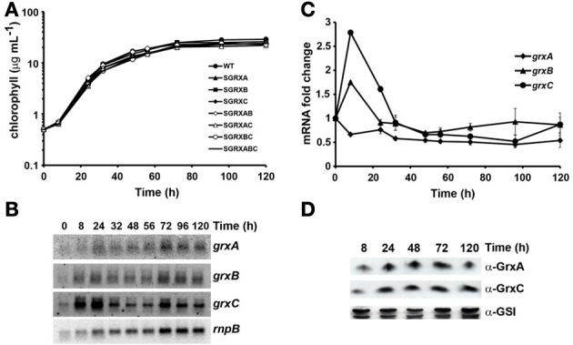
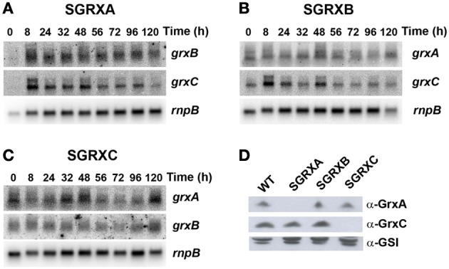
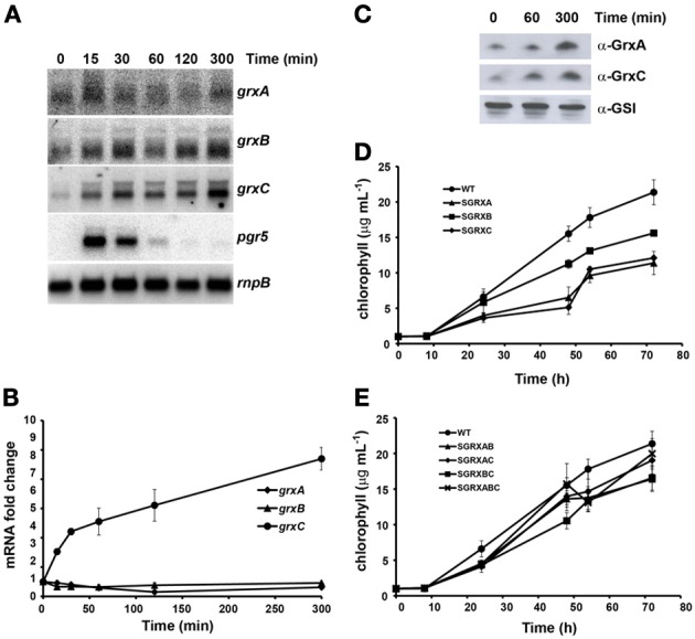
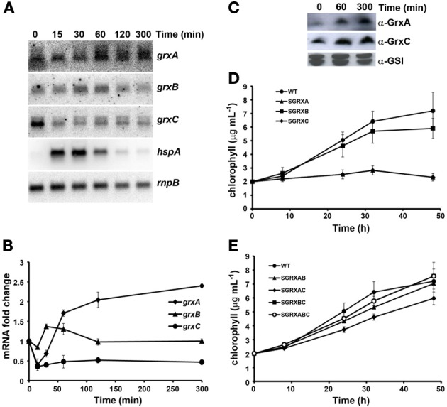
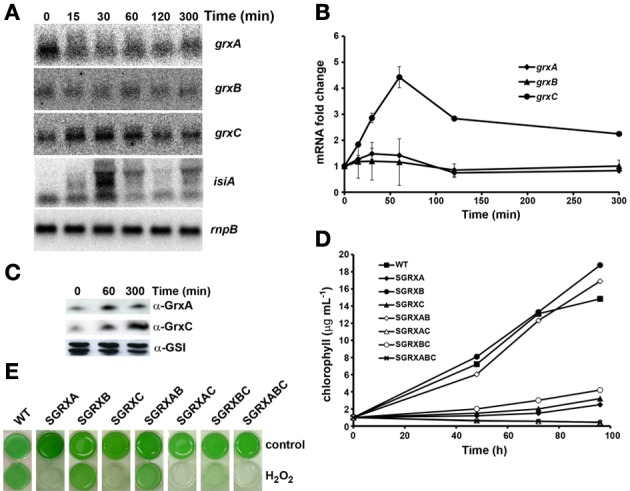
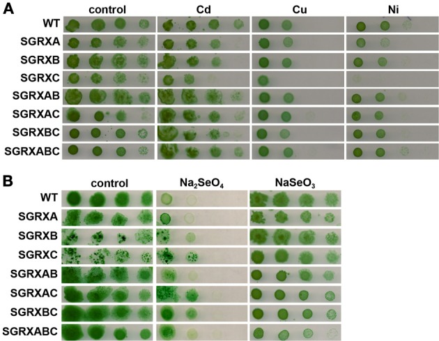
Glutaredoxins are small redox proteins able to reduce disulfides and mixed disulfides between GSH and proteins. Synechocystis sp. PCC 6803 contains three genes coding for glutaredoxins: ssr2061 (grxA) and slr1562 (grxB) code for dithiolic glutaredoxins while slr1846 (grxC) codes for a monothiolic glutaredoxin. We have analyzed the expression of these glutaredoxins in response to different stresses, such as high light, H2O2 and heat shock. Analysis of the mRNA levels showed that grxA is only induced by heat while grxC is repressed by heat shock and is induced by high light and H2O2. In contrast, grxB expression was maintained almost constant under all conditions. Analysis of GrxA and GrxC protein levels by western blot showed that GrxA increases in response to high light, heat or H2O2 while GrxC is only induced by high light and H2O2, in accordance with its mRNA levels. In addition, we have also generated mutants that have interrupted one, two, or three glutaredoxin genes. These mutants were viable and did not show any different phenotype from the WT under standard growth conditions. Nevertheless, analysis of these mutants under several stress conditions revealed that single grxA mutants grow slower after H2O2, heat and high light treatments, while mutants in grxB are indistinguishable from WT. grxC mutants were hypersensitive to treatments with H2O2, heat, high light and metals. A double grxAgrxC mutant was found to be even more sensitive to H2O2 than each corresponding single mutants. Surprisingly a mutation in grxB suppressed totally or partially the phenotypes of grxA and grxC mutants except the H2O2 sensitivity of the grxC mutant. This suggests that grxA and grxC participate in independent pathways while grxA and grxB participate in a common pathway for H2O2 resistance. The data presented here show that glutaredoxins are essential for stress adaptation in cyanobacteria, although their targets and mechanism of action remain unidentified.

Keywords: cyanobacteria; glutaredoxin; heat shock; high light; metal resistance; oxidative stress; redox regulation; stress.


Figures

Figure 1
Figure 1
Glutaredoxins are not essential under standard growth conditions. (A) Semi-logarithmic representation of growth of Synechocystis glutaredoxin mutants strains under standard conditions. WT (•), SGRXA (▲), SGRXB (■), SGRXC (♦), SGRXAB (◊), SGRXAC (Δ), SGRXBC (○), and SGRXABC (−) strains were inoculated at 0.5 μg chlorophyll mL−1 and growth was monitored by measuring chlorophyll content. (B) Northern blot analysis of grxA, grxB, and grxC expression along the growth curve. Total RNA was isolated from WT cells grown in BG11C at the indicated times. The filter was hybridized with grxA, grxB, and grxC probes and subsequently stripped and re-hybridized with an rnpB probe as a control. (C) Quantification of relative mRNA levels of grxA, grxB, and grxC during the growth curve. Radioactive signals were quantified and normalized to the rnpB signal. Plots of relative mRNA levels vs. time were drawn; data represent average of 3 independent experiments and error bars represent SE. grxA (♦), grxB (▲), and grxC (•). (D) Western blot analysis of GrxA and GrxC levels during the growth curve. WT cells were grown in BG11C medium and cells were harvested at the indicated times. Fifteen microgram of total protein from soluble extracts were separated by 15% SDS-PAGE and subjected to western blot to detect GrxA, GrxC, and GSI.
Figure 2
Figure 2
Glutaredoxin gene expression is not altered in single glutaredoxin mutants. (A) Northern blot analysis of grxB and grxC expression during different stages of the growth curve in SGRXA. Total RNA was isolated from SGRXA cells grown in BG11C at the indicated times. The filter was hybridized with grxB and grxC probes and subsequently stripped and re-hybridized with an rnpB probe as a control. (B) Northern blot analysis of grxA and grxC expression during different states of the growth curve in SGRXB. Total RNA was isolated from SGRXB cells grown in BG11C at the indicated times. The filter was hybridized with a grxA and grxC probes and subsequently stripped and re-hybridized with an rnpB probe as a control. (C) Northern blot analysis of grxA and grxB expression during different states of the growth curve in SGRXC. Total RNA was isolated from SGRXC cells grown in BG11C at the indicated times. The filter was hybridized with a grxA and grxB probes and subsequently stripped and re-hybridized with an rnpB probe as a control. (D) Western blot analysis of GrxA and GrxC levels in glutaredoxin mutants. WT, SGRXA, SGRXB, and SGRXC cells were grown in BG11C medium and cells were harvested at exponential phase (3–5 μg chl mL−1). Fifteen microgram of total protein from soluble extracts were separated by 15% SDS-PAGE and subjected to western blot to detect GrxA, GrxC, and GSI.
Figure 3
Figure 3
Effect of high light on glutaredoxin gene expression and growth. (A) Northern blot analysis of grxA, grxB, grxC, and pgr5 expression in response to a shift from 50 to 500 μE·m−2·s−1 light intensity. Total RNA was isolated from exponentially growing WT cells at the indicated times after the shift. The filter was hybridized with grxA, grxB, grxC, and pgr5 probes and subsequently stripped and re-hybridized with an rnpB probe as a control. (B) Quantification of relative mRNA levels of grxA, grxB, and grxC in response to a shift from 50 to 500 μE·m·s−1 light intensity. Radioactive signals were quantified and normalized to the rnpB signal. Plots of relative mRNA levels vs. time were drawn; data represent average of 3 independent experiments and error bars represent SE. grxA (♦), grxB (▲), and grxC (•). (C) Western blot analysis of GrxA and GrxC levels in response to a shift from 50 to 500 μE·m−2·s−1. WT cells were grown in BG11C medium and samples were collected at the indicated times after the shift. 15 μg of total protein from soluble extracts were separated by 15% SDS-PAGE and subjected to western blot to detect GrxA, GrxC, and GSI. (D) Growth of glutaredoxin single mutants strains after a shift to high light. WT (•), SGRXA (▲), SGRXB (■), SGRXC (♦) were grown until the exponential phase, diluted to 1 μg chlorophyll mL−1 and shifted to high light intensity. Growth was monitored by measuring chlorophyll content. (E) Growth of glutaredoxin double and triple mutants strains after a shift to high light. WT (•), SGRXAB (▲), SGRXAC (♦), SGRXBC (■), and SGRXABC (x) were grown until the exponential phase, diluted to 1 μg chlorophyll mL−1 and shifted to high light intensity. Growth was monitored by measuring chlorophyll content.
Figure 4
Figure 4
Effect of heat shock on glutaredoxin gene expression and growth. (A) Northern blot analysis of grxA, grxB, grxC, and hspA expression in response to heat shock. Total RNA was isolated from exponentially growing WT cells at the indicated times after the shift from 30 to 42°C. The filter was hybridized with grxA, grxB, grxC, and hspA probes and subsequently stripped and re-hybridized with an rnpB probe as a control. (B) Quantification of relative mRNA levels of grxA, grxB, and grxC in to response to heat shock. Radioactive signals were quantified and normalized to the rnpB signal. Plots of relative mRNA levels vs. time were drawn; data represent average of 3 independent experiments and error bars represent SE. grxA (♦), grxB (▲), and grxC (•). (C) Western blot analysis of GrxA and GrxC levels in response to heat shock. WT cells grown in BG11C and samples were collected at the indicated times after a shift from 30 to 42°C. Fifteen microgram of total protein from soluble extracts were separated by 15% SDS-PAGE and subjected to western blot to detect GrxA, GrxC, and GSI. (D) Growth of glutaredoxin single mutants strains after heat shock. WT (•), SGRXA (▲), SGRXB (■), and SGRXC (♦) were grown until the exponential phase, diluted to 2 μg chlorophyll mL−1 and shifted from 30 to 42°C. Growth was monitored by measuring chlorophyll content. (E) Growth of glutaredoxin double and triple mutant strains after heat shock. WT (•), SGRXAB (▲), SGRXAC (♦), SGRXBC (■), and SGRXABC (○) were grown until the exponential phase, diluted to 2 μg chlorophyll mL−1 and shifted from 30 to 42°C. Growth was monitored by measuring chlorophyll content.
Figure 5
Figure 5
Effect of H2O2 addition on glutaredoxin gene expression and growth. (A) Northern blot analysis of grxA, grxB, grxC, and isiA expression in response to H2O2 addition. Total RNA was isolated from WT cells grown in BG11C at the indicated times after a 1 mM H2O2 addition. The filter was hybridized with grxA, grxB, grxC, and isiA probes and subsequently stripped and re-hybridized with an rnpB probe as a control. (B) Quantification of relative mRNA levels of grxA, grxB, and grxC in response to H2O2 addition. Radioactive signals were quantified and normalized to the rnpB signal. Plots of relative mRNA levels vs. time were drawn; data represent average of 3 independent experiments and error bars represent SE. grxA (♦), grxB (▲), and grxC (•). (C) Western blot analysis of GrxA and GrxC levels in response to hydrogen peroxide. WT cells grown in BG11C and samples were collected at the indicated times after 1mM H2O2 addition. Fifteen microgram of total protein from soluble extracts were separated by 15% SDS-PAGE and subjected to western blot to detect GrxA, GrxC, and GSI. (D) Growth of glutaredoxin mutants strains after H2O2 addition. WT (■), SGRXA (♦), SGRXB (•), SGRXC(▲), SGRXAB (◊), SGRXAC (Δ), SGRXBC (○), and SGRXABC (x) were grown until the exponential phase, diluted to 1 μg chlorophyll mL−1 and 1 mM H2O2 was added. Growth was monitored by measuring chlorophyll content. (E) Photograph of glutaredoxin mutants grown in liquid BG11C or BG11C supplemented with 1 mM H2O2 for 5 days.
Figure 6
Figure 6
Effect of different metals on glutaredoxin mutants growth. (A) Sensitivity of glutaredoxin mutants to metals. Tolerance of WT, SGRXA, SGRXB, SGRXC, SGRXAB, SGRXAC, SGRXBC, and SGRXABC strains to cadmium, copper, and nickel was examined. Tenfold serial dilutions of a 1 μg chlorophyll mL−1 of exponentially growing cells suspension were spotted onto BG11C supplemented with 2 μM Cd, 3 μM Cu, and 5 μM Ni. Plates were photographed after 5 days of growth. (B) Sensitivity of glutaredoxin mutants to selenium. Tolerance of WT, SGRXA, SGRXB, SGRXC, SGRXAB, SGRXAC, SGRXBC, and SGRXABC strains to selenate (Na2SeO4) or selenite (Na2SeO3) was examined. Ten-fold serial dilutions of a 1 μg chlorophyll mL−1 of exponentially growing cells suspension were spotted onto BG11C supplemented with 30 μM Na2SeO3 or 30 μM Na2SeO4. Plates were photographed after 5 days of growth.

Similar articles

Cited by

References

    1. Allakhverdiev S. I., Nishiyama Y., Miyairi S., Yamamoto H., Inagaki N., Kanesaki Y., et al. (2002). Salt stress inhibits the repair of photodamaged photosystem II by suppressing the transcription and translation of psbA genes in synechocystis. Plant Physiol. 130, 1443–1453 10.1104/pp.011114 - DOI - PMC - PubMed
    1. Bandyopadhyay S., Gama F., Molina-Navarro M. M., Gualberto J. M., Claxton R., Naik S. G., et al. (2008). Chloroplast monothiol glutaredoxins as scaffold proteins for the assembly and delivery of [2Fe-2S] clusters. EMBO J. 27, 1122–1133 10.1038/emboj.2008.50 - DOI - PMC - PubMed
    1. Bedhomme M., Adamo M., Marchand C. H., Couturier J., Rouhier N., Lemaire S. D., et al. (2012). Glutathionylation of cytosolic glyceraldehyde-3-phosphate dehydrogenase from the model plant Arabidopsis thaliana is reversed by both glutaredoxins and thioredoxins in vitro. Biochem. J. 445, 337–347 10.1042/BJ20120505 - DOI - PubMed
    1. Belli G., Molina M. M., Garcia-Martinez J., Perez-Ortin J. E., Herrero E. (2004). Saccharomyces cerevisiae glutaredoxin 5-deficient cells subjected to continuous oxidizing conditions are affected in the expression of specific sets of genes. J. Biol. Chem. 279, 12386–12395 10.1074/jbc.M311879200 - DOI - PubMed
    1. Benyamina S. M., Baldacci-Cresp F., Couturier J., Chibani K., Hopkins J., Bekki A., et al. (2013). Two Sinorhizobium meliloti glutaredoxins regulate iron metabolism and symbiotic bacteroid differentiation. Environ. Microbiol. 15, 795–810 10.1111/j.1462-2920.2012.02835.x - DOI - PubMed