. 2013 Oct 24;8(10):e76520.
doi: 10.1371/journal.pone.0076520. eCollection 2013.

Extracellular vesicles derived from gut microbiota, especially Akkermansia muciniphila, protect the progression of dextran sulfate sodium-induced colitis

Affiliations

Extracellular vesicles derived from gut microbiota, especially Akkermansia muciniphila, protect the progression of dextran sulfate sodium-induced colitis

Chil-Sung Kang et al. PLoS One. .

Abstract

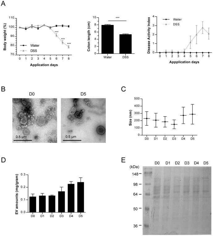
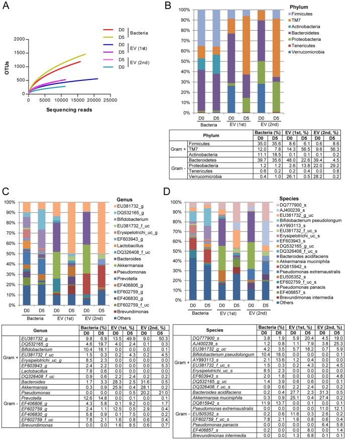
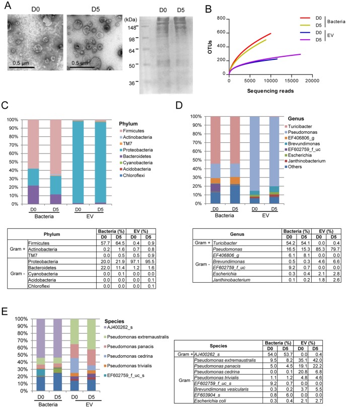
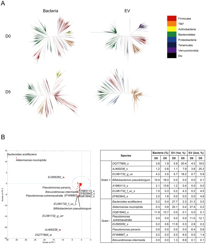
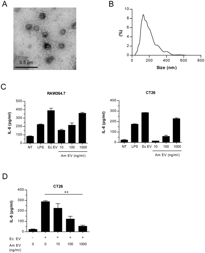
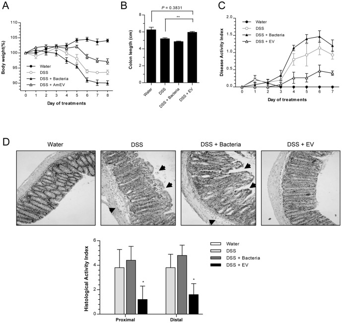
Gut microbiota play an important part in the pathogenesis of mucosal inflammation, such as inflammatory bowel disease (IBD). However, owing to the complexity of the gut microbiota, our understanding of the roles of commensal and pathogenic bacteria in the maintenance of immune homeostasis in the gut is evolving only slowly. Here, we evaluated the role of gut microbiota and their secreting extracellular vesicles (EV) in the development of mucosal inflammation in the gut. Experimental IBD model was established by oral application of dextran sulfate sodium (DSS) to C57BL/6 mice. The composition of gut microbiota and bacteria-derived EV in stools was evaluated by metagenome sequencing using bacterial common primer of 16S rDNA. Metagenomics in the IBD mouse model showed that the change in stool EV composition was more drastic, compared to the change of bacterial composition. Oral DSS application decreased the composition of EV from Akkermansia muciniphila and Bacteroides acidifaciens in stools, whereas increased EV from TM7 phylum, especially from species DQ777900_s and AJ400239_s. In vitro pretreatment of A. muciniphila-derived EV ameliorated the production of a pro-inflammatory cytokine IL-6 from colon epithelial cells induced by Escherichia coli EV. Additionally, oral application of A. muciniphila EV also protected DSS-induced IBD phenotypes, such as body weight loss, colon length, and inflammatory cell infiltration of colon wall. Our data provides insight into the role of gut microbiota-derived EV in regulation of intestinal immunity and homeostasis, and A. muciniphila-derived EV have protective effects in the development of DSS-induced colitis.


Conflict of interest statement

Competing Interests: The authors have declared that no competing interests exist.

Figures

Figure 1
Figure 1. Characterization of stool EV from a DSS-induced colitis mouse model.
For all figures, C57BL/6 mice (each group = 5) were ingested by water or 3% DSS. (A) Body weight changes (%, left panel), colon length (middle panel), and disease activity index including body weight, colon length, diarrhea, and stool blood (right panel). Water: water-administered group; DSS: 3% DSS-administered group; *, p<0.05; ***, p<0.001. (B) TEM images (×80 k) of EV isolated from stools on days 0 and 5. D0; stools before DSS application; D5: stools 5 days after 3% DSS application. (C) EV sizes (d.nm) measured by nanoparticle tracking analysis (NTA) on day 0 through day 5. (D) EV extraction amounts (protein concentration per gram stool) from stools D0 – D5. (E) Protein profiles in EV (D0 – D5) through SDS-PAGE by using coomassie brilliant blue G250 dye.
Figure 2
Figure 2. The composition of bacterial and bacteria-derived EV in stools following 3% DSS administration.
For all figures, stool bacteria and EV were isolated from mice before (D0) and 5 days (D5) after 3% DSS oral administration (each group = 5). As for EV metagenomics, two independent experiments were performed. (A) Operational taxonomic units (OTUs) using Roche 454 GS FLX Titanium. For figures (B)–(D), the proportion of stool bacteria and bacterial EV is displayed at the phylum (B), genus (C), and species (D) levels. As for genus and species, the proportion less than 1% occupancy is noted as others.
Figure 3
Figure 3. The composition of bacteria and bacteria-derived EV in small intestinal fluids following 3% DSS administration.
For all figures, bacteria and EV were isolated from small intestinal fluids of mice before (D0) and 5 days (D5) after 3% DSS oral administration (each group = 5). (A) EV TEM images (×100 k, left panel) and protein profiles through SDS-PAGE by using coomassie brilliant blue G250 dye (right panel). (B) Operational taxonomic units (OTUs) of bacteria and bacterial EV using Roche 454 GS FLX Titanium. For figures (C)–(E), the proportion of bacteria and bacterial EV in small intestinal fluids is displayed at the phylum (C), genus (D), and species (E) levels. As for genus and species, the proportion less than 1% occupancy is noted as others.
Figure 4
Figure 4. Change of stool bacteria and bacterial EV following 3% DSS administration.
For all figures, stool bacteria and EV were isolated from mice before (D0) and 5 days (D5) after 3% DSS oral administration (each group = 5). As for EV metagenomics, two independent experiments were performed. (A) Phylogenetic trees of stool bacteria and bacterial EV. (B) Principal component analysis (PCA) of bacterial EV (left panel) and changes of candidate bacteria and bacterial EV following 3% DSS administration (right panel). Blue circle means 95% confidence interval, and green cross means the starting point to diverge each species.
Figure 5
Figure 5. Characterization and immunogenicity of A. muciniphila-derived EV.
(A) A TEM image (×100 k) showing a spherical shape of A. muciniphila EV. (B) Size (d.nm) of A. muciniphila EV measured by NTA. (C) Levels of a pro-inflammatory cytokine IL-6. IL-6 was measured 12 h after A. muciniphila-derived EV treated to peritoneal macrophage cell line (Raw264.7, left panel) and colon epithelial cell line (CT26, right panel). LPS: lipopolysaccharide, 75 ng/ml, Ec EV: E. coli-derived EV, 100 ng/ml; Am EV: A. muciniphila–derived EV. (D) In vitro anti-inflammatory effect of A. muciniphila EV. IL-6 levels were measured in supernatants of colon epithelial cells (CT26) after A. muciniphila EV were pre-treated to CT26 for 12 h and then 100 ng/ml of E. coli EV treated for 12 h. **, p<0.01 by ANOVA and test of linearity.
Figure 6
Figure 6. In vivo protective effect of A. muciniphila-derived EV on the development of 2% DSS-induced colitis.
For all figures, mice (each group = 5) were ingested by A. muciniphila (Bacteria, 5.0×10⁸) or A. muciniphila-derived EV (Am EV, 100 µg), concomitantly with water or 2% DSS. (A) Body weight changes (%). (B) Colon lengths. **, p<0.01. (C) Changes of disease activity index, reflected by body weight, colon length, diarrhea, and stool blood. (D) Histology (upper panel) and histological activity index (lower panel) of colon. Arrows indicate injury area, and histological activity index was measured by epithelial barrier disruption and infiltration depth of inflammatory cells. *. p<0.05 vs. the DSS and DSS+Bacteria groups.

References

    1. Bouma G, Strober W (2003) The immunological and genetic basis of inflammatory bowel disease. Nat Rev Immunol 3: 521–533. - PubMed
    1. Round JL, Mazmanian SK (2009) The gut microbiota shapes intestinal immune responses during health and disease. Nat Rev Immunol 9: 313–323. - PMC - PubMed
    1. Xavier RJ, Podolsky DK (2007) Unravelling the pathogenesis of inflammatory bowel disease. Nature 448: 427–434. - PubMed
    1. Slack E, Hapfelmeier S, Stecher B, Velykoredko Y, Stoel M, et al. (2009) Innate and adaptive immunity cooperate flexibly to maintain host-microbiota mutualism. Science 325: 617–620. - PMC - PubMed
    1. Hooper LV, Gordon JI (2001) Commensal host-bacterial relationships in the gut. Science 292: 1115–1118. - PubMed

Publication types

Substances

LinkOut - more resources