

Neonatal pain-related stress predicts cortical thickness at age 7 years in children born very preterm
- PMID: 24204657
- PMCID: PMC3800011
- DOI: 10.1371/journal.pone.0076702
Neonatal pain-related stress predicts cortical thickness at age 7 years in children born very preterm
Abstract
Background: Altered brain development is evident in children born very preterm (24-32 weeks gestational age), including reduction in gray and white matter volumes, and thinner cortex, from infancy to adolescence compared to term-born peers. However, many questions remain regarding the etiology. Infants born very preterm are exposed to repeated procedural pain-related stress during a period of very rapid brain development. In this vulnerable population, we have previously found that neonatal pain-related stress is associated with atypical brain development from birth to term-equivalent age. Our present aim was to evaluate whether neonatal pain-related stress (adjusted for clinical confounders of prematurity) is associated with altered cortical thickness in very preterm children at school age.
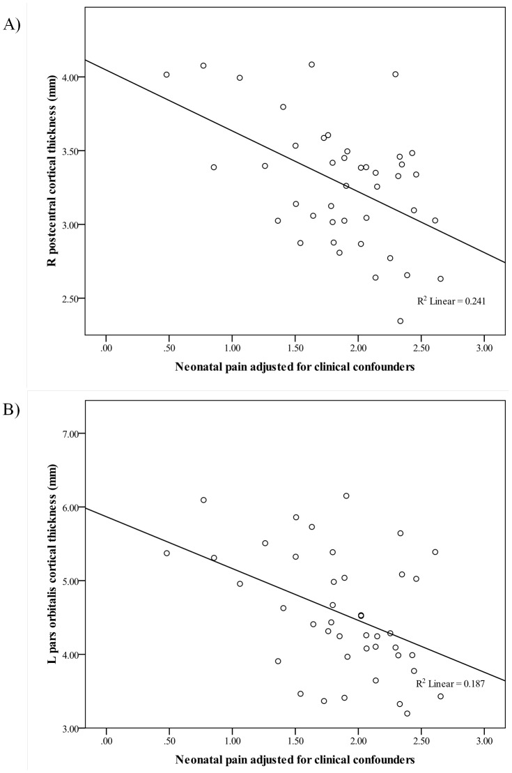
Methods: 42 right-handed children born very preterm (24-32 weeks gestational age) followed longitudinally from birth underwent 3-D T1 MRI neuroimaging at mean age 7.9 yrs. Children with severe brain injury and major motor/sensory/cognitive impairment were excluded. Regional cortical thickness was calculated using custom developed software utilizing FreeSurfer segmentation data. The association between neonatal pain-related stress (defined as the number of skin-breaking procedures) accounting for clinical confounders (gestational age, illness severity, infection, mechanical ventilation, surgeries, and morphine exposure), was examined in relation to cortical thickness using constrained principal component analysis followed by generalized linear modeling.
Results: After correcting for multiple comparisons and adjusting for neonatal clinical factors, greater neonatal pain-related stress was associated with significantly thinner cortex in 21/66 cerebral regions (p-values ranged from 0.00001 to 0.014), predominately in the frontal and parietal lobes.
Conclusions: In very preterm children without major sensory, motor or cognitive impairments, neonatal pain-related stress appears to be associated with thinner cortex in multiple regions at school age, independent of other neonatal risk factors.
Conflict of interest statement
Figures
References
-
- Miller SP, Vigneron DB, Henry RG, Bohland MA, Ceppi-Cozzio C, et al. (2002) Serial quantitative diffusion tensor MRI of the premature brain: development in newborns with and without injury. J Magn Reson Imaging 16: 621–632. - PubMed
Publication types
MeSH terms
Grants and funding
LinkOut - more resources
Full Text Sources
Other Literature Sources
Medical
