. 2013 Oct 29;8(10):e76973.
doi: 10.1371/journal.pone.0076973. eCollection 2013.

Essential role of the m2R-RGS6-IKACh pathway in controlling intrinsic heart rate variability

Affiliations

Essential role of the m2R-RGS6-IKACh pathway in controlling intrinsic heart rate variability

Ekaterina Posokhova et al. PLoS One. .

Abstract

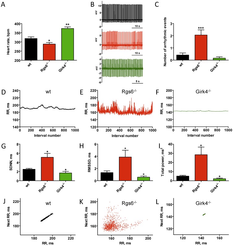
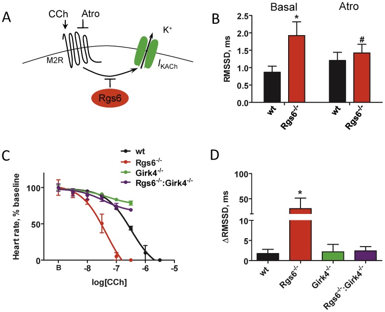
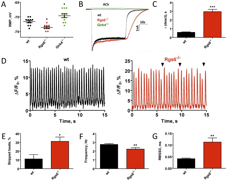
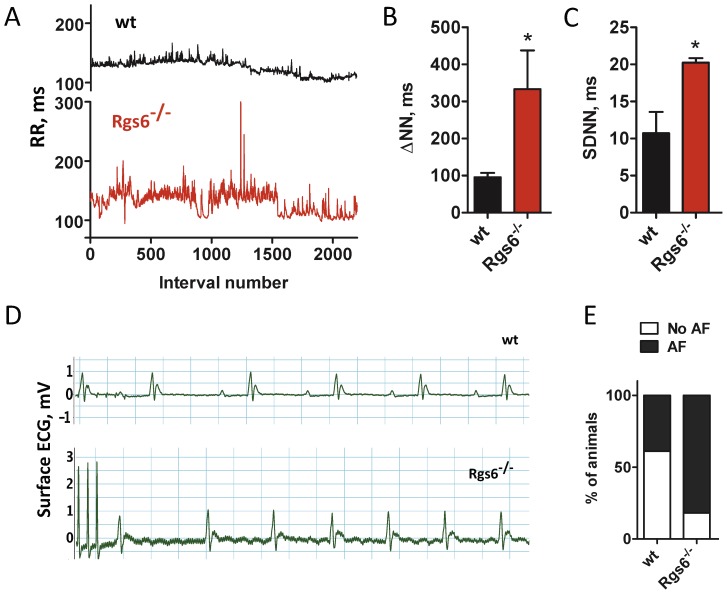
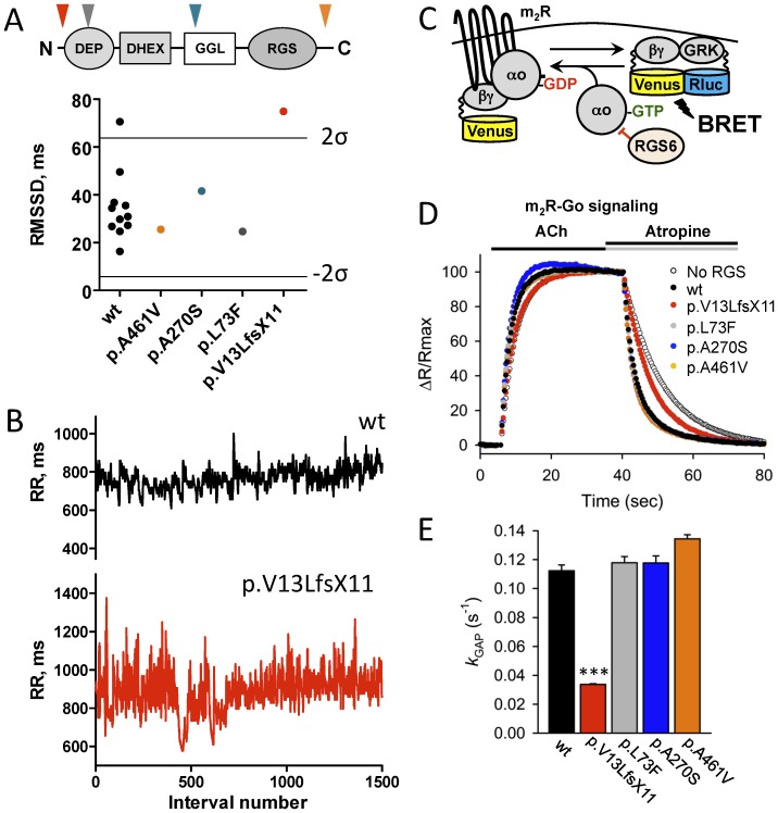
Normal heart function requires generation of a regular rhythm by sinoatrial pacemaker cells and the alteration of this spontaneous heart rate by the autonomic input to match physiological demand. However, the molecular mechanisms that ensure consistent periodicity of cardiac contractions and fine tuning of this process by autonomic system are not completely understood. Here we examined the contribution of the m2R-I(KACh) intracellular signaling pathway, which mediates the negative chronotropic effect of parasympathetic stimulation, to the regulation of the cardiac pacemaking rhythm. Using isolated heart preparations and single-cell recordings we show that the m2R-I(KACh) signaling pathway controls the excitability and firing pattern of the sinoatrial cardiomyocytes and determines variability of cardiac rhythm in a manner independent from the autonomic input. Ablation of the major regulator of this pathway, Rgs6, in mice results in irregular cardiac rhythmicity and increases susceptibility to atrial fibrillation. We further identify several human subjects with variants in the RGS6 gene and show that the loss of function in RGS6 correlates with increased heart rate variability. These findings identify the essential role of the m2R-I(KACh) signaling pathway in the regulation of cardiac sinus rhythm and implicate RGS6 in arrhythmia pathogenesis.


Conflict of interest statement

Competing Interests: The authors have declared that no competing interests exist.

Figures

Figure 1
Figure 1. Rgs6 and Girk4 have opposite effects on HRV in isolated hearts.
A, Average HR in hearts isolated from wild-type (wt, n = 36), Rgs6 −/− (n = 52), and Girk4 −/− (n = 19) mice. B, ECG traces recorded in isolated wild-type (black), Rgs6−/− (red), and Girk4−/− (green) hearts. Note rhythm irregularity in Rgs6 −/− hearts. C, Quantification of sinoatrial dysfunction events. D–F, Representative tachograms of baseline ECG in wild-type (black), Rgs6 −/− (red), and Girk4 −/− (green) hearts. G–I, Key HRV parameters in the time and frequency domains from ECG recordings. J–L, Non-linear HRV analysis by Poincare plots for wild-type (J), Rgs6−/− (K), and Girk4−/− (L) hearts. Symbols: * P<0.05, ** P<0.01, ***P<0.001 vs. wild-type.
Figure 2
Figure 2. The effects of the Rgs6 on HRV are mediated by the IKACh and are influenced by the m2R activity.
A, Schematic representation of the pathway targeted both genetically and pharmacologically. Abbreviations are: atropine (Atro), carbamylcholine (CCh). B, Effect of m2R blockade by atropine on HRV in wild-type (black; n = 7) and Rgs6−/− hearts (red; n = 10). No significant effect of drug was observed in wild-type hearts. C, Increased sensitivity of Rgs6−/− hearts to m2R stimulation and its rescue by IKACh (Girk4) ablation. Increasing concentrations of CCh were applied to isolated perfused hearts (n = 4–6 per genotype). D, m2R stimulation non-proportionately increased HRV in Rgs6−/− hearts. Hearts (n = 3–6 per genotype) were perfused with CCh (∼IC10 concentration) identified from dose-response studies, followed by measurement of changes in the RMSSD parameters. Symbols: * P<0.05 vs wild-type, #P<0.05 vs treatment.
Figure 3
Figure 3. Ablation of Rgs6 reduces excitability of sinoatrial cells and disrupts their automaticity.
A, Resting membrane potential measured immediately after obtaining whole-cell access in wild-type (wt), Rgs6−/−, and Girk4−/− SAN cells. B, Inward currents evoked by application of acetylcholine (ACh, 100 µM) in SAN cells from wild-type (black), Rgs6−/− (red) and Girk4−/− (green, no current) mice. C, Summary of steady-state ACh-induced deactivation kinetics of IKACh in wild-type and Rgs6−/− SAN cells (n = 11–15 cells/genotype). D, Representative traces of spontaneous calcium oscillations recorded from wild-type (black; n = 14) and Rgs6−/− (red, n = 20) SAN cells. Arrows show skipped beats. E, Quantification of SAN arrhythmic events defined as more than 15% change in duration of peak-to-peak interval of spontaneous calcium oscillations in wild-type (n = 11) and Rgs6−/− (n = 17) SAN cells. F, Reduced frequency of spontaneous calcium oscillations recorded in Rgs6−/− SAN cardiomyocytes as compared to wild-type (n = 14–20 cells/genotype). G, Increased variability of spontaneous calcium oscillations in Rgs6−/− SAN cells as determined by increase in RMSSD values (n = 14–20 cells per genotype). Symbols: *P<0.05; **P<0.01; ***P<0.001.
Figure 4
Figure 4. Inactivation of Rgs6 disrupts cardiac rhythm in mice.
A, Representative tachograms of RR intervals from wild-type (black) and Rgs6−/− (red) mice recorded by ECG radiotelemetry. B and C, Summary of HRV analysis in conscious, freely-moving mice. D, Burst pacing induced AF in Rgs6−/− but not in wild-type mice. Note an irregular rhythm with no discernible P waves in the Rgs6−/− recording. E, Quantification of AF induction probability. Symbols: *, P<0.05.
Figure 5
Figure 5. Abnormal sinus arrhythmia in a human subject with dysfunctional RGS6.
A, HRV measured in humans carrying variants in RGS6 and 11 age-matched control subjects (wt, black). Lines represent upper (2σ) and lower (−2 σ) 95% confidence thresholds as determined by the 2σ rule. Insert: domain structure of RGS6 protein. Arrows show localization of corresponding variants. B, Representative tachograms of RR intervals from a control subject (black) and a subject heterozygous for the p.Val13LeufsX11 variant in the RGS6 gene (red) determined from continuous Holter recordings. C, Schematics of the assay design to study effects of mutations on the RGS6 function. Stimulation of the m2R by ACh results in the dissociation of Gμo from the heterotrimer. Released Gβγ subunits tagged with Venus become available for the interaction with Nluc8-tagged GRK reporter producing the BRET signal. D. Representative responses to sequential application of ACh (10 µM) and atropine (1 mM) recorded in the presence of the indicated constructs. The BRET signals averaged from 4 experiments were plotted as individual data points. E, Catalytic activity of RGS6 defined by the k GAP parameter. To determine the k GAP values, the deactivation rate constant measured in the absence of RGS6 was subtracted from values measured in the presence of RGS6. Symbols: ***, p<0.001 (n = 4).

References

    1. Podrid PJ, Kowey PR (2001) Cardiac Arrhythmia: Mechanisms, Diagnosis, and Management: Lippincott Williams & Wilkins. 973 p.
    1. Mangoni ME, Nargeot J (2008) Genesis and regulation of the heart automaticity. Physiol Rev 88: 919–982. - PubMed
    1. Akselrod S, Gordon D, Ubel FA, Shannon DC, Berger AC, et al. (1981) Power spectrum analysis of heart rate fluctuation: a quantitative probe of beat-to-beat cardiovascular control. Science 213: 220–222. - PubMed
    1. Lombardi F, Stein PK (2011) Origin of heart rate variability and turbulence: an appraisal of autonomic modulation of cardiovascular function. Front Physiol 2: 95. - PMC - PubMed
    1. Gehrmann J, Hammer PE, Maguire CT, Wakimoto H, Triedman JK, et al. (2000) Phenotypic screening for heart rate variability in the mouse. Am J Physiol Heart Circ Physiol 279: H733–740. - PubMed

Publication types

MeSH terms

Substances