. 2013 Oct 16;8(10):e77262.
doi: 10.1371/journal.pone.0077262. eCollection 2013.

Effect of Iboga alkaloids on µ-opioid receptor-coupled G protein activation

Affiliations

Effect of Iboga alkaloids on µ-opioid receptor-coupled G protein activation

Tamara Antonio et al. PLoS One. .

Abstract

Objective: The iboga alkaloids are a class of small molecules defined structurally on the basis of a common ibogamine skeleton, some of which modify opioid withdrawal and drug self-administration in humans and preclinical models. These compounds may represent an innovative approach to neurobiological investigation and development of addiction pharmacotherapy. In particular, the use of the prototypic iboga alkaloid ibogaine for opioid detoxification in humans raises the question of whether its effect is mediated by an opioid agonist action, or if it represents alternative and possibly novel mechanism of action. The aim of this study was to independently replicate and extend evidence regarding the activation of μ-opioid receptor (MOR)-related G proteins by iboga alkaloids.

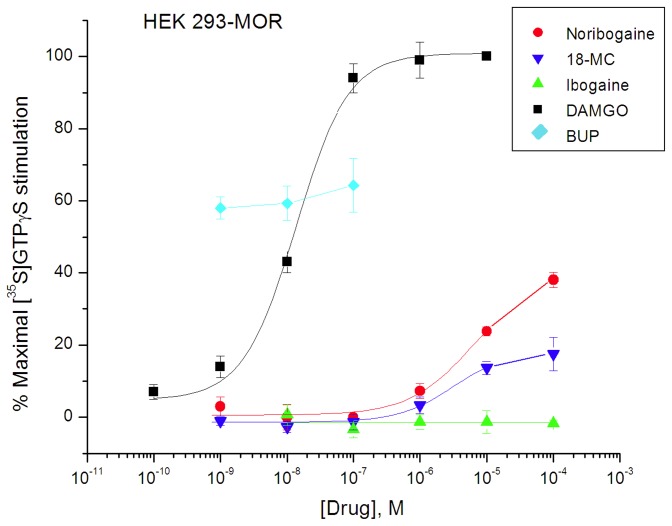
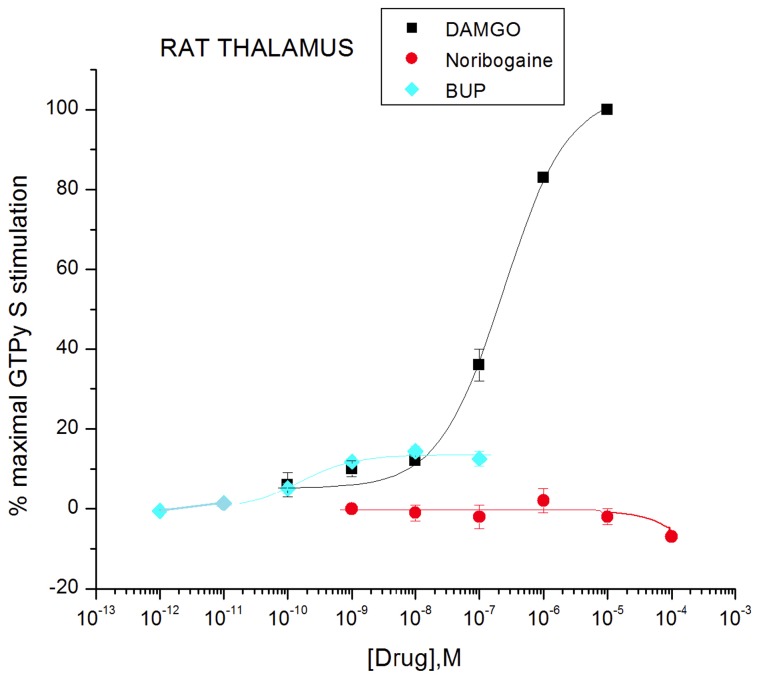
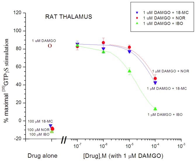
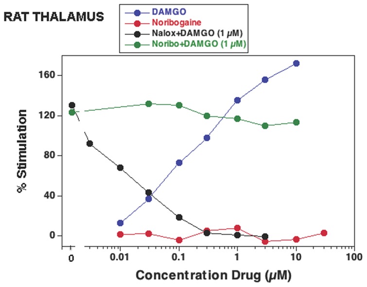
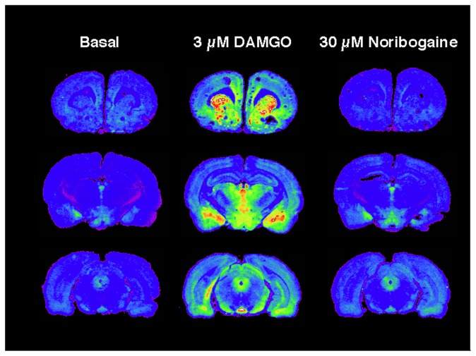
Methods: Ibogaine, its major metabolite noribogaine, and 18-methoxycoronaridine (18-MC), a synthetic congener, were evaluated by agonist-stimulated guanosine-5´-O-(γ-thio)-triphosphate ([(35)S]GTPγS) binding in cells overexpressing the recombinant MOR, in rat thalamic membranes, and autoradiography in rat brain slices.

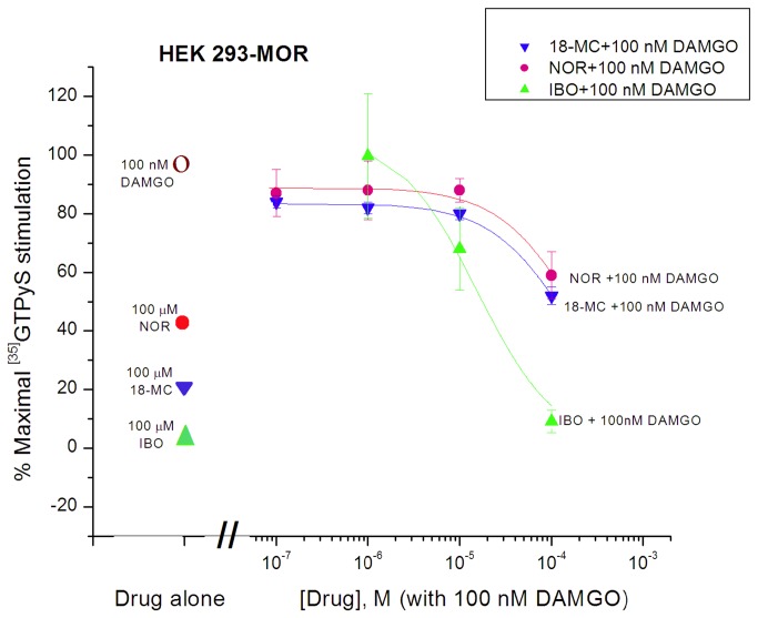
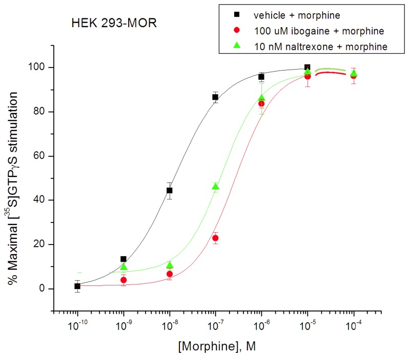
Results and significance: In rat thalamic membranes ibogaine, noribogaine and 18-MC were MOR antagonists with functional Ke values ranging from 3 uM (ibogaine) to 13 uM (noribogaine and 18MC). Noribogaine and 18-MC did not stimulate [(35)S]GTPγS binding in Chinese hamster ovary cells expressing human or rat MORs, and had only limited partial agonist effects in human embryonic kidney cells expressing mouse MORs. Ibogaine did not did not stimulate [(35)S]GTPγS binding in any MOR expressing cells. Noribogaine did not stimulate [(35)S]GTPγS binding in brain slices using autoradiography. An MOR agonist action does not appear to account for the effect of these iboga alkaloids on opioid withdrawal. Taken together with existing evidence that their mechanism of action also differs from that of other non-opioids with clinical effects on opioid tolerance and withdrawal, these findings suggest a novel mechanism of action, and further justify the search for alternative targets of iboga alkaloids.


Conflict of interest statement

Competing Interests: The authors have declared that no competing interests exist.

Figures

Figure 1
Figure 1. Structures of the iboga alkaloid ibogamine parent skeleton and ibogaine, noribogaine, and 18-MC.
Figure 2
Figure 2. Effect of ibogaine, noribogaine, and 18-MC on [35S]GTPγS binding in HEK 293-mMOR cells compared with full agonist DAMGO and partial agonist buprenorphine (BUP) (Reith lab).
Cell suspension aliquots were incubated with indicated drug for 15 min and subsequently with an additional concentration of 0.08 nM of [35S]GTPγS at 30°C. Data are expressed as % of maximal stimulation by 10 µM DAMGO and presented as mean ± SEM (vertical bar) for 3 - 4 independent experiments assayed in triplicate.
Figure 3
Figure 3. Antagonism of DAMGO (100 nM)-induced [35S]GTPγS binding in HEK 293-mMOR cells by ibogaine, noribogaine, and 18-MC (Reith lab).
Degree of stimulation by drug alone, i.e.,100 nM DAMGO, 100 µM noribogaine (NOR), 100 µM 18-MC, or 100 µM ibogaine (IBO) is indicated by symbols on the left. The colored curves represent the effect of increasing the concentrations of ibogaine, noribogaine, or 18-MC co-incubated with 100 nM DAMGO. Otherwise as in Figure 2.
Figure 4
Figure 4. Rightward shift in morphine curves for stimulation of [35S]GTPγS binding in HEK 293-mMOR cells by ibogaine (100 µM) or naltrexone (10 nM) (Reith lab).
The indicated fixed concentration of ibogaine was co-present with increasing concentrations of morphine (colored curves). Otherwise as in Figure 2 .
Figure 5
Figure 5. Effect of ibogaine and 18-MC on [35S]GTPγS binding in Sprague-Dawley rat thalamic membranes compared with DAMGO (Reith lab).
Tissue suspension aliquots were incubated with indicated drug and 0.09 nM of [35S]GTPγS for 1 h at 30°C. Data are expressed as % of maximal stimulation by 10 µM DAMGO and presented as mean ± SEM (vertical bar) for 3 - 4 independent experiments assayed in triplicate.
Figure 6
Figure 6. Effect of noribogaine on [35S]GTPγS binding in Sprague-Dawley rat thalamic membranes compared with DAMGO and buprenorphine (BUP) (Reith lab).
Otherwise as in Figure 5.
Figure 7
Figure 7. Antagonism of DAMGO (1 µM)-induced [35S]GTPγS binding in Sprague-Dawley rat thalamic membranes by ibogaine, noribogaine, and 18-MC (Reith lab).
Degree of stimulation by drug alone, i.e., 1 µM DAMGO, 100 µM 18-MC, 100 µM noribogaine (NOR), or 100 µM ibogaine (IBO) is indicated by the symbols on the left. The colored curves represent the effect of increasing the concentration of the respective iboga alkaloids co-incubated with 1 µM DAMGO (5 independent experiments). Otherwise as in Figures 5 and 6.
Figure 8
Figure 8. Effect on [35S]GTPγS binding of noribogaine (Noribo) by itself and in combination with DAMGO (1 µM) in Sprague-Dawley rat thalamic membranes (Childers lab).
Tissue suspension aliquots were incubated with the indicated drug and 0.05 nM of [35S]GTPγS for 2 h at 30°C. DAMGO and naloxone (Nalox) were used as controls. Data are expressed as % of baseline and are from a representative experiment performed three times.
Figure 9
Figure 9. Effect of noribogaine (30 µM) compared with DAMGO (3 µM) on [35S]GTPγS binding measured by autoradiography in brain slices from Sprague-Dawley rats (Childers lab).
Coronal sections were incubated with 0.04 nM of [35S]GTPγS for 2 h at 30°C with or without 3 µM DAMGO or 30 µM noribogaine. Basal binding was deducted to obtain net agonist-stimulated binding. Results shown are from a representative experiment, carried out three times in sections from three individual rats.

References

    1. Bartlett MF, Dickel DF, Taylor WI (1958) The alkaloids of Tabernanthe-Iboga. Part VI. The Structures of ibogamine, ibogaine, tabernanthine and voacangine. J Am Chem Soc 80: 126-136. doi:10.1021/ja01534a036. - DOI
    1. Jana GK, Paul S, Sinha S (2011) Progress in the synthesis of iboga-alkaloids and their congeners. Org Prep Proced Int 43: 541-573. doi:10.1080/00304948.2011.629563. - DOI
    1. Fernandez JW (1982) Bwiti: An Ethnography of Religious Imagination in Africa. Princeton, New Jersey: Princeton University Press. 731 pp.
    1. Alper KR, Lotsof HS, Kaplan CD (2008) The ibogaine medical subculture. J Ethnopharmacol 115: 9-24. doi:10.1016/j.jep.2007.08.034. PubMed: 18029124. - DOI - PubMed
    1. Alper KR, Lotsof HS, Frenken GM, Luciano DJ, Bastiaans J (1999) Treatment of acute opioid withdrawal with ibogaine. Am J Addict 8: 234-242. doi:10.1080/105504999305848. PubMed: 10506904. - DOI - PubMed

Publication types

MeSH terms